. 2013 Jan;65(1):236-46.
doi: 10.1002/art.37706.

Interleukin-13-producing CD8+ T cells mediate dermal fibrosis in patients with systemic sclerosis

Affiliations

Interleukin-13-producing CD8+ T cells mediate dermal fibrosis in patients with systemic sclerosis

Patrizia Fuschiotti et al. Arthritis Rheum. 2013 Jan.

Abstract

Objective: Fibrosis is a major contributor to morbidity and mortality in systemic sclerosis (SSc). T cells are the predominant inflammatory infiltrate in affected tissue and are thought to produce cytokines that drive the synthesis of extracellular matrix (ECM) proteins by fibroblasts, resulting in excessive fibrosis. We have previously shown that aberrant interleukin-13 (IL-13) production by peripheral blood effector CD8+ T cells from SSc patients correlates with the extent of skin fibrosis. The present study was undertaken to investigate the role of IL-13 production by CD8+ T cells in dermal fibrosis, an early and specific manifestation of SSc.

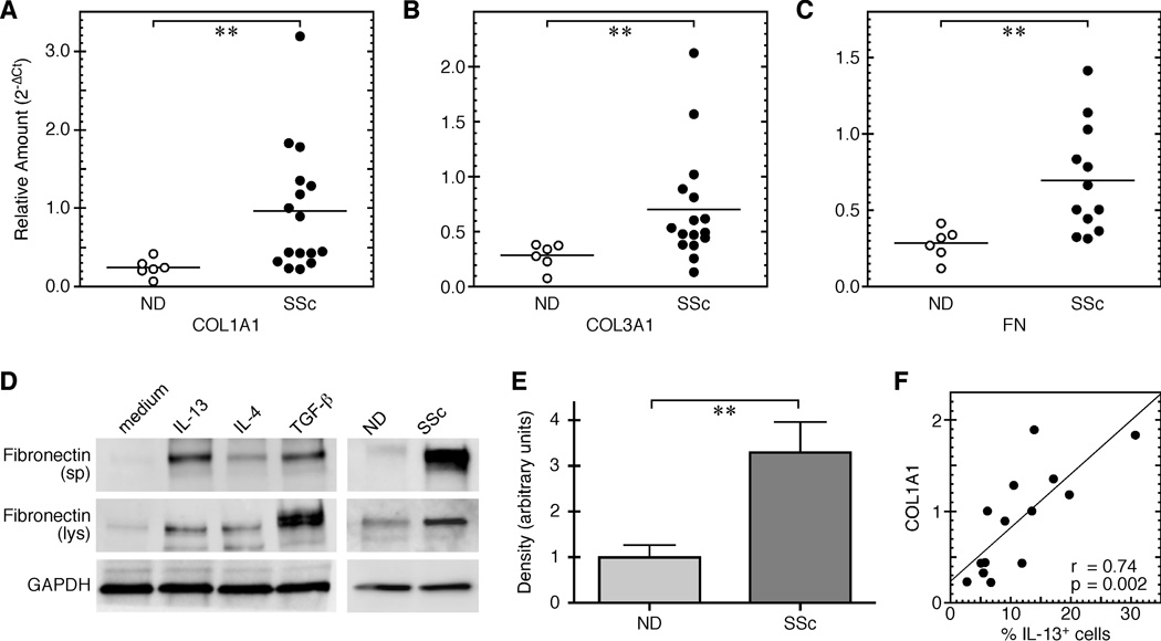
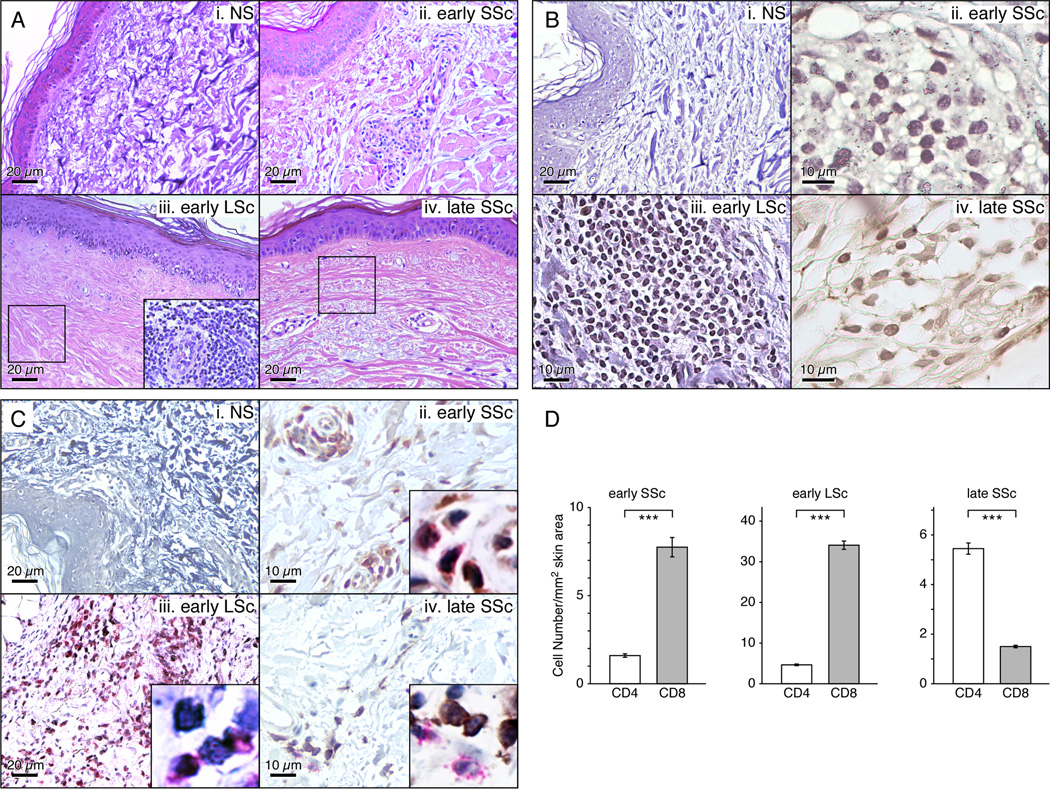
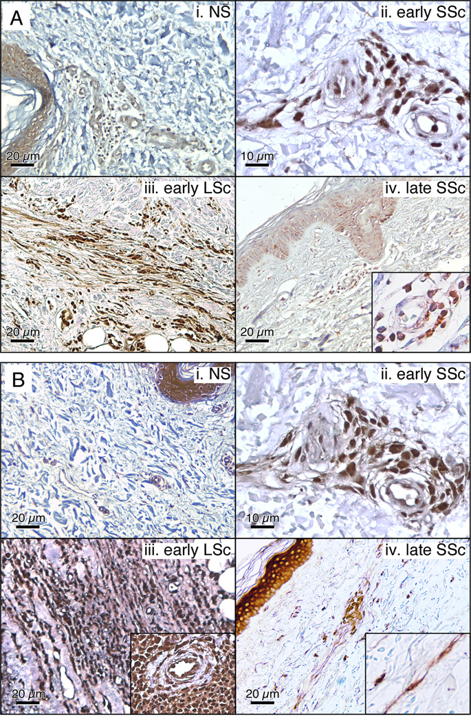
Methods: ECM protein production by normal dermal fibroblasts cocultured with SSc CD8+ T cell supernatants was determined by quantitative polymerase chain reaction and Western blotting. Skin-homing receptor expression and IL-13 production by CD8+ T cells in the peripheral blood of SSc patients were measured by flow cytometry. IL-13+ and CD8+ cells in sclerotic skin were identified by immunohistochemistry.

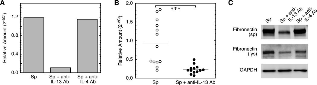
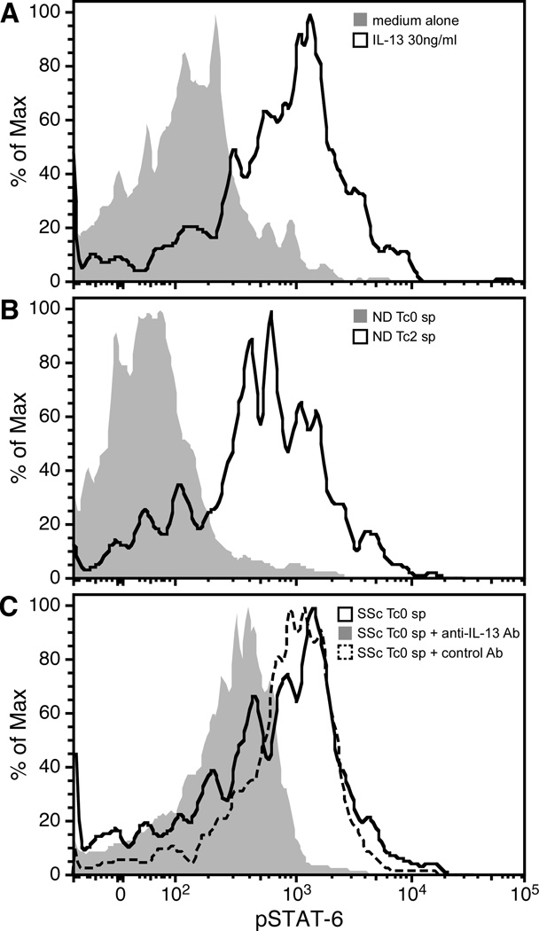
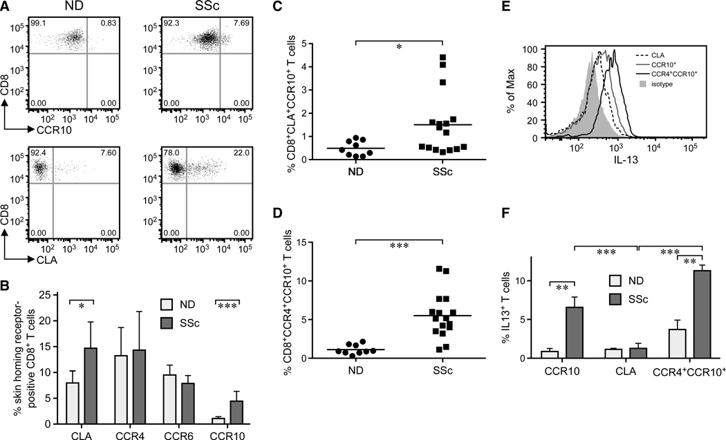
Results: IL-13-producing circulating CD8+ T cells from patients with SSc expressed skin-homing receptors and induced a profibrotic phenotype in normal dermal fibroblasts, which was inhibited by an anti-IL-13 antibody. High numbers of CD8+ T cells and IL-13+ cells were found in the skin lesions of SSc patients, particularly during the early inflammatory phase of the disease.

Conclusion: These findings show that IL-13-producing CD8+ T cells are directly involved in modulating dermal fibrosis in SSc. The demonstration that CD8+ T cells homing to the skin early in the course of SSc are associated with accumulation of IL-13 is an important mechanistic contribution to the understanding of the pathogenesis of dermal fibrosis in SSc and may represent a potential target for therapeutic intervention.


Conflict of interest statement

The authors declare that no competing financial interests or conflicts exist.

Figures

Figure 1
Figure 1. CD8+ T-cell-supernatant from SSc patients induces extracellular matrix protein (ECM) production by normal dermal fibroblasts in vitro
ECM production was determined in normal dermal fibroblasts co-cultured for 24 hours with CD8+ T-cell supernatants from SSc patients and normal donors (ND). Each symbol represents one patient or ND. Fibroblast mRNA expression of (A) COL1A1, SSc/ND number=16/6, (B) COL3A1, n=16/6, and (C) FN, n=12/6, was quantified by qPCR. Data were normalized by GAPDH. A horizontal line indicates mean response. The Mann-Whitney test was used for significance. (D) Representative Western blot for FN protein expression in fibroblast culture media (sp) and fibroblast lysates (lys) following incubation with CD8+ T-cell supernatants from one SSc patient and one ND (right panel) out of six of each tested. Fibroblasts stimulated with IL-13, IL-4, or TGF-β were used as positive controls (left panel). GAPDH protein was a loading control in lys. (E) Densitometric quantification of secreted FN protein measured as in (D). The difference between the means of 13 SSc CD8+ T-cell supernatants and 12 from NDs is significant (P=0.001; Mann-Whitney test). (F) IL-13 production by SSc CD8+ T cells correlates with COL1A1 mRNA induction in fibroblasts (n=14, Spearman’s rank correlation coefficient, P= 0.002).
Figure 2
Figure 2. An anti-IL-13 antibody inhibits COL1A1 production by CD8+ T-cell-supernatant from SSc patients in normal dermal fibroblasts in vitro
COL1A1 mRNA levels were determined in fibroblasts following 24-hour stimulation with CD8+ T cell supernatants from SSc patients with or without pre-incubation with a neutralizing anti-IL-13 antibody or a control anti-IL-4-antibody. The number of transcripts was normalized to human GAPDH. A representative example (A) and multiple CD8+ T-cell supernatants (B, n=13, P=0.0003 by Mann-Whitney test) are shown. (C) Detection of FN in culture supernatants or cell lysates by Western blot in dermal fibroblasts cultured as described in (A). A representative experiment out of five independent experiments from different SSc patients is shown.
Figure 3
Figure 3. STAT-6 phosphorylation (pSTAT-6) is induced in normal dermal fibroblasts after incubation with CD8+ T-cell supernatants from SSc patients
Normal dermal fibroblasts, serum starved for 18 hours, were co-cultured for 24 hrs with CD8+ T-cell supernatants from SSc and NDs as described above and pSTAT-6 in the fibroblasts was determined by intracellular staining as described in the Material and Methods section. A representative example out of five independent experiments giving similar results. (A) pSTAT-6 determination in fibroblasts incubated with medium alone or IL-13 (30ng/ml). (B) CD8+ T cells from one normal donor were cultured in Tc0 or Tc2 culture conditions as previously described . The supernatants were collected and added to the fibroblasts. pSTAT-6 was determined after 24 hours. (C) Co-culture of one SSc CD8+ T-cell supernatant untreated (black line) or pre-incubated with an anti-IL-13 antibody (grey shade) or an unrelated control antibody (dot line) with skin fibroblasts.
Figure 4
Figure 4. Skin-homing receptor expression by peripheral blood CD8+ T cells from SSc patients and age-matched NDs
Freshly isolated CD8+ T cells were stained by multicolor flow cytometry for skin-homing molecules CLA, CCR4, CCR6 and CCR10. Lymphocyte population was gated according to light scatter characteristics (FSC/SSC) and CD8+ T cells identified for CD8 and CD3 positivity. Each symbol represents one patient or ND. (A) Two-color plots of CCR10 or CLA versus CD8 from one SSc patient and one ND. (B) Frequencies of skin-homing receptor-positive CD8+ T cells show a higher percentage of CD8+CLA+ T cells in SSc patients (n=16) compared to ND (n=9) (P=0.031) and a higher frequency of CCR10+ T cells (P<0.0001). Statistics by the unpaired two-tailed Student’s t test. Values represent the mean percentage of positive cells±SD. A higher proportion of CD8+CCR10+ cells in SSc patients co-express CLA (C) and CCR4 (D) compared to controls (P=0.03 and P<0.0001, respectively: Mann-Whitney test). A horizontal line indicates mean response. (E, F) IL-13 production by skin-homing SSc CD8+ T cell subsets. (E) A representative example, and (F) IL-13 frequency in multiple SSc patients (n=10, P=0.002: Kruskal-Wallis test). IL-13 production was determined by ICC . Mean percentage of positive cells±SD is shown.
Figure 5
Figure 5. Immunohistochemical localization of IL-13 and CD8+ T cells in scleroderma skin biopsies
(A) Comparative histological analysis of skin samples from normal donors and early or late scleroderma patients (H&E, 200X). IL-13 expression (B), cell infiltrate characterization (C) in normal skin and samples of patients with early or late scleroderma. (B) is peroxidase anti-peroxidase (brown stain) and (C) is peroxidase and alkaline phosphatase staining identifying CD4+ (black LSc (200X) and brown early and late SSc (400X)) and CD8+ (red) T cells, respectively. Insets 1000X. (D) Quantification of the cellular infiltrate in tissue sections by microscopy is expressed as mean±SD of cell numbers/mm2 skin area. Statistics by Student’s t test.
Figure 6
Figure 6. Immunohistochemical localization of IL-13 receptors in scleroderma skin biopsies
IL-13Rα1 (A) and IL-13Rα2 (B) expression in normal skin and samples of patients with early or late scleroderma, using peroxidase anti-peroxidase (brown stain, 200X). Insets 500X.

Similar articles

Cited by

References

    1. Gabrielli A, Avvedimento EV, Krieg T. Scleroderma. The New England journal of medicine. 2009;360:1989–2003. - PubMed
    1. Varga J, Abraham D. Systemic sclerosis: a prototypic multisystem fibrotic disorder. J Clin Invest. 2007;117:557–567. - PMC - PubMed
    1. Jinnin M. Mechanisms of skin fibrosis in systemic sclerosis. J Dermatol. 2010;37:11–25. - PubMed
    1. Roumm AD, Whiteside TL, Medsger TA, Jr, Rodnan GP. Lymphocytes in the skin of patients with progressive systemic sclerosis. Quantification, subtyping, and clinical correlations. Arthritis Rheum. 1984;27:645–653. - PubMed
    1. Fleischmajer R, Perlish JS, Reeves JR. Cellular infiltrates in scleroderma skin. Arthritis Rheum. 1977;20:975–984. - PubMed

Publication types

MeSH terms