. 2013 Jan;65(1):130-8.
doi: 10.1002/art.37709.

Efficacy of B cell depletion therapy for murine joint arthritis flare is associated with increased lymphatic flow

Affiliations

Efficacy of B cell depletion therapy for murine joint arthritis flare is associated with increased lymphatic flow

Jie Li et al. Arthritis Rheum. 2013 Jan.

Abstract

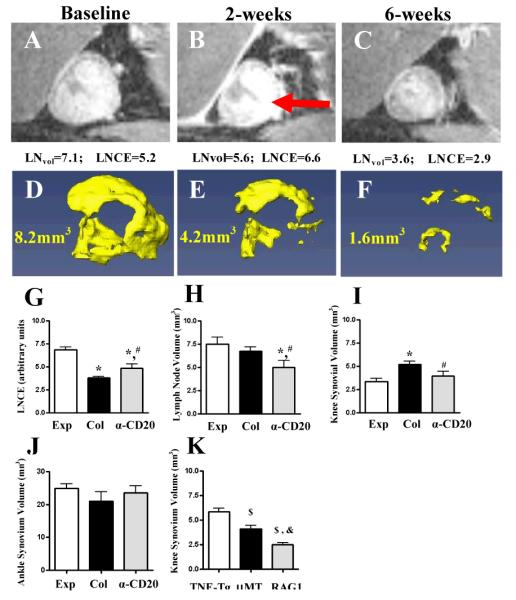
Objective: B cell depletion therapy ameliorates rheumatoid arthritis by mechanisms that are incompletely understood. Arthritis flare in tumor necrosis factor (TNF)-transgenic mice is associated with efferent lymph node (LN) "collapse," triggered by B cell translocation into lymphatic spaces and decreased lymphatic drainage. The aim of this study was to examine whether the efficacy of B cell depletion therapy is associated with restoration of lymphatic drainage due to removal of obstructing nodal B cells.

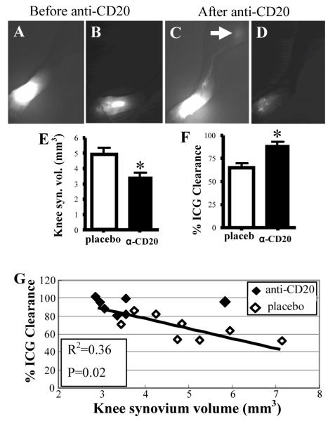
Methods: We used contrast-enhanced magnetic resonance imaging, indocyanine green near-infrared imaging, and intravital immunofluorescence imaging to longitudinally assess synovitis, lymphatic flow, and cell migration in lymphatic vessels in TNF-transgenic mice. We conducted tests to determine whether the efficacy of B cell depletion therapy is associated with restoration of lymphatic draining and cell egress from arthritic joints.

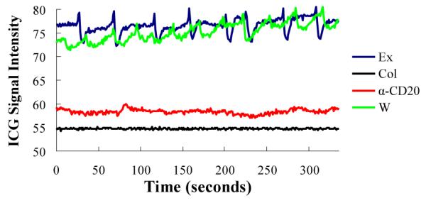
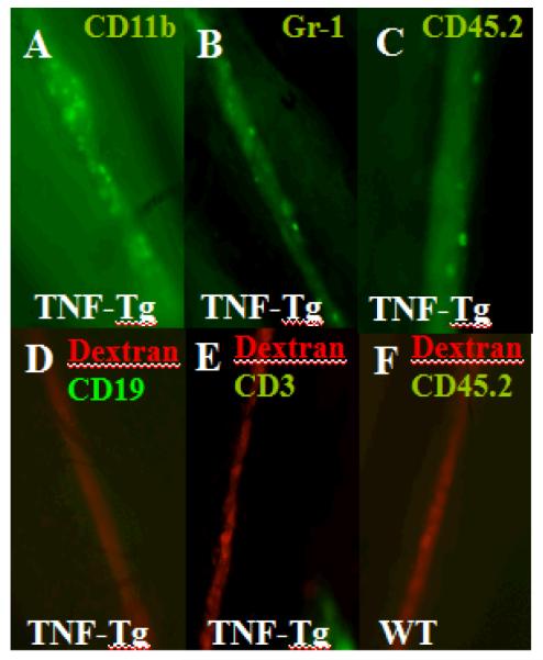
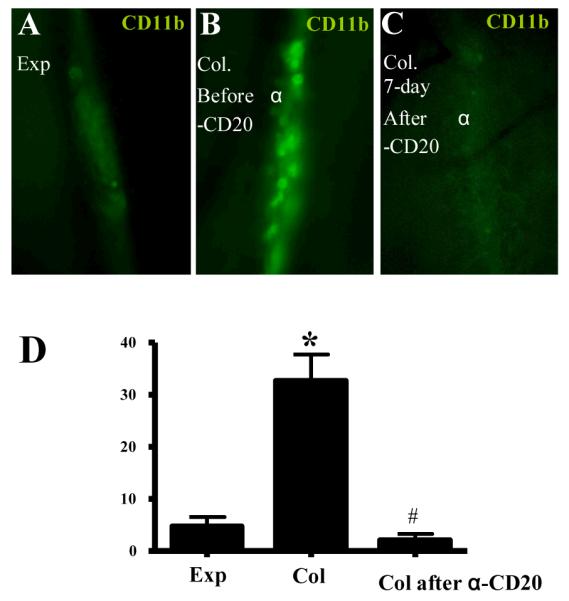
Results: Unlike active lymphatics to normal and prearthritic knees, afferent lymphatic vessels to collapsed LNs in inflamed knees do not pulse. Intravital immunofluorescence imaging demonstrated that CD11b+ monocyte/macrophages in lymphatic vessels afferent to expanding LNs travel at high velocity (mean±SD 186±37 μm/second), while these cells are stationary in lymphatic vessels afferent to collapsed popliteal LNs. B cell depletion therapy for arthritis flares in TNF-transgenic mice significantly decreased knee synovium volume (by 50% from the baseline level) and significantly increased lymphatic clearance compared with placebo (P<0.05). This increased lymphatic drainage restored macrophage egress from inflamed joints without recovery of the lymphatic pulse.

Conclusion: These results support a novel mechanism in which B cell depletion therapy for joint arthritis flares lessens inflammation by increasing lymphatic drainage and subsequent migration of cells and cytokines from the synovial space.


Figures

Figure 1
Figure 1
LNCE is enhanced during effective anti-CD20 therapy for flaring knee synovitis. Untreated TNF-Tg mice with expanding (Exp) and collapsed (Col) (n=12 knees in each group) were identified by longitudinal CE-MRI (36). A separate Col group was treated with anti-CD20 (a-CD20) (n=11 knees, from 4 TNF-Tg mice with bilateral Col PLN and 3 with unilateral Col PLN). A-F, Longitudinal CE-MRI of a Col PLN (A-C), and reconstructed 3D images of the adjacent synovium (D-F), from a representative flaring knee at baseline (A, D), 2 weeks after treatment (B, E), and 6 weeks after treatment (C, F) are shown. The PLN displayed enhanced LNCE at 2 weeks (red arrow in B). G-J, LNCE (G), LNvol (H), knee synovium volume (I), and ankle synovium volume (J) were quantified in Exp, Col and Col PLN 6 weeks after anti-CD20 treatment. K, Decreased knee flare in B cell deficient TNF-Tg mice. CE-MRI was performed on 6-month-old TNF-Tg (n=8 legs), TNF-Tg × μMT−/− (n=4 legs) and TNF-Tg × RAG1−/− (n=6 legs) mice with equivalent ankle synovitis, and their knee synovial volume is shown. All data represent mean±s.d. *p<0.05 vs. Exp, # p<0.05 vs. Col. $p<0.01 vs. TNF-Tg, &p<0.01 vs. TNF-Tg × μMT−/− .
Figure 2
Figure 2
Effective BCDT for knee flare increases lymphatic flow in the lower limb. CE-MRI and NIR-ICG imaging was performed to assess synovitis and lymphatic flow in anti-CD20 (n=7 legs, from 3 TNF-Tg mice with bilateral Col PLN and 1 with unilateral Col PLN) vs. placebo (n=8 legs, from 1 mouse with bilateral Col PLN and 6 with unilateral Col PLN) treated TNF-Tg mice. A-D, NIR-ICG imaging of a representative responder before (A, B), and after treatment (C, D) are shown to illustrate the lymphatic clearance of ICG 30mins (A, C) and 24hrs (B, D) after footpad injection. Note the lack of ICG migration in lymphatic vessels to the PLN at 30mins (A), and the large residual amount of ICG in the foot at 24hrs pre-treatment (B). In contrast, there is apparent ICG in efferent lymphatics to the PLN (arrow) at 30mins (C), and scant residual ICG at 24hrs (D) post-treatment. E and F, significant anti-CD20 affects on knee synovitis (E) and improved lymphatic flow (F) are presented as the mean ± SD.* p<0.05 vs. placebo. G, A linear regression of knee synovial volume vs. %ICG clearance data was performed demonstrating a significant negative correlation.
Figure 3
Figure 3
Anti-CD20 therapy does not restore lymphatic pulsing. Lymphatic pulsing frequency was quantified by real time video analysis of NIR-ICG imaging of a region of interest (ROI) of the draining lymphatic vessel afferent to PLN as previously described(22). Shown are the signal intensities minus background intensity of representative ROIs plotted over time for the four groups studied in which the mean ± SD pulsing frequencies were: WT (WT mice; green line) = 1.4 ± 0.40, Exp (TNF-Tg mice with expanding PLN; blue line) =1.4 ± 0.03, Col (TNF-Tg mice with collapsed PLN; black line) =0 and a-CD20 (TNF-Tg mice with collapsed PLN + 6-weeks of anti-CD20 therapy; red line) = 0 pulses/min. (n≥4 in each group).
Figure 4
Figure 4
Presence of non-migrating CD11b+ and Gr-1+ cells and absence of B and T cells in draining lymphatic vessels afferent to collapsed PLN. Intravital immunofluorescent microscopy was performed on draining lymphatic vessels afferent to PLN by injecting FITC conjugated anti-CD11b, anti-Gr-1, anti-CD19, anti-CD3, or anti-CD45.2 antibody into the footpad of TNF-Tg or WT mice 2hrs prior to imaging (n=5). Immediately prior to imaging Texas red-conjugated dextran beads were subcutaneously injected into the footpad to mark the draining lymphatic vessel, and an incision was made behind the knee to expose the PLN and afferent lymphatics to the 5x objective lens of a fluorescent microscope (Axio Imager M1m, Zeiss). Immunofluorescent photographs of a representative lymphatic vessel afferent to a collapsed PLN showing the: CD11b+ (A) and Gr-1+ (B) and hematopoietic (CD45.2+) (C) cells. Representative micrographs illustrating the lack of CD19+ (D) and CD3+ (E) cells in lymphatic vessels afferent to collapsed PLN, and absence of hematopoietic (CD45.2+) cells in lymphatic vessels afferent to WT PLN (F).
Figure 5
Figure 5
Anti-CD20 treatment restores CD11b+ cell flow in lymphatic vessels afferent to Col PLN. A-C, Time lapse intravital immunofluorescent microscopy (1 frame/min) of CD11b+ cells in lymphatic vessels afferent to PLN in TNF-Tg mice was performed as described in Figure 4. Representative micrographs of a lymphatic vessel afferent to an Exp PLN (A), Col PLN (B), and the same Col PLN 7days after anti-CD20 therapy (C), are shown to illustrate the resident CD11b+ cells. D, The number of CD11b+ cells/mm lymphatic vessel/min was quantified during a 40 min imaging session of lymphatic vessels afferent to Exp PLN, or a Col PLN before and after 6-weeks of anti-CD20 therapy. The data are graphed as the mean ± SD. n=5 in each group, * p< 0.01 vs. Exp, # p<0.01 vs. Col.

Similar articles

Cited by

References

    1. Firestein GS. Evolving concepts of rheumatoid arthritis. Nature. 2003;423(6937):356–61. - PubMed
    1. Keffer J, Probert L, Cazlaris H, Georgopoulos S, Kaslaris E, Kioussis D, et al. Transgenic mice expressing human tumour necrosis factor: a predictive genetic model of arthritis. EMBO J. 1991;10(13):4025–31. - PMC - PubMed
    1. Maini RN, Elliott MJ, Brennan FM, Williams RO, Chu CQ, Paleolog E, et al. Monoclonal anti-TNF alpha antibody as a probe of pathogenesis and therapy of rheumatoid disease. Immunol Rev. 1995;144:195–223. - PubMed
    1. Lipsky PE, van der Heijde DM, St Clair EW, Furst DE, Breedveld FC, Kalden JR, et al. Anti-Tumor Necrosis Factor Trial in Rheumatoid Arthritis with Concomitant Therapy Study Group Infliximab and methotrexate in the treatment of rheumatoid arthritis. N Engl J Med. 2000;343(22):1594–602. - PubMed
    1. Moreland LW, Baumgartner SW, Schiff MH, Tindall EA, Fleischmann RM, Weaver AL, et al. Treatment of rheumatoid arthritis with a recombinant human tumor necrosis factor receptor (p75)-Fc fusion protein. N Engl J Med. 1997;337(3):141–7. - PubMed

Publication types