

Animal models of Alzheimer disease
- PMID: 23002015
- PMCID: PMC3543097
- DOI: 10.1101/cshperspect.a006320
Animal models of Alzheimer disease
Abstract
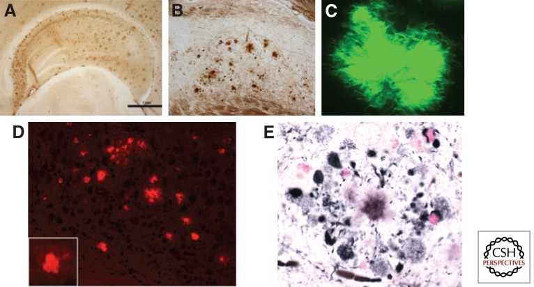
Significant insights into the function of genes associated with Alzheimer disease and related dementias have occurred through studying genetically modified animals. Although none of the existing models fully reproduces the complete spectrum of this insidious human disease, critical aspects of Alzheimer pathology and disease processes can be experimentally recapitulated. Genetically modified animal models have helped advance our understanding of the underlying mechanisms of disease and have proven to be invaluable in the preclinical evaluation of potential therapeutic interventions. Continuing refinement and evolution to yield the next generation of animal models will facilitate successes in producing greater translational concordance between preclinical studies and human clinical trials and eventually lead to the introduction of novel therapies into clinical practice.
Figures

References
-
- Adlard PA, Cherny RA, Finkelstein DI, Gautier E, Robb E, Cortes M, Volitakis I, Liu X, Smith JP, Perez K, et al. 2008. Rapid restoration of cognition in Alzheimer’s transgenic mice with 8-hydroxy quinoline analogs is associated with decreased interstitial Aβ. Neuron 59: 43–55 - PubMed
-
- Billings LM, Oddo S, Green KN, McGaugh JL, LaFerla FM 2005. Intraneuronal Aβ causes the onset of early Alzheimer’s disease-related cognitive deficits in transgenic mice. Neuron 45: 675–688 - PubMed
Publication types
MeSH terms
Substances
LinkOut - more resources
Full Text Sources
Other Literature Sources
Medical