. 2012 Nov;40(21):11100-14.
doi: 10.1093/nar/gks867. Epub 2012 Sep 23.

Rbg1-Tma46 dimer structure reveals new functional domains and their role in polysome recruitment

Affiliations

Rbg1-Tma46 dimer structure reveals new functional domains and their role in polysome recruitment

Sandrea M Francis et al. Nucleic Acids Res. 2012 Nov.

Abstract

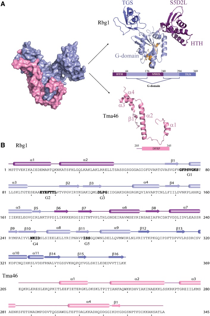
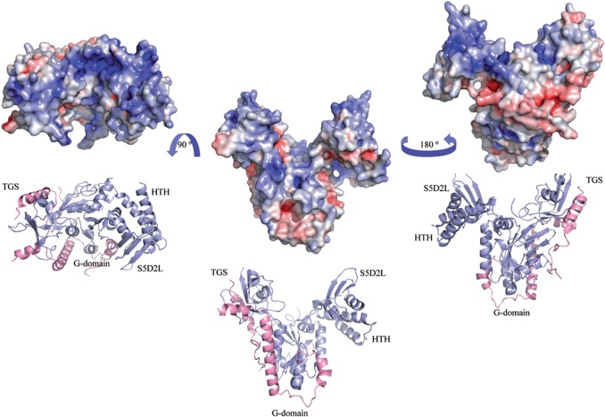
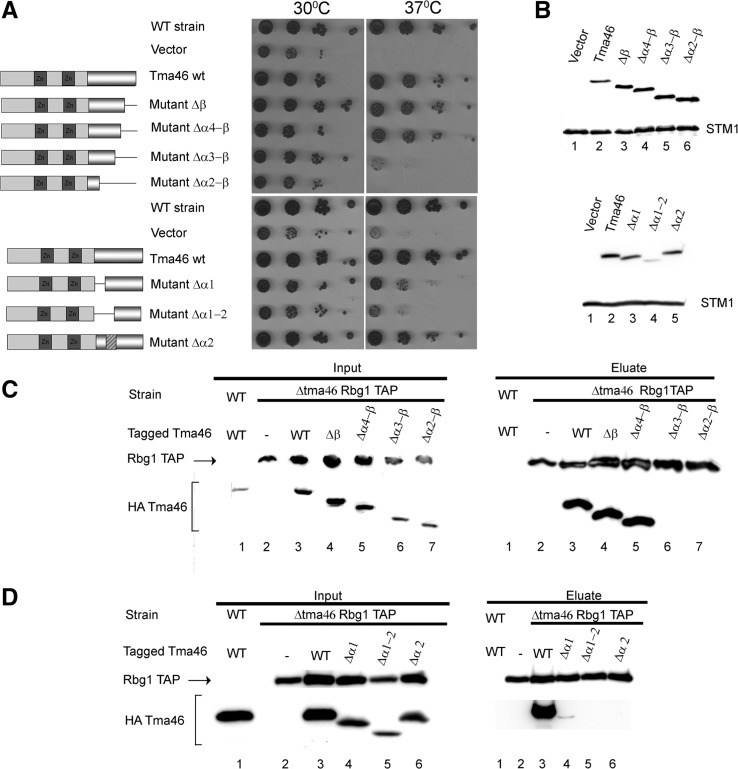
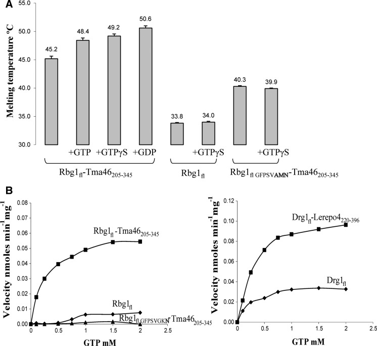
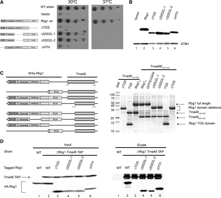
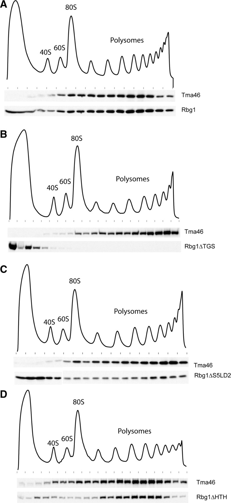
Developmentally Regulated GTP-binding (DRG) proteins are highly conserved GTPases that associate with DRG Family Regulatory Proteins (DFRP). The resulting complexes have recently been shown to participate in eukaryotic translation. The structure of the Rbg1 GTPase, a yeast DRG protein, in complex with the C-terminal region of its DFRP partner, Tma46, was solved by X-ray diffraction. These data reveal that DRG proteins are multimodular factors with three additional domains, helix-turn-helix (HTH), S5D2L and TGS, packing against the GTPase platform. Surprisingly, the S5D2L domain is inserted in the middle of the GTPase sequence. In contrast, the region of Tma46 interacting with Rbg1 adopts an extended conformation typical of intrinsically unstructured proteins and contacts the GTPase and TGS domains. Functional analyses demonstrate that the various domains of Rbg1, as well as Tma46, modulate the GTPase activity of Rbg1 and contribute to the function of these proteins in vivo. Dissecting the role of the different domains revealed that the Rbg1 TGS domain is essential for the recruitment of this factor in polysomes, supporting further the implication of these conserved factors in translation.


Figures

Figure 1.
Figure 1.
Structure of the Rbg1fl–Tma46205–345 complex with sequence information. (A) A surface representation of the Tma46 C-terminal fragment (pink) enveloping Rbg1 (pale blue) is shown on the left. The individual components are shown color-coded on the right: the Tma46 C-terminal fragment (pink) and Rbg1 with the G-domain (pale blue, this includes the short β sheet formed by β5 and β9 connecting the S5D2L domain), the protuberance formed by the HTH and S5D2L domains (purple) and the TGS domain (blue). The GTP-binding pocket is also represented with the five G motifs colored as orange. A schematic domain organization of the structurally solved complex is also shown. (B) The component sequences and secondary structure elements of the crystallized complex are represented with the G-motifs (G1–G5) given in bold letters. Domain boundaries are indicated in the same color scheme as in Figure 1A.
Figure 2.
Figure 2.
Electrostatic surface representation. The solvent-accessible surface electrostatic potential of the Rbg1fl–Tma46205–345 complex as calculated by APBS (Pymol) is shown as a surface alongside the cartoon representation. The potential is given with the negative (red) and positive (blue) contour levels in the range from −8.0 to +8.0 kBT respectively. The left figure shows the positively-charged surface formed partly by the G, HTH, S5D2L and TGS domains.
Figure 3.
Figure 3.
Analysis of Tma46 mutants. (A) Complementation assay for Tma46 function. The ability of plasmid-encoded Tma46 mutants to complement the growth phenotype of a triple Δtma46Δgir2Δslh1 strain was assayed by spotting serial dilution on selective plates and incubating at 30°C or 37°C for 3 days. The structure of the various mutants is shown schematically on the left. Dark grey boxes indicate the Tma46 Zn-fingers, while the pseudo-cylinder represents the C-terminal region interacting with Rbg1. The hatched box indicates the alanine linker. WT strain indicates the original wild-type parental strain without mutation. (B) Mutant protein accumulation. The level of accumulation of the mutant proteins in cells shown on panel A grown at 30°C was assessed by detecting the HA tag by western blotting. Uniform loading is supported by analysis of the levels of the endogenous Stm1 protein. (C) Effect of C-terminal Tma46 truncation on its binding to Rbg1 in yeast. Extracts prepared from Δtma46 strains carrying TAP-tagged Rbg1 and the various HA-Tma46 mutants grown at 30°C were used for immunoprecipitation on IgG beads. As control for the specificity of the coprecipitation a wild-type strain expressing wild-type Tma46 tagged with HA was used. Proteins present in extracts (Input) and (co)precipitated factors (Eluate) were analyzed by western blotting. (D) Effect of deletion of helices α1 and α1+α2 of Tma46 on binding to Rbg1. Samples were prepared as in panel C.
Figure 4.
Figure 4.
GTP binding and hydrolysis. (A) Presence of 0.2 mM GTP, GTPγS or GDP (in 4-fold excess over the protein concentration) causes an increase in melting temperature of the Rbg1fl–Tma46205–345 complex in the thermal-shift assay indicative of nucleotide binding. In contrast, addition of a 10-fold excess of GTPγS (0.5 mM) does not increase the melting temperature of Rbg1 alone. Also shown is the lack of increase in protein unfolding temperature for a Rbg1fl–Tma46205–345 G1 motif mutant (GFPSVAMN) in presence of 10× GTPγS. Note also that Rbg1fl melts at a much lower temperature than Rbg1fl–Tma46205–345. (B) The GTP hydrolytic activity of Rbg1/Drg1, alone and in complex with Tma46205–345/Lerepo4220–396, respectively, is represented as a graph with increasing substrate concentration in the x axis. A catalytic mutant Rbg1fl VFPSVGKN in complex with Tma46205–345 was used as a negative control.
Figure 5.
Figure 5.
Analysis of Rbg1 domains. (A) Complementation assay for Rbg1 function. The ability of plasmid-encoded Rbg1 mutants to complement the growth phenotype of a triple Δrbg1Δrbg2Δslh1 strain was assayed by spotting serial dilution on selective plates and incubating at 30°C or 37°C for 4 days. The structure of the various mutants is shown schematically on the left. WT strain indicates the original wild-type parental strain without mutation. (B) Mutant protein accumulation. The level of accumulation of the mutant proteins in cells shown on panel A grown at 30°C was assessed by detecting the HA tag by western blotting. Uniform loading is supported by analysis of the levels of the endogenous Stm1 protein. (C) Interaction between recombinant Tma46 and Rbg1 mutants. Plasmids harboring operons encoding His6-tagged Rbg1 [wild-type, point mutant (GFPSVGKN) or mutant deleted for specific domains] together with Tma46 (either amino-acids 154–345 or 205–345) were used to express protein in E. coli. Recombinant proteins purified on Ni–NTA agarose were detected by Coomassie staining. Organization of the different operons is shown on the left. (D) Effect of C-terminal Tma46 truncation on binding to Rbg1 in yeast. Extracts prepared from Δrbg1 strains carrying TAP-tagged Tma46 and various HA-Rbg1 mutants grown at 30°C were used for immunoprecipitation on IgG beads. As control for the specificity of the coprecipitation a wild-type strain expressing wild-type Rbg1 tagged with HA was used. Proteins present in extracts (Input) and (co)precipitated factors (Eluate) were analyzed by western blotting.
Figure 6.
Figure 6.
Polysome association of Rbg1 mutants. Polysomes extracts were prepared from cells expressing TAP-tagged Tma46 and HA-tagged Rbg1 (wild-type or domain deletion mutants). Polysomes were resolved by density sedimentation in 10–50% sucrose gradient. The UV absorbance trace (254 nm) obtained by continuous monitoring during fractionation is shown with the position of the 40S, 60S, 80S and polysomes peaks indicated. Fractions were analyzed by western blotting to detect the TAP and HA tags. (A) Distribution of Tma46-TAP and wild-type HA-Rbg1. (B) Distribution of Tma46-TAP and HA-Rbg1 ΔTGS. (C) Distribution of Tma46-TAP and HA-Rbg1 ΔS5D2L. (D) Distribution of Tma46-TAP and HA-Rbg1 ΔHTH. Previously reported control analyses demonstrate that Rbg1 association with polysome is specific (15).

References

    1. Leipe DD, Wolf YI, Koonin EV, Aravind L. Classification and evolution of P-loop GTPases and related ATPases. J. Mol. Biol. 2002;317:41–72. - PubMed
    1. Paduch M, Jelen F, Otlewski J. Structure of small G proteins and their regulators. Acta Biochim. Pol. 2001;48:829–850. - PubMed
    1. Gelperin D, Horton L, Beckman J, Hensold J, Lemmon SK. Bms1p, a novel GTP-binding protein, and the related Tsr1p are required for distinct steps of 40S ribosome biogenesis in yeast. RNA. 2001;7:1268–1283. - PMC - PubMed
    1. Wegierski T, Billy E, Nasr F, Filipowicz W. Bms1p, a G-domain-containing protein, associates with Rcl1p and is required for 18S rRNA biogenesis in yeast. RNA. 2001;7:1254–1267. - PMC - PubMed
    1. Polkinghorne A, Ziegler U, Gonzalez-Hernandez Y, Pospischil A, Timms P, Vaughan L. Chlamydophila pneumoniae HflX belongs to an uncharacterized family of conserved GTPases and associates with the Escherichia coli 50S large ribosomal subunit. Microbiology. 2008;154:3537–3546. - PubMed

Publication types

Substances

Associated data