. 2012 Nov;40(21):10980-94.
doi: 10.1093/nar/gks834. Epub 2012 Sep 23.

Genome-wide activation of latent donor splice sites in stress and disease

Affiliations

Genome-wide activation of latent donor splice sites in stress and disease

Yuval Nevo et al. Nucleic Acids Res. 2012 Nov.

Abstract

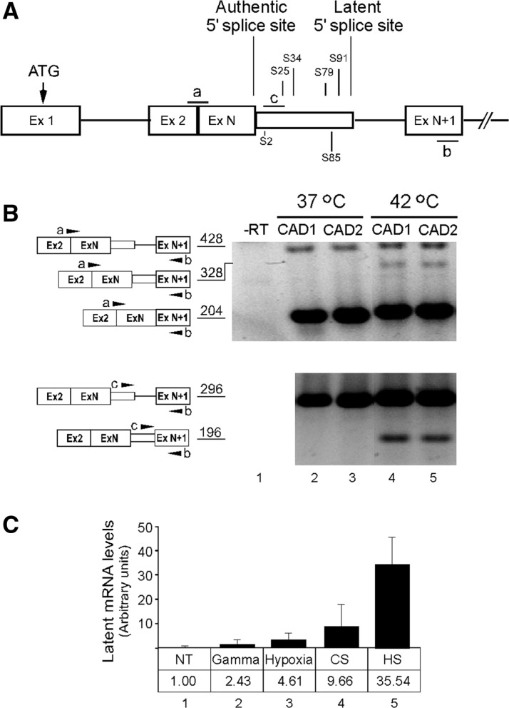
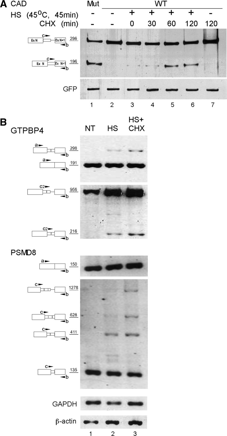
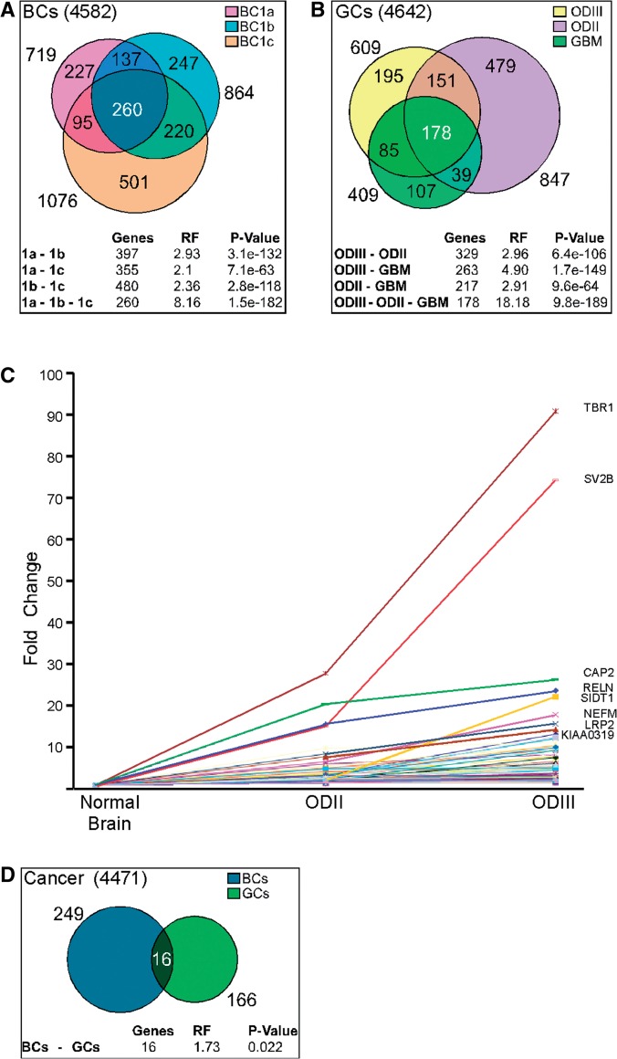
Sequences that conform to the 5' splice site (5'SS) consensus are highly abundant in mammalian introns. Most of these sequences are preceded by at least one in-frame stop codon; thus, their use for splicing would result in pre-maturely terminated aberrant mRNAs. In normally grown cells, such intronic 5'SSs appear not to be selected for splicing. However, under heat shock conditions aberrant splicing involving such latent 5'SSs occurred in a number of specific gene transcripts. Using a splicing-sensitive microarray, we show here that stress-induced (e.g. heat shock) activation of latent splicing is widespread across the human transcriptome, thus highlighting the possibility that latent splicing may underlie certain diseases. Consistent with this notion, our analyses of data from the Gene Expression Omnibus (GEO) revealed widespread activation of latent splicing in cells grown under hypoxia and in certain cancers such as breast cancer and gliomas. These changes were found in thousands of transcripts representing a wide variety of functional groups; among them are genes involved in cell proliferation and differentiation. The GEO analysis also revealed a set of gene transcripts in oligodendroglioma, in which the level of activation of latent splicing increased with the severity of the disease.


Figures

Figure 1.
Figure 1.
Latent splicing is elicited under stress conditions—the CAD case. (A) Schematic representation of CAD1 and CAD2 minigene constructs. Both constructs share the same start codon (ATG) but differ in the position of the stop codons between the authentic and latent 5′SSs (designated S; numbers represent distance in nucleotide from the authentic 5′SS; stop codons for CAD1 and CAD2 are marked above and below the latent exon, respectively). Open boxes, exons; lines, introns; narrow box, latent exon; a, b, c, mark the position of the PCR primers. (B) RT–PCR analysis of RNA extracted from human 293 T cells transiently transfected with either CAD1 or CAD2 and grown at 37°C (lanes 1–3) or heat-treated at 42°C for 45 min (lanes 4–5). PCRs were performed with primer pair a + b (upper panel) and primer pair b + c (lower panel). The PCR products and their sizes (nt) are depicted on the left. (C) Relative levels of latent CAD mRNAs expressed from the endogenous CAD gene in 165-28 cells in response to different environmental stress conditions. NT, no treatment; Gamma, 30 Gy; Hypoxia, 500 µM DFO; CS, cold shock (10°C for 4 h); HS, heat shock (45°C for 45 min). All measurements were normalized to the basal level detected for the control of untreated cells (designated as 1.00). Error bars represent standard errors of three to five independent experiments.
Figure 2.
Figure 2.
Latent 5′SSs are highly abundant in the human genome. Flow chart of global computational and experimental analyses of latent splicing. Boxes on the left describe the computational search for latent 5′SSs; boxes on the right describe the experimental chip-aided analysis of elevation of latent splicing after heat shock. Rectangles, program inputs; rounded rectangles, programs; octagons, program outputs; arrows, direction of information flow.
Figure 3.
Figure 3.
Validation, by RT–PCR, of microarray-detected heat-induced elevation in the level of latent splicing of mRNAs expressed from eight genes in HEK 293 T cells. Right panels: Curves corresponding to the levels of probesets signals along the gene were created using the Partek Genomics Suite software (red, heat-treated cells; blue, untreated cells). Arrows point at probesets that recognized latent exons. Schematics of the genes are drawn above the expression curves. Boxes, exons; lines, introns; narrow box, latent exon; arrowheads mark the positions of the PCR primers, which are identical to the primers indicated on the left panel. Left panels: RT–PCR analyses with primer-pairs flanking the latent sites, as indicated in the schematic drawings of the PCR products on the left (numbers indicate sizes of the PCR products). GTPBP4, GTP-binding protein 4; M6PR, mannose-6-phosphate receptor; C1R, complement component 1, r subcomponent; TMED10, transmembrane emp24-like trafficking protein 10; VIPR2, vasoactive intestinal peptide receptor 2; PSMD8, proteasome 26 S subunit, non-ATPase, 8; DERL2, derlin 2; and LPCAT4, lysophosphatidylcholine acyltransferase 4; C, untreated cells; HS, heat-treated cells. All PCR products were verified by sequencing.
Figure 3.
Figure 3.
Validation, by RT–PCR, of microarray-detected heat-induced elevation in the level of latent splicing of mRNAs expressed from eight genes in HEK 293 T cells. Right panels: Curves corresponding to the levels of probesets signals along the gene were created using the Partek Genomics Suite software (red, heat-treated cells; blue, untreated cells). Arrows point at probesets that recognized latent exons. Schematics of the genes are drawn above the expression curves. Boxes, exons; lines, introns; narrow box, latent exon; arrowheads mark the positions of the PCR primers, which are identical to the primers indicated on the left panel. Left panels: RT–PCR analyses with primer-pairs flanking the latent sites, as indicated in the schematic drawings of the PCR products on the left (numbers indicate sizes of the PCR products). GTPBP4, GTP-binding protein 4; M6PR, mannose-6-phosphate receptor; C1R, complement component 1, r subcomponent; TMED10, transmembrane emp24-like trafficking protein 10; VIPR2, vasoactive intestinal peptide receptor 2; PSMD8, proteasome 26 S subunit, non-ATPase, 8; DERL2, derlin 2; and LPCAT4, lysophosphatidylcholine acyltransferase 4; C, untreated cells; HS, heat-treated cells. All PCR products were verified by sequencing.
Figure 4.
Figure 4.
Latent mRNAs that escape suppression of splicing are subjected to the NMD pathway. (A) HEK 293 T cells were co-transfected with the WT CAD1 plasmid (lanes 2–7) and GFP. Twenty-four hours post-transfection, cells were heated at 45°C for 45 min, followed by treatment with CHX as indicated. Total RNA was isolated and analyzed by RT–PCR. To mark the location of latent mRNA, we show the splicing pattern of CAD mRNA expressed from a mutant construct lacking in-frame stop codons upstream of the latent site (lane 1). Note that treatment with CHX for 120 min without heat shock did not elicit latent splicing (lane 7). (B) RT–PCR analysis of the splicing patterns of RNAs expressed from the endogenous genes GTP binding protein 4 (GTPBP4) and proteasome 26 S subunit non-ATPase 8 (PSMD8) in untreated HEK 293 T cells (NT); heat-treated cells (HS) and cells treated with both heat and CHX (HS + CHX). The levels of GAPDH and β-actin were used as a loading control. The PCR products and their sizes (nt) are depicted on the left.
Figure 5.
Figure 5.
Overlap between activated latent 5′SSs in cancerous states and correlation between the level of latent splicing and the severity of oligodendroglioma. (A) Sets of activated latent 5′SSs in MCF-7 cells, taken from three different sources and compared to MCF-10 A cells, are represented by overlapping circles. BC1a, MCF-7 from (38); BC1b, MCF-7 from (40); BC1c, MCF-7 from (39). The sizes of the different intersections are given in their proper places, while the total size of each set is given outside its relevant circle. The table shows the significance of the intersections between each pair and between all three sets, compared to a random selection of equally sized sets. RF, representation factor, is the ratio between the observed size and the randomly expected one; P-value is the actual probability of having equally sized or larger intersections (at random selection). (B) A similar representation as in a, for the overlap between latent 5′SSs that are activated in glioblastoma (GBM), oligodendroglioma Grade II (ODII) and oligodendroglioma Grade III (ODIII), compared to normal brain (41). (C) Correlation between the level of activation of latent splicing and the severity of oligodendroglioma. The graph depicts fold-changes in the level of latent splicing in 125 gene transcripts whose latent splicing expression increased from oligodendroglioma Grade II to Grade III. Names of the top-scoring gene transcripts are indicated. (D) A similar representation as in (A) and (B), for the overlap between latent 5′SSs that are activated in breast cancer and in glial tumors.

References

    1. Wang Z, Burge CB. Splicing regulation: from a parts list of regulatory elements to an integrated splicing code. RNA. 2008;14:802–813. - PMC - PubMed
    1. Miriami E, Motro U, Sperling J, Sperling R. Conservation of an open-reading frame as an element affecting 5′ splice site selection. J. Struct. Biol. 2002;140:116–122. - PubMed
    1. Li B, Wachtel C, Miriami E, Yahalom G, Friedlander G, Sharon G, Sperling R, Sperling J. Stop codons affect 5′ splice site selection by surveillance of splicing. Proc. Natl Acad. Sci. USA. 2002;99:5277–5282. - PMC - PubMed
    1. Miriami E, Sperling R, Sperling J, Motro U. Regulation of splicing: the importance of being translatable. RNA. 2004;10:1–4. - PMC - PubMed
    1. Sperling J, Sperling R. Nuclear surveillance of RNA polymerase II transcripts. RNA Biol. 2008;5:220–224. - PubMed

Publication types

Substances