

Biocompatible, pH-sensitive AB(2) Miktoarm Polymer-Based Polymersomes: Preparation, Characterization, and Acidic pH-Activated Nanostructural Transformation
- PMID: 23002330
- PMCID: PMC3447630
- DOI: 10.1039/C2JM33750A
Biocompatible, pH-sensitive AB(2) Miktoarm Polymer-Based Polymersomes: Preparation, Characterization, and Acidic pH-Activated Nanostructural Transformation
Abstract
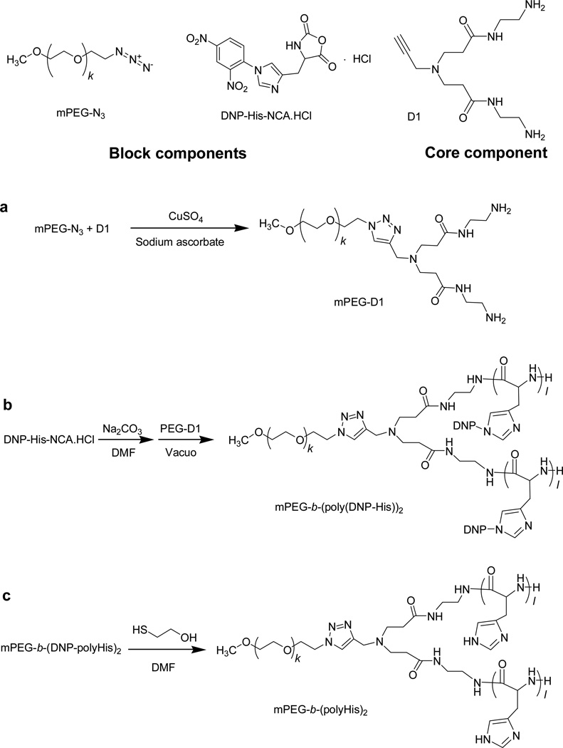
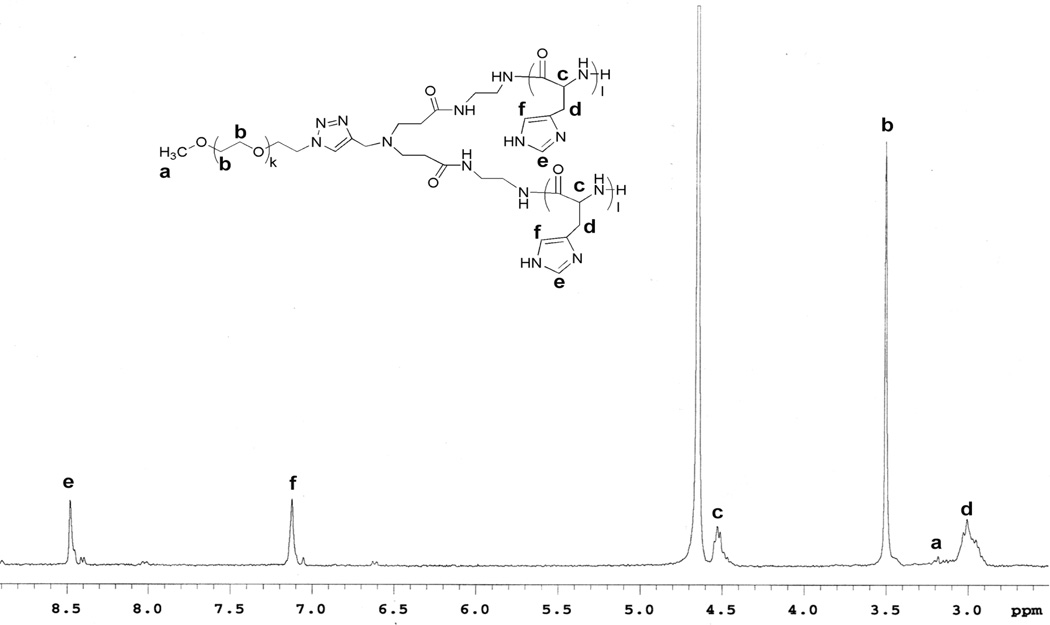
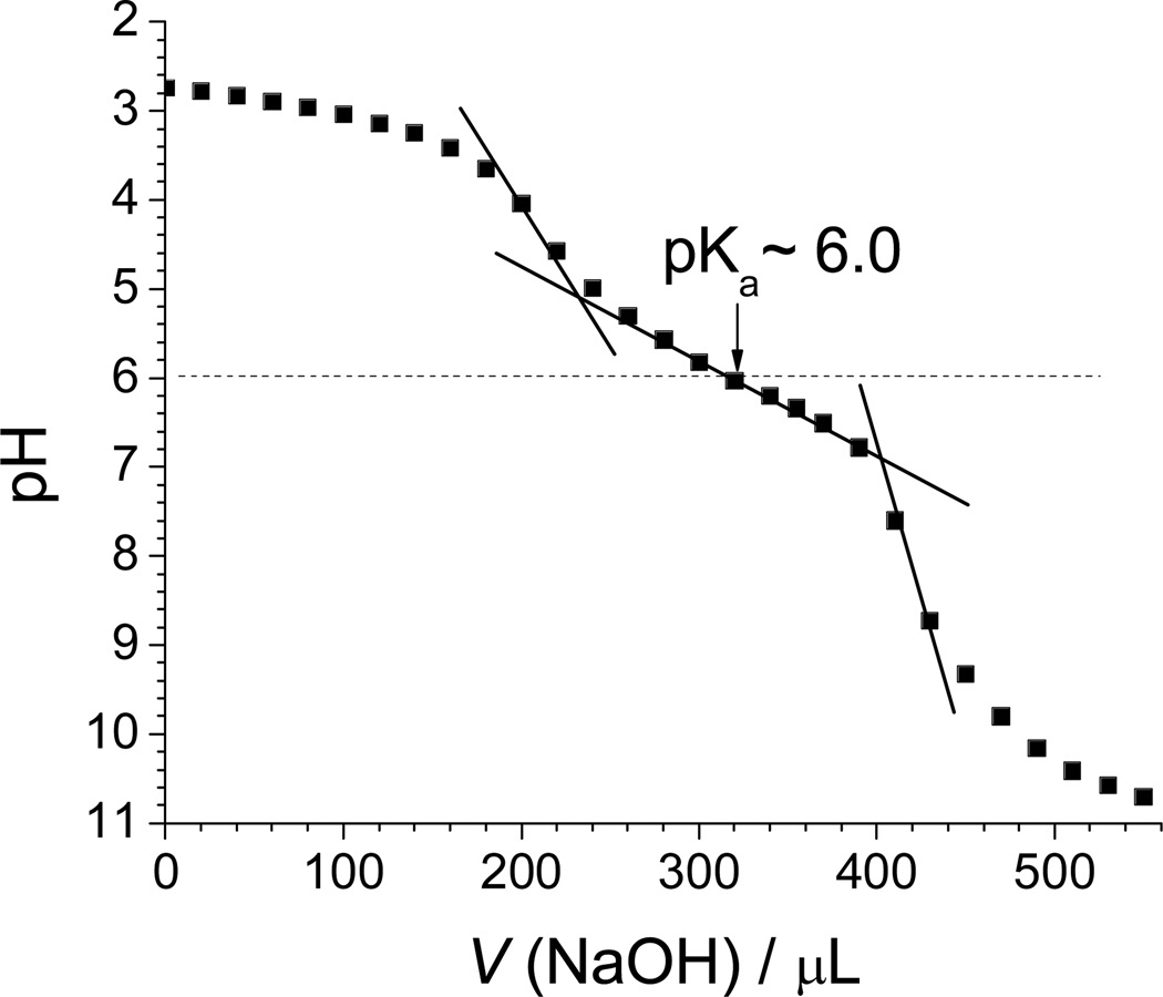
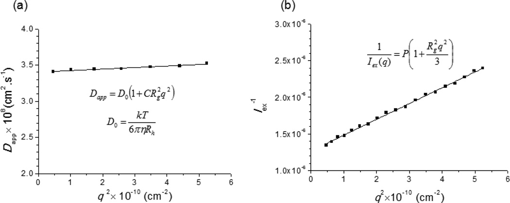
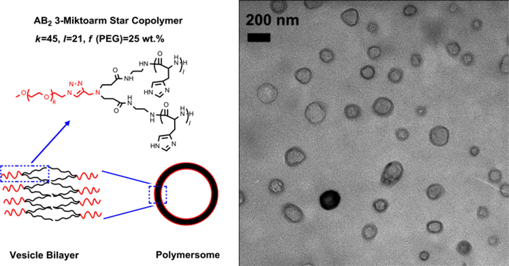
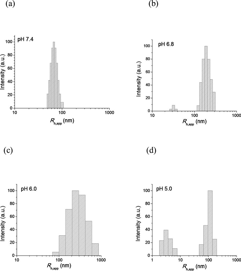
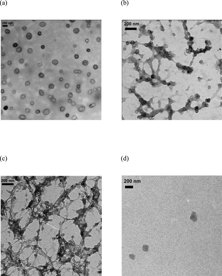
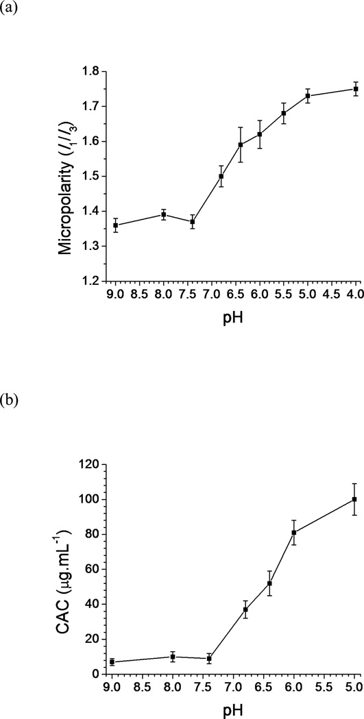
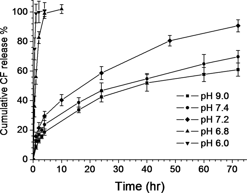
Motivated by the limitations of liposomal drug delivery systems, we designed a novel histidine-based AB(2)-miktoarm polymer (mPEG-b-(polyHis)(2)) equipped with a phospholipid-mimic structure, low cytotoxicity, and pH-sensitivity. Using "core-first" click chemistry and ring-opening polymerization, mPEG(2kDa)-b-(polyHis(29kDa))(2) was successfully synthesized with a narrow molecular weight distribution (1.14). In borate buffer (pH 9), the miktoarm polymer self-assembled to form a nano-sized polymersome with a hydrodynamic radius of 70.2 nm and a very narrow size polydispersity (0.05). At 4.2 µmol/mg polymer, mPEG(2kDa)-b-(polyHis(29kDa))(2) strongly buffered against acidification in the endolysosomal pH range and exhibited low cytotoxicity on a 5 d exposure. Below pH 7.4 the polymersome transitioned to cylindrical micelles, spherical micelles, and finally unimers as the pH was decreased. The pH-induced structural transition of mPEG(2kDa)-b-(polyHis(29kDa))(2) nanostructures may be caused by the increasing hydrophilic weight fraction of mPEG(2kDa)-b-(polyHis(29kDa))(2) and can help to disrupt the endosomal membrane through proton buffering and membrane fusion of mPEG(2kDa)-b-(polyHis(29kDa))(2). In addition, a hydrophilic model dye, 5(6)-carboxyfluorescein encapsulated into the aqueous lumen of the polymersome showed a slow, sustained release at pH 7.4 but greatly accelerated release below pH 6.8, indicating a desirable pH sensitivity of the system in the range of endosomal pH. Therefore, this polymersome that is based on a biocompatible histidine-based miktoarm polymer and undergoes acid-induced transformations could serve as a drug delivery vehicle for chemical and biological drugs.
Figures
Similar articles
-
Effects of cholesterol incorporation on the physicochemical, colloidal, and biological characteristics of pH-sensitive AB₂ miktoarm polymer-based polymersomes.Colloids Surf B Biointerfaces. 2014 Apr 1;116:128-37. doi: 10.1016/j.colsurfb.2013.12.041. Epub 2013 Dec 30. Colloids Surf B Biointerfaces. 2014. PMID: 24463148 Free PMC article.
-
Evaluation of zwitterionic polymersomes spontaneously formed by pH-sensitive and biocompatible PEG based random copolymers as drug delivery systems.Colloids Surf B Biointerfaces. 2016 Mar 1;139:107-16. doi: 10.1016/j.colsurfb.2015.11.042. Epub 2015 Nov 26. Colloids Surf B Biointerfaces. 2016. PMID: 26704991
-
Poly(L-histidine)-PEG block copolymer micelles and pH-induced destabilization.J Control Release. 2003 Jul 31;90(3):363-74. doi: 10.1016/s0168-3659(03)00205-0. J Control Release. 2003. PMID: 12880703
-
Poly(l-histidine) based triblock copolymers: pH induced reassembly of copolymer micelles and mechanism underlying endolysosomal escape for intracellular delivery.Biomacromolecules. 2014 Nov 10;15(11):4032-45. doi: 10.1021/bm5010756. Epub 2014 Oct 29. Biomacromolecules. 2014. PMID: 25308242
-
Hallmarks of Polymersome Characterization.ACS Mater Au. 2024 Dec 23;5(2):223-230. doi: 10.1021/acsmaterialsau.4c00107. eCollection 2025 Mar 12. ACS Mater Au. 2024. PMID: 40093839 Free PMC article. Review.
Cited by
-
Extracellularly activatable nanocarriers for drug delivery to tumors.Expert Opin Drug Deliv. 2014 Oct;11(10):1601-1618. doi: 10.1517/17425247.2014.930434. Epub 2014 Jun 20. Expert Opin Drug Deliv. 2014. PMID: 24950343 Free PMC article. Review.
-
pH-Sensitive Micelles Based on Star Copolymer Ad-(PCL-b-PDEAEMA-b-PPEGMA)₄ for Controlled Drug Delivery.Polymers (Basel). 2018 Apr 14;10(4):443. doi: 10.3390/polym10040443. Polymers (Basel). 2018. PMID: 30966478 Free PMC article.
-
Co-Delivery of Imiquimod and Plasmid DNA via an Amphiphilic pH-Responsive Star Polymer that Forms Unimolecular Micelles in Water.Polymers (Basel). 2016 Nov 16;8(11):397. doi: 10.3390/polym8110397. Polymers (Basel). 2016. PMID: 30974677 Free PMC article.
-
Effects of cholesterol incorporation on the physicochemical, colloidal, and biological characteristics of pH-sensitive AB₂ miktoarm polymer-based polymersomes.Colloids Surf B Biointerfaces. 2014 Apr 1;116:128-37. doi: 10.1016/j.colsurfb.2013.12.041. Epub 2013 Dec 30. Colloids Surf B Biointerfaces. 2014. PMID: 24463148 Free PMC article.
-
Polymersome-based drug-delivery strategies for cancer therapeutics.Ther Deliv. 2015;6(4):521-34. doi: 10.4155/tde.14.125. Ther Deliv. 2015. PMID: 25996048 Free PMC article. Review.
References
Grants and funding
LinkOut - more resources
Full Text Sources
Other Literature Sources