. 2013 Mar 15;22(6):845-54.
doi: 10.1089/scd.2012.0395. Epub 2012 Nov 7.

Exosomes derived from human umbilical cord mesenchymal stem cells alleviate liver fibrosis

Affiliations

Exosomes derived from human umbilical cord mesenchymal stem cells alleviate liver fibrosis

Tingfen Li et al. Stem Cells Dev. .

Abstract

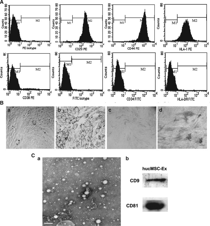
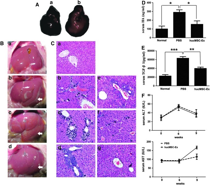
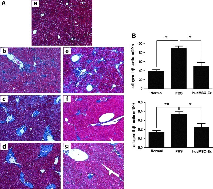
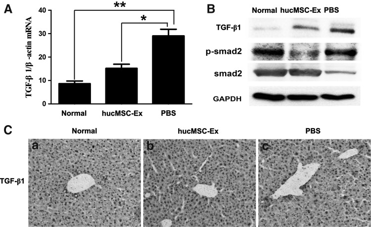
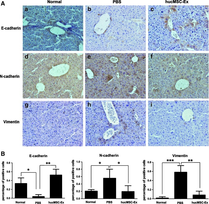
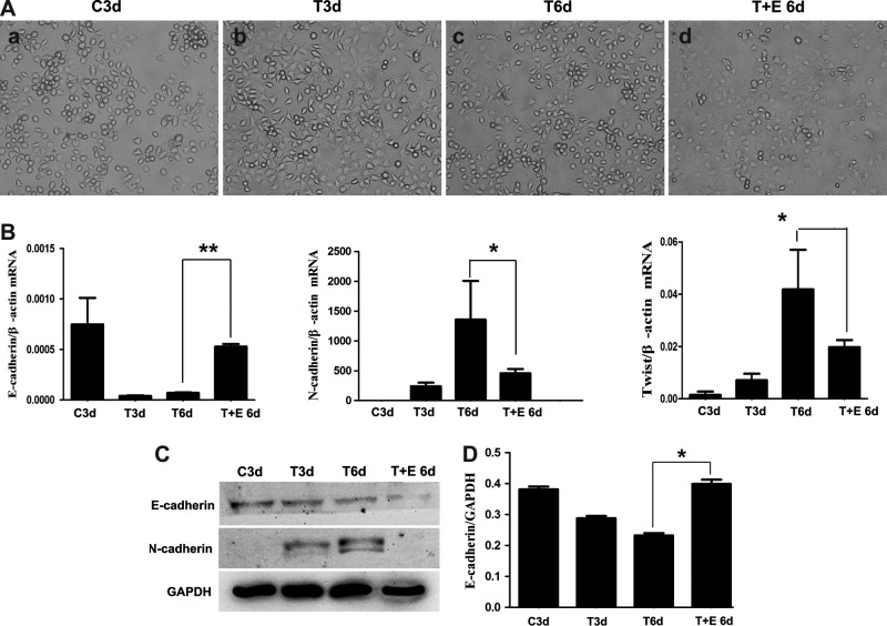
Mesenchymal stem cells (MSCs) have been considered as an attractive tool for the therapy of diseases. Exosomes excreted from MSCs can reduce myocardial ischemia/reperfusion damage and protect against acute tubular injury. However, whether MSC-derived exosomes can relieve liver fibrosis and its mechanism remain unknown. Previous work showed that human umbilical cord-MSCs (hucMSCs) transplanted into acutely injured and fibrotic livers could restore liver function and improve liver fibrosis. In this study, it was found that transplantation of exosomes derived from hucMSC (hucMSC-Ex) reduced the surface fibrous capsules and got their textures soft, alleviated hepatic inflammation and collagen deposition in carbon tetrachloride (CCl4)-induced fibrotic liver. hucMSC-Ex also significantly recovered serum aspartate aminotransferase (AST) activity, decreased collagen type I and III, transforming growth factor (TGF)-β1 and phosphorylation Smad2 expression in vivo. In further experiments, we found that epithelial-to-mesenchymal transition (EMT)-associated markers E-cadherin-positive cells increased and N-cadherin- and vimentin-positive cells decreased after hucMSC-Ex transplantation. Furthermore, the human liver cell line HL7702 underwent typical EMT after induction with recombinant human TGF-β1, and then hucMSC-Ex treatment reversed spindle-shaped and EMT-associated markers expression in vitro. Taken together, these results suggest that hucMSC-Ex could ameliorate CCl4-induced liver fibrosis by inhibiting EMT and protecting hepatocytes. This provides a novel approach for the treatment of fibrotic liver disease.


Figures

FIG. 1.
FIG. 1.
Identification of human umbilical cord-mesenchymal stem cells (hucMSCs) and exosomes derived from hucMSC (hucMSC-Ex). (A) Flow cytometry analysis of the surface markers in hucMSCs. (B) Adipogenic and osteogenic differentiation of hucMSCs (100×). (a, b) Results of Oil-Red-O staining detection in hucMSCs cultures grown for 14 days; (a) hucMSCs cultured in the regular medium; (b) hucMSCs cultured in adipogenic medium; (c, d) Results of neutrophil alkaline phosphatase (NAP) with NAP staining kit detection in hucMSCs cultures grown for 14 days; (c) hucMSCs cultured in the regular medium; (d) hucMSCs cultured in osteogenic medium. (C) Identification of hucMSCs-Ex. (a) Transmission electron micrograph of hucMSC-Ex, Scale bar=200 nm; (b) Detection of hucMSC-Ex CD9 and CD81 expression by western blotting.
FIG. 2.
FIG. 2.
hucMSC-Ex promoted the recovery of carbon tetrachloride (CCl4)-induced mouse liver injury. (A) hucMSC-Ex location in mouse liver by an in vivo imaging system. (a) The phosphate-buffered saline (PBS) group liver; (b) The cell dye-labeled hucMSC-Ex administrated into mouse liver showed red fluorescence. (B) Pictures of mouse livers. (a) normal group; (b) CCl4 group, 6 weeks after CCl4 injury; (c) PBS group 3 weeks after PBS treatment; (d) hucMSCs-Ex group 3 weeks after hucMSC-Ex transplantation. The arrows indicated the liver damage/fibrotic areas. (C) Hematoxylin and eosin dyeing of liver sections (200×). (a) normal group; (b) PBS group, 1 week; (c) PBS group, 2 weeks; (d) PBS group, 3 weeks; (e) hucMSC-Ex group, 1 week; (f) hucMSC-Ex group, 2 weeks; (g) hucMSC-Ex group, 3 weeks. (D) Serum hyaluronic acid (HA) level 3 weeks after hucMSC-Ex transplantation. (E) Serum transforming growth factor (TGF)-β1 level 3 weeks after hucMSC-Ex transplantation. (F) Serum alanine aminotransferase (ALT) and aspartate aminotransferase (AST) levels after hucMSC-Ex transplantation. *P<0.05, **P<0.01, and ***P<0.001.
FIG. 3.
FIG. 3.
hucMSC-Ex reduced collagen deposition and inhibited the expression of collagen mRNA. (A) Masson's trichrome stain of liver sections (100×). (a) normal group; (b) PBS group, 1 week; (c) PBS group, 2 weeks; (d) PBS group, 3 weeks; (e) hucMSC-Ex group, 1 week; (f) hucMSC-Ex group, 2 weeks; (g) hucMSC-Ex group, 3 weeks. (B) Quantitative analyses of type I and III collagen mRNA expression 3 weeks after hucMSC-Ex transplantation. *P<0.05 and **P<0.01.
FIG. 4.
FIG. 4.
hucMSC-Ex inactivated TGF-β1/Smad signaling pathway. (A) Quantitative reverse transcription–polymerase chain reaction (qRT-PCR) analyses of TGF-β1 expression 3 weeks after hucMSC-Ex transplantation. (B) Western blotting analysis of TGF-β1 and Smad2 expression 3 weeks after hucMSC-Ex transplantation. (C) Immunohistochemical analysis of TGF-β1 expression 3 weeks after hucMSC-Ex transplantation (200×). (a) normal group; (b) hucMSC-Ex group; (c) PBS group. *P<0.05 and **P<0.01.
FIG. 5.
FIG. 5.
hucMSC-Ex inhibited epithelial-to-mesenchymal transition (EMT). (A) Immunohistochemistry analysis of E-cadherin, N-cadherin, and vimentin 3 weeks after hucMSC-Ex transplantation (200×). (a, d, g) normal group; (b, e, h) PBS group; (c, f, i) hucMSC-Ex group. (B) Positive cells analysis of immunohistochemical sections. (n=3). *P<0.05, **P<0.01, and ***P<0.001.
FIG. 6.
FIG. 6.
hucMSC-Ex reversed TGF-β1-induced EMT in human hepatocyte cell line HL7702 cells. (A) The morphology of HL7702 cells treated with TGF-β1 for 3 days followed by hucMSC-Ex for 3 days (200×). (a) HL7702 cells, 3 days (C3d); (b) TGF-β1+HL7702, 3 days (T3d); (c) TGF-β1+HL7702, 6 days (T6d); (d) TGF-β1+HL7702+hucMSC-Ex, 6 days (T+E 6d). (B) Quantitative RT-PCR analyses of E-cadherin, N-cadherin, and Twist mRNA expression. (C) Western blotting analyses of E-cadherin and N-cadherin expression in cells. (D) Density analysis of western blot bands. *P<0.05 and **P<0.01.

References

    1. Snowdon VK. Fallowfield JA. Models and mechanisms of fibrosis resolution. Alcohol Clin Exp Res. 2011;35:794–799. - PubMed
    1. Ghatak S. Biswas A. Dhali GK. Chowdhury A. Boyer JL. Santra A. Oxidative stress and hepatic stellate cell activation are key events in arsenic induced liver fibrosis in mice. Toxicol Appl Pharmacol. 2011;251:59–69. - PMC - PubMed
    1. Domitrović R. Jakovac H. Effects of standardized bilberry fruit extract on resolution of CCl4-induced liver fibrosis in mice. Food Chem Toxicol. 2011;49:848–854. - PubMed
    1. Malhi H. Gores GJ. Cellular and molecular mechanisms of liver injury. Gastroenterology. 2008;134:1641–1654. - PMC - PubMed
    1. Zeisberg M. Yang C. Martino M. Duncan MB. Rieder F. Tanjore H. Kalluri R. Fibroblasts derive from hepatocytes in liver fibrosis via epithelial to mesenchymal transition. J Biol Chem. 2007;282:23337–23347. - PubMed

Publication types

MeSH terms