Review
. 2012 Oct;167(4):699-719.
doi: 10.1111/j.1476-5381.2012.02025.x.

Mitochondria in traumatic brain injury and mitochondrial-targeted multipotential therapeutic strategies

Affiliations
Review

Mitochondria in traumatic brain injury and mitochondrial-targeted multipotential therapeutic strategies

Gang Cheng et al. Br J Pharmacol. 2012 Oct.

Abstract

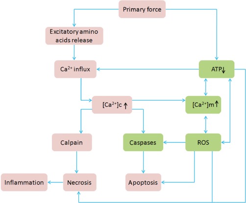
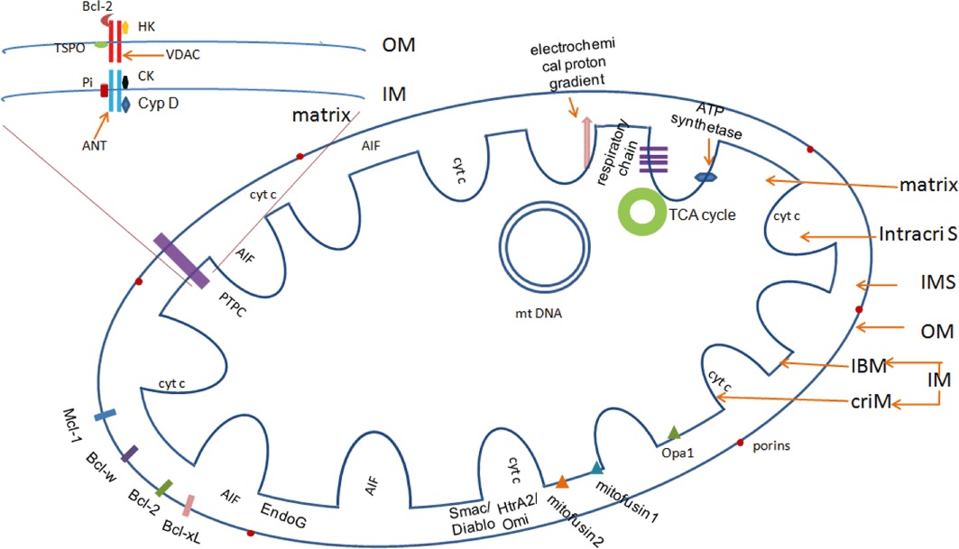
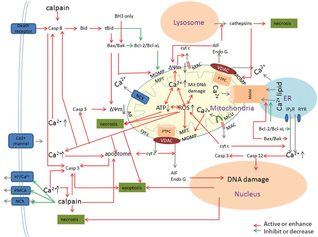
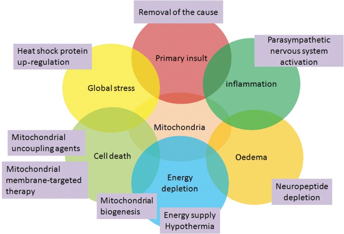
Traumatic brain injury (TBI) is a major health and socioeconomic problem throughout the world. It is a complicated pathological process that consists of primary insults and a secondary insult characterized by a set of biochemical cascades. The imbalance between a higher energy demand for repair of cell damage and decreased energy production led by mitochondrial dysfunction aggravates cell damage. At the cellular level, the main cause of the secondary deleterious cascades is cell damage that is centred in the mitochondria. Excitotoxicity, Ca(2+) overload, reactive oxygen species (ROS), Bcl-2 family, caspases and apoptosis inducing factor (AIF) are the main participants in mitochondria-centred cell damage following TBI. Some preclinical and clinical results of mitochondria-targeted therapy show promise. Mitochondria- targeted multipotential therapeutic strategies offer new hope for the successful treatment of TBI and other acute brain injuries.


Figures

Figure 1
Figure 1
Pathological changes of TBI. The primary force (trauma) will cause direct tissue damage, impaired regulation of CBF and metabolism and excessive release of excitatory neurotransmitters. This results in an abnormally high level of free intracellular and mitochondrial Ca2+. The consequent Ca2+ overload leads to self-digesting (catabolic) intracellular processes that involve overproduction of free radicals, activation of cell death signalling pathways and up-regulation of inflammatory mediators. Together, these events lead ultimately to cell death. Note: the green boxes show the main pathological changes in mitochondria. [Ca2+]c indicates intracellular Ca2+ concentration. [Ca2+]m indicates mitochondrial Ca2+ concentration.
Figure 2
Figure 2
The structure of a mitochondrion. According to the ‘crista junction’ model, each mitochondrion includes three membrane systems and three compartments. The outer membrane (OM) contains large numbers of porins that allow free diffusion of small molecules. In physiological conditions, there are many anti-apoptotic Bcl-2 family members attached to the OM. The inner membrane (IM) contains compartmentalized proteins with different functions, such as those that perform the redox reactions of oxidative phosphorylation, ATP synthase, mitochondria fusion and fission protein. There is an electrochemical proton gradient across the IM. The permeability transition pore complex (PTPC) is a supramolecular channel that is assembled at the junctions between the IM and the OM, which is proposed to be composed of the adenine nucleotide translocase (ANT), cyclophilin D (Cyp D), voltage-dependent anion channels (VDAC), creatine kinase (CK), hexokinase (HK), the translocator protein TSPO and the mitochondrial phosphate carrier Pi. The IMS contains many proteins that play important roles in cell death, such as cyt c and AIF. Enzymes that carry out the citric acid cycle and mitochondrial DNA (mtDNA) are located in the matrix.
Figure 3
Figure 3
The role of mitochondria in cell death in TBI pathology. In TBI pathology, mitochondria provide the main platform of many intertwined factors that direct the cell to live or die. Three major pathways – the extrinsic pathway, the mitochondrial pathway and the caspase-12-mediated ER apoptotic pathway participate in cell death. In the extrinsic apoptotic pathway, mitochondria amplify the apoptotic signal or are essential for execution of the apoptotic program. The BH3-only protein Bid is one of the major links between extrinsic and mitochondrial apoptosis. The mitochondrial apoptotic pathway is classified as caspase-dependent or caspase-independent. In a caspase-dependent pathway, cyt c is the essential component whereas AIF is critically involved in the caspase-independent pathway. There are synergistic effects between mitochondrial membrane, Ca2+ and ROS in mediating cell damage after TBI. MOMP and MPT are two models of the formation of pores in the mitochondrial membranes. The Bcl-2 family is a critical regulator of mitochondria and cell fate. There are many amplifying loops among different pro-apoptotic effectors. In addition, there is coordination among mitochondria, ER and lysosome. In pathological conditions, Ca2+ exchanges between mitochondria and ER mediated by MAM is an important apoptotic control point. The release of cathepsin proteases from lysosome has a positive effect on mitochondria-mediated cell death.
Figure 4
Figure 4
Mitochondrial-targeted multipotential therapeutic strategies. TBI is a pathological process with many intertwined participants that are involved in mitochondria. Combined strategies that target different pathological process will obtain the best outcome in mitochondrial-targeted multipotential therapy.

References

    1. Albensi BC, Sullivan PG, Thompson MB, Scheff SW, Mattson MP. Cyclosporin ameliorates traumatic brain-injury-induced alterations of hippocampal synaptic plasticity. Exp Neurol. 2000;162:385–389. - PubMed
    1. Alessandri B, Rice AC, Levasseur J, DeFord M, Hamm RJ, Bullock MR. Cyclosporin A improves brain tissue oxygen consumption and learning/memory performance after lateral fluid percussion injury in rats. J Neurotrauma. 2002;19:829–841. - PubMed
    1. Anderson S, Bankier AT, Barrell BG, de Bruijn MH, Coulson AR, Drouin J, et al. Sequence and organization of the human mitochondrial genome. Nature. 1981;290:457–465. - PubMed
    1. Andreyev AY, Kushnareva YE, Starkov AA. Mitochondrial metabolism of reactive oxygen species. Biochemistry (Mosc) 2005;70:200–214. - PubMed
    1. Ankarcrona M, Dypbukt JM, Bonfoco E, Zhivotovsky B, Orrenius S, Lipton SA, et al. Glutamate-induced neuronal death: a succession of necrosis or apoptosis depending on mitochondrial function. Neuron. 1995;15:961–973. - PubMed

Substances