. 2012 Sep 25:12:219.
doi: 10.1186/1471-2180-12-219.

Characterization of EssB, a protein required for secretion of ESAT-6 like proteins in Staphylococcus aureus

Affiliations

Characterization of EssB, a protein required for secretion of ESAT-6 like proteins in Staphylococcus aureus

Yi-Hsing Chen et al. BMC Microbiol. .

Abstract

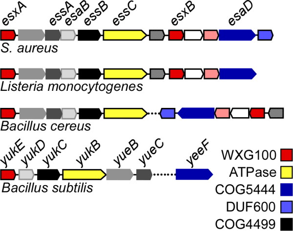
Background: Staphylococcus aureus secretes EsxA and EsxB, two small polypeptides of the WXG100 family of proteins. Genetic analyses have shown that production and secretion of EsxA and EsxB require an intact ESAT-6 Secretion System (ESS), a cluster of genes that is conserved in many Firmicutes and encompasses esxA and esxB . Here, we characterize EssB, one of the proteins encoded by the ESS cluster. EssB is highly conserved in Gram-positive bacteria and belongs to the Cluster of Orthologous Groups of protein COG4499 with no known function.

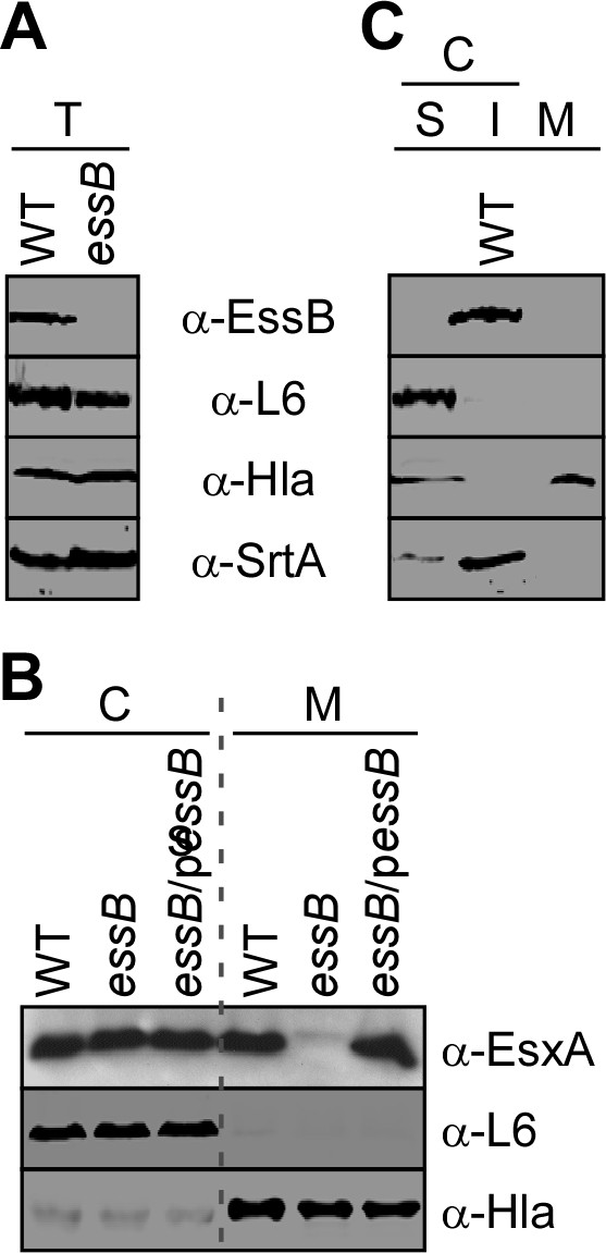
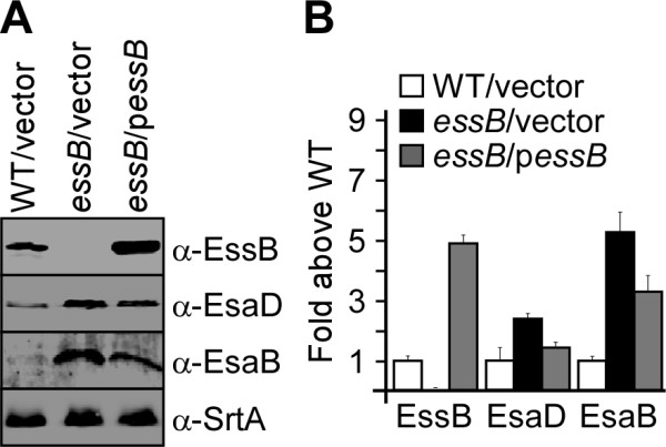
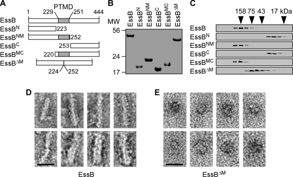
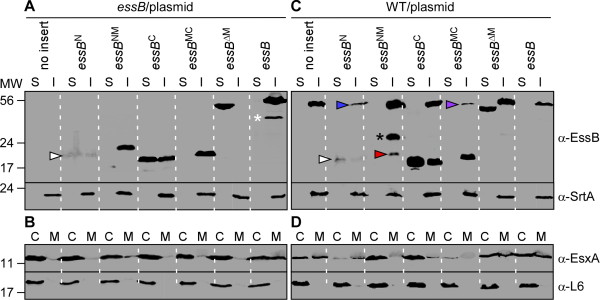
Results: By generating an internal deletion in essB , we demonstrate that EssB is required for secretion of EsxA. We use a polyclonal antibody to identify EssB and show that the protein fractionates with the plasma membrane of S. aureus . Yet, when produced in Escherichia coli, EssB remains mostly soluble and the purified protein assembles into a highly organized oligomer that can be visualized by electron microscopy. Production of truncated EssB variants in wild-type S. aureus confers a dominant negative phenotype on EsxA secretion.

Conclusions: The data presented here support the notion that EssB may oligomerize and interact with other membrane components to form the WXG100-specific translocon in S. aureus .


Figures

Figure 1
Figure 1
Schematic of ESS gene clusters in Gram-positive bacteria. Comparison of the S. aureus ESS locus with Listeria monocytogenes (strain EGD-e ), Bacillus cereus “cytotoxicus” (strain NVH391-98) and B. subtilis (subsp. subtilis strain 168) . Genes sharing sequence homology are depicted with the same color. Proteins with defined conserved domains are indicated as follows: WXG100 family of proteins (red), FtsK SpoIIIE-like ATPases (yellow), Cluster of Orthologous Groups of proteins COG5444 (dark blue), COG4499 (black) and proteins with a Domain of Unknown Function DUF600 (light blue). Dashed lines between blocks of genes indicate that the genes are not found in close proximity but elsewhere on the same chromosome. The nomenclature for the S. aureus cluster has been described [20]. The genetic organization is conserved in S. aureus strains. Gene names for B. subtilis (subsp. Subtilis strain 168) are annotated as described in the National Center for Biotechnology Information databank.
Figure 2
Figure 2
Identification and characterization of EssB. (A) S. aureus USA300 (WT) or isogenic mutant essB were examined for production (T: total culture extracts) and subcellular localization of EssB (C: cell extracts followed by 100,000 x g sedimentation and separation of soluble, S and insoluble I proteins; M: medium). Proteins in each fraction were precipitated with trichloroacetic acid, separated by SDS-PAGE and detected by immunoblotting with specific antibodies [α-EssB, as well as α-L6, α-Hla, α-SrtA, as cytoplasmic, secreted and membrane protein controls, respectively]. (B) Plasmid complementation analysis of bacterial cultures separated between cells (C) and medium (M). S. aureus USA300 (WT) or essB mutants harboring or not a complementing plasmid (p essB ) were examined for their ability to secrete EsxA in the culture medium. Samples were analyzed as in panel A.
Figure 3
Figure 3
Loss of EssB affects production of EsaB and EsaD. (A) Total culture lysates of WT (USA300) and essB mutant with the empty vector control (vector) or complementing plasmid (p essB ) were examined for the production of EssB, EsaB and EsaD using polyclonal antibodies and as described in the legend of Figure 2A. The experiment was repeated three times in duplicate and bands corresponding to immune reactive species were scanned and quantified using a Li-Cor Biosystems Odyssey imager. Quantification of the data is shown in panel B.
Figure 4
Figure 4
Purification and characterization of recombinant EssB and truncated variants. (A) Diagrammatic representation of full length EssB and truncated variants produced in E. coli. Numbers indicate amino acid positions in the primary sequence and the grey box labeled PTMD depicts the hydrophobic sequence. (B) Coomassie gel of purified recombinant proteins as shown in panel A. Proteins were purified from E. coli by affinity chromatography and affinity tags were removed. (C) Size exclusion chromatography of full length EssB and truncated variants shown in panel B. Proteins (~100 μg) were loaded onto a SuperdexTM 75 10/300 GL and fractions (0.5 ml) were collected and analyzed by SDS-PAGE. Proteins in the gel were visualized by Coomassie staining. Masses of protein standards used for calibration are shown above the gels (158, 75, 43, 17 kDa) and correspond to the exclusion volumes of Aldolase, Conalbumin, Ovalbumin and Myoglobin, respectively. (D-E) TEM of purified recombinant EssB (D) and EssBΔM (E). The proteins were allowed to bind to glow discharged grids and were negatively stained using 2% uranyl acetate. This analysis reveals a rod-like structure for EssB and more spherical, aggregated-like structure for EssBΔM. Scale bar = 20 nm.
Figure 5
Figure 5
Complementation and dominant negative activity of truncated EssB variants. (A-B) Complementation studies. S. aureus USA300 lacking functional essB was transformed with vector carrying either no insert, or various truncated variants of EssB or full length EssB. (A) The subcellular localization of EssB immune reactive species was assessed by subjecting cell lysates to ultracentrifugation to separate soluble (S) and (I) insoluble proteins and proteins in both extracts were resolved by SDS-PAGE followed by immunoblotting with specific antibodies (α-SrtA is used for subcelluar fractionation control of an insoluble membrane protein). (B) Cultures were examined for production and secretion of EsxA. Cultures were spun to separate proteins in cells (C) from secreted protein in the medium (M). α-L6 is used for fractionation control of a cytosolic protein. (C-D) Dominant negative studies. Truncated variants of EssB were examined for protein localization (C) and EsxA secretion (D) as described in panel A. All plasmids were transformed in wild-type strain USA300 (WT). All truncated variants with the exception of EssBΔM lacking PTMD prevented secretion of EsxA. The data for a duplicate of three independent experiments are shown. Arrows indicate proteins with correct mass found in reduced abundance (white arrow: EssBN; red arrow: EssBNM; blue and purple arrows: endogenous EssB). Protein products with aberrant mass are depicted with asterisks.

Similar articles

Cited by

References

    1. Dalbey RE, Wickner W. Leader peptidase catalyzes the release of exported proteins from the outer surface of the Escherichia coli plasma membrane. J Biol Chem. 1985;260:15925–15931. - PubMed
    1. Emr SD, Hanley-Way S, Silhavy TJ. Suppressor mutations that restore export of a protein with a defective signal sequence. Cell. 1981;23:79–88. doi: 10.1016/0092-8674(81)90272-5. - DOI - PubMed
    1. Oliver DB, Beckwith J. E. coli mutant pleiotropically defective in the export of secreted proteins. Cell. 1981;25:765–772. doi: 10.1016/0092-8674(81)90184-7. - DOI - PubMed
    1. Hartl FU, Lecker S, Schiebel E, Hendrick JP, Wickner W. The binding cascade of SecB to SecA to SecY/E mediates preprotein targeting to the E. coli plasma membrane. Cell. 1990;63:269–279. doi: 10.1016/0092-8674(90)90160-G. - DOI - PubMed
    1. Gorlich D, Rapoport TA. Protein translocation into proteoliposomes reconstituted from purified components of the endoplasmic reticulum membrane. Cell. 1993;75:615–630. doi: 10.1016/0092-8674(93)90483-7. - DOI - PubMed

Publication types

MeSH terms

LinkOut - more resources