Cerebral sparganosis in children: epidemiological, clinical and MR imaging characteristics
- PMID: 23006504
- PMCID: PMC3484034
- DOI: 10.1186/1471-2431-12-155
Cerebral sparganosis in children: epidemiological, clinical and MR imaging characteristics
Abstract
Background: Cerebral sparganosis in children is an extremely rare disease of central nervous system, and caused by a tapeworm larva from the genus of Spirometra. In this study, we discussed and summarized epidemiological, clinical and MR imaging characteristics of eighteen children with cerebral sparganosis for a better diagnosis and treatment of the disease.
Methods: Eighteen children with cerebral sparganosis verified by pathology, serological tests and MR presentations were retrospectively investigated, and the epidemiologic and clinical characteristics of the disease were studied.
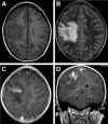
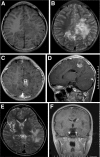
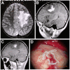
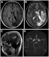
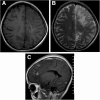
Results: Twenty-seven lesions were found in the eighteen children. Twelve lesions in twelve patients were solitary while the lesions in the rest six patients were multiple and asymmetrical. The positions of the lesions were: seven in frontal, eleven in parietal, four in temporal and two in occipital lobes, one in basal ganglia, one in cerebella hemisphere and one in pons. The lesions were presented as slight hypointensity on T1-weighted images but moderate hyperintensity on T2-weighted images with perilesional brain parenchyma edema. Enhanced MR scans by using Gadopentetic Acid Dimeglumine Salt were performed in the patients, and the images demonstrated abnormal enhancements with the patterns of a peripheral ring, or a tortuous beaded, or a serpiginous tubular shape. Follow-up MR scans were preformed for eight patients, and three out of the eight cases exposed migrations and changes in shapes of the lesion areas.
Conclusions: The MR presentations in our study in general were similar to those in previous studies. However serpiginous tubular and comma-shaped enhancements of lesions have not been previously reported. The enhanced MR imaging and follow-up MR scans with the positive results from serological tests are the most important methods for the clinical diagnosis of cerebral sparganosis in children.
Figures

Similar articles
-
[MRI diagnosis of cerebral sparganosis in children].Zhongguo Dang Dai Er Ke Za Zhi. 2008 Aug;10(4):481-4. Zhongguo Dang Dai Er Ke Za Zhi. 2008. PMID: 18706167 Chinese.
-
MR spectroscopy and MR perfusion character of cerebral sparganosis: a case report.Br J Radiol. 2010 Feb;83(986):e31-4. doi: 10.1259/bjr/73038348. Br J Radiol. 2010. PMID: 20139254 Free PMC article.
-
Cerebral sparganosis: MR imaging versus CT features.Radiology. 1993 Sep;188(3):751-7. doi: 10.1148/radiology.188.3.8351344. Radiology. 1993. PMID: 8351344
-
Cerebral sparganosis: case report and review of the European cases.Acta Neurochir (Wien). 2015 Sep;157(8):1339-43; discussion 1343. doi: 10.1007/s00701-015-2466-9. Epub 2015 Jun 18. Acta Neurochir (Wien). 2015. PMID: 26085111 Review.
-
Cerebral sparganosis: case report and review.Rev Infect Dis. 1991 Jan-Feb;13(1):155-9. doi: 10.1093/clinids/12.5.155. Rev Infect Dis. 1991. PMID: 2017616 Review.
Cited by
-
Surgical treatment of a patient with live intracranial sparganosis for 17 years.BMC Infect Dis. 2022 Apr 9;22(1):353. doi: 10.1186/s12879-022-07293-7. BMC Infect Dis. 2022. PMID: 35397512 Free PMC article.
-
Cerebral Sparganosis - An Unusual Parasitic Infection Mimicking Cerebral Tuberculosis: Isolation of a Live Plerocercoid Larva of Spirometra mansoni.Ann Indian Acad Neurol. 2024 Jul 1;27(4):443-447. doi: 10.4103/aian.aian_2_24. Epub 2024 Apr 17. Ann Indian Acad Neurol. 2024. PMID: 38819429 Free PMC article. No abstract available.
-
Diagnosis of gastrointestinal parasites in reptiles: comparison of two coprological methods.Acta Vet Scand. 2014 Aug 12;56(1):44. doi: 10.1186/s13028-014-0044-4. Acta Vet Scand. 2014. PMID: 25299119 Free PMC article.
-
Orbital sparganosis in an 8-year boy: a case report.BMC Ophthalmol. 2018 Jan 22;18(1):13. doi: 10.1186/s12886-018-0675-8. BMC Ophthalmol. 2018. PMID: 29357839 Free PMC article.
-
Follow-up study of high-dose praziquantel therapy for cerebral sparganosis.PLoS Negl Trop Dis. 2019 Jan 14;13(1):e0007018. doi: 10.1371/journal.pntd.0007018. eCollection 2019 Jan. PLoS Negl Trop Dis. 2019. PMID: 30640909 Free PMC article.
References
-
- Chen H, Wu JS, Zhang FL. et al.Clinicopathological analysis of cerebral sparganosis: eight cases report. Chin J Nerv Ment Dis. 1999;25:18–20.
-
- Takeuchi K. A case with plerocercoid which is parasitic in the brain. Nippon Byoyi Gakkai Kaishi. 1918;7:611–20.
-
- Tung CC, Lin JW, Chou FF. et al.Sparganosis in male breast. J Formos Med Assoc. 2005;104:127–8. - PubMed
-
- Zhou YQ, Ma YH, Huang HG. et al.Misdiagnosis in 12 cases of mansoni sparganosis. Clinical Misdiagnosis & Mistherapy. 1997;10:48–49.
Publication types
MeSH terms
LinkOut - more resources
Full Text Sources
Medical

