. 2012 Sep 24:10:200.
doi: 10.1186/1479-5876-10-200.

Overexpression of FoxM1 is associated with tumor progression in patients with clear cell renal cell carcinoma

Affiliations

Overexpression of FoxM1 is associated with tumor progression in patients with clear cell renal cell carcinoma

Yi-Jun Xue et al. J Transl Med. .

Abstract

Background: Fork head box M1 (FoxM1) is a proliferation-associated transcription factor essential for cell cycle progression. Numerous studies have documented that FoxM1 has multiple functions in tumorigenesis and its elevated levels are frequently associated with cancer progression. The present study was conducted to investigate the expression of FoxM1 and its prognostic significance in clear cell renal cell carcinoma (ccRCC). Meanwhile, the function of FoxM1 in human ccRCC was further investigated in cell culture models.

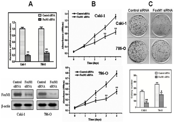
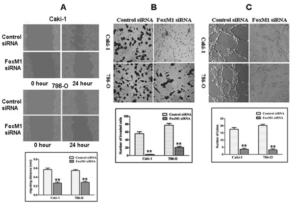
Methods: Real-time quantitative PCR, western blot and immunohistochemistry were used to explore FoxM1 expression in ccRCC cell lines and primary ccRCC clinical specimens. FoxM1 expression was knocked down by small interfering RNA (siRNA) in Caki-1 and 786-O cells; proliferation, colony formation, cell cycle, migration, invasion, and angiogenesis were assayed.

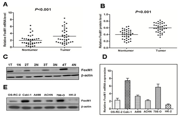
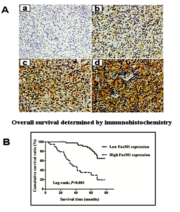
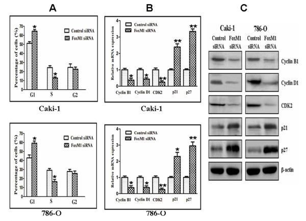
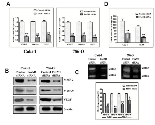
Results: FoxM1 expression was up-regulated in the majority of the ccRCC clinical tissue specimens at both mRNA and protein levels. Clinic pathological analysis showed that FoxM1 expression was significantly correlated with primary tumor stage (P <0.001), lymph node metastasis (P = 0.01), distant metastasis (P = 0.01), TNM stage (P < 0.001) and histological grade (P = 0.003). The Kaplan-Meier survival curves revealed that high FoxM1 expression was associated with poor prognosis in ccRCC patients (P < 0.001). FoxM1 expression was an independent prognostic marker of overall ccRCC patient survival in a multivariate analysis (P = 0.008). Experimentally, we found that down-regulation of FoxM1 inhibited cell proliferation and induced cell cycle arrest with reduced expression of cyclin B1, cyclin D1, and Cdk2, and increased expression of p21 and p27. Also, down-regulation of FoxM1 reduced expression and activity of matrix metalloproteinase-2 (MMP-2), MMP-9 and vascular endothelial growth factor (VEGF), resulting in the inhibition of migration, invasion, and angiogenesis.

Conclusions: These results suggest that FoxM1 expression is likely to play important roles in ccRCC development and progression, and that FoxM1 is a prognostic biomarker and a promising therapeutic target for ccRCC.


Figures

Figure 1
Figure 1
The expression of FoxM1 mRNA and protein in the human ccRCC surgical specimens and RCC cell lines, as evaluated by real-time quantitative PCR and western blot.A, The relative mRNA expression of FoxM1 was higher in 39 ccRCC tumor tissues than in matched adjacent nontumorous tissues (P < 0.001). B, The FoxM1 protein expression was higher in the tumor tissues than in matched adjacent nontumorous tissues (P < 0.001). C, Expression of FoxM1 protein in four representative pairs of ccRCC tissues is presented. N, nontumorous tissues; T, ccRCC tissues. D, The FoxM1 mRNA expression in human RCC cell lines was higher in the OS-RC-2, Caki-1, A498, ACHN and 786-O cells, particularly in the Caki-1 and 786-O cells, compared with the normal proximal tubule epithelial cell line HK-2. E, The FoxM1 protein expression was elevated in the OS-RC-2, Caki-1, A498, ACHN and 786-O cells compared to the normal proximal tubule epithelial cell line HK-2.
Figure 2
Figure 2
FoxM1 protein expression and patient survival.A, Immunohistochemical analysis of FoxM1 protein expression in 83 cases of ccRCC tissues: – in a, 1+ in b, 2+ in c, and 3+ in d. Magnification, all × 200. B, Overall survival analysis using the Kaplan–Meier method revealed that patients with high FoxM1 expression had obviously lower overall survival rates than did those with low FoxM1 expression.
Figure 3
Figure 3
Effects of FoxM1 depletion on cell growth.A, FoxM1 mRNA levels were down-regulated by FoxM1 siRNA. B, FoxM1 protein levels were down-regulated by siRNA. C, Inhibition of cancer cell proliferation by FoxM1 siRNA tested by MTT assay. D, Inhibition of cancer cell colony formation capacity by FoxM1 siRNA. Experiments were repeated at least three times, and representative data are presented; bars, SD.*, P < 0.05; **, P < 0.01, relative to control.
Figure 4
Figure 4
Effect of FoxM1 deletion on cell cycle.A, The cell cycle distribution was analyzed using propidium iodide staining and flow cytometry. B and C, The expression level of several known cell cycle regulatory factors as detected by real-time quantitative PCR (B) and Western blotting (C), respectively. The experiments were repeated thrice. *, P < 0.05; **, P < 0.01, relative to control.
Figure 5
Figure 5
Effect of FoxM1 deletion on the expression of various cell cycle regulatory factors and MMP-2, MMP-9, and VEGF.A and B, real-time quantitative PCR and Western blot analysis showed that FoxM1 siRNA inhibited the expression of MMP-2, MMP-9, and VEGF at mRNA and protein levels in Caki-1 and 786-O cells. C, FoxM1 siRNA inhibited the activity of MMP-2 and MMP-9 based on gelatin zymography assay in Caki-1 and 786-O cells. D, FoxM1 siRNA inhibited the activity of VEGF in Caki-1 and 786-O cells. The experiments were repeated thrice. *, P < 0.05; **, P < 0.01, relative to control.
Figure 6
Figure 6
FoxM1 siRNA decreased migration and invasion of Caki-1 and 786-O cells, and reduced the HUVECs tube formation.A, Scratch migration assay showing that FoxM1 siRNA decreased cell migration. B, Matrigel invasion assay showing that FoxM1 siRNA-transfected cells resulted in low penetration through the Matrigel-coated membrane, compared with control cells. C, Conditioned media from FoxM1 siRNA-transfected cells were able to significantly reduce the tube formation of HUVECs compared with the medium from control cells. The experiments were repeated thrice. **, P < 0.01, relative to control.

References

    1. Rini BI, Rathmell WK, Godley P. Renal cell carcinoma. Curr Opin Oncol. 2008;20:300–306. doi: 10.1097/CCO.0b013e3282f9782b. - DOI - PubMed
    1. Siegel R, Ward E, Brawley O, Jemal A. Cancer statistics, 2011: the impact of eliminating socioeconomic and racial disparities on premature cancer deaths. CA Cancer J Clin. 2011;61:212–236. doi: 10.3322/caac.20121. - DOI - PubMed
    1. Gupta K, Miller JD, Li JZ, Russell MW, Charbonneau C. Epidemiologic and socioeconomic burden of metastatic renal cell carcinoma (mRCC): a literature review. Cancer Treat Rev. 2008;34:193–205. doi: 10.1016/j.ctrv.2007.12.001. - DOI - PubMed
    1. Rini BI, Campbell SC, Escudier B. Renal cell carcinoma. Lancet. 2009;373:1119–1132. doi: 10.1016/S0140-6736(09)60229-4. - DOI - PubMed
    1. Cindolo L, Patard JJ, Chiodini P, Schips L, Ficarra V, Tostain J, de La Taille A, Altieri V, Lobel B, Zigeuner RE, Artibani W, Guillé F, Abbou CC, Salzano L, Gallo C. Comparison of predictive accuracy of four prognostic models for nonmetastatic renal cell carcinoma after nephrectomy: A multicenter European study. Cancer. 2005;104:1362–1371. doi: 10.1002/cncr.21331. - DOI - PubMed