

Cell interaction study method using novel 3D silica nanoneedle gradient arrays
- PMID: 23006558
- PMCID: PMC3513632
- DOI: 10.1016/j.colsurfb.2012.07.044
Cell interaction study method using novel 3D silica nanoneedle gradient arrays
Abstract
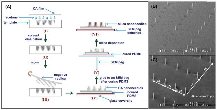
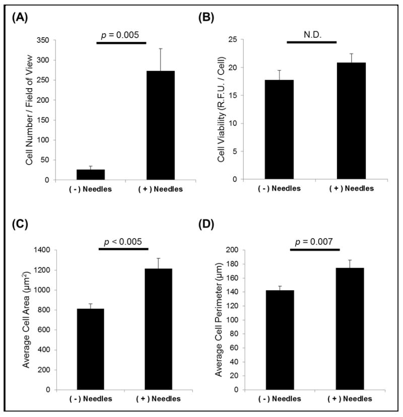
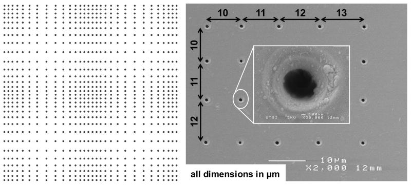
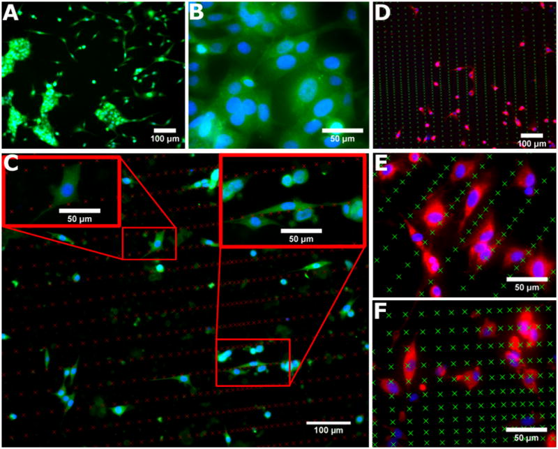
Understanding cellular interactions with culture substrate features is important to advance cell biology and regenerative medicine. When surface topographical features are considerably larger in vertical dimension and are spaced at least one cell dimension apart, the features act as 3D physical barriers that can guide cell adhesion, thereby altering cell behavior. In the present study, we investigated competitive interactions of cells with neighboring cells and matrix using a novel nanoneedle gradient array. A gradient array of nanoholes was patterned at the surface of fused silica by single-pulse femtosecond laser machining. A negative replica of the pattern was extracted by nanoimprinting with a thin film of polymer. Silica was deposited on top of the polymer replica to form silica nanoneedles. NIH 3T3 fibroblasts were cultured on silica nanoneedles and their behavior was studied and compared with those cultured on a flat silica surface. The presence of silica nanoneedles was found to enhance the adhesion of fibroblasts while maintaining cell viability. The anisotropy in the arrangement of silica nanoneedles was found to affect the morphology and spreading of fibroblasts. Additionally, variations in nanoneedle spacing regulated cell-matrix and cell-cell interactions, effectively preventing cell aggregation in areas of tightly-packed nanoneedles. This proof-of-concept study provides a reproducible means for controlling competitive cell adhesion events and offers a novel system whose properties can be manipulated to intimately control cell behavior.
Copyright © 2012 Elsevier B.V. All rights reserved.
Figures
Similar articles
-
Silica coating of polymer nanowires produced via nanoimprint lithography from femtosecond laser machined templates.Nanotechnology. 2012 Mar 16;23(10):105304. doi: 10.1088/0957-4484/23/10/105304. Epub 2012 Feb 24. Nanotechnology. 2012. PMID: 22361985
-
Intracellular delivery of molecules using microfabricated nanoneedle arrays.Biomed Microdevices. 2016 Feb;18(1):10. doi: 10.1007/s10544-016-0038-2. Biomed Microdevices. 2016. PMID: 26797026
-
Rapid Cell Patterning Induced by Differential Topography on Silica Nanofractal Substrates.Small. 2015 Nov 11;11(42):5642-6. doi: 10.1002/smll.201502085. Epub 2015 Sep 16. Small. 2015. PMID: 26376008
-
Tutorial: using nanoneedles for intracellular delivery.Nat Protoc. 2021 Oct;16(10):4539-4563. doi: 10.1038/s41596-021-00600-7. Epub 2021 Aug 23. Nat Protoc. 2021. PMID: 34426708 Review.
-
Controlled silica synthesis inspired by diatom silicon biomineralization.J Nanosci Nanotechnol. 2005 Jan;5(1):68-78. doi: 10.1166/jnn.2005.0l0. J Nanosci Nanotechnol. 2005. PMID: 15762163 Review.
Cited by
-
Cell microenvironment engineering and monitoring for tissue engineering and regenerative medicine: the recent advances.Biomed Res Int. 2014;2014:921905. doi: 10.1155/2014/921905. Epub 2014 Jul 20. Biomed Res Int. 2014. PMID: 25143954 Free PMC article. Review.
-
Engineered Microsystems for Spheroid and Organoid Studies.Adv Healthc Mater. 2021 Jan;10(2):e2001284. doi: 10.1002/adhm.202001284. Epub 2020 Nov 13. Adv Healthc Mater. 2021. PMID: 33185040 Free PMC article. Review.
-
Ultrathin Polymer Membranes with Patterned, Micrometric Pores for Organs-on-Chips.ACS Appl Mater Interfaces. 2016 Aug 31;8(34):22629-36. doi: 10.1021/acsami.6b05754. Epub 2016 Aug 22. ACS Appl Mater Interfaces. 2016. PMID: 27513606 Free PMC article.
-
Femtosecond laser-patterned nanopore arrays for surface-mediated peptide treatment.Nanomedicine. 2014 Jan;10(1):11-4. doi: 10.1016/j.nano.2013.09.002. Epub 2013 Oct 1. Nanomedicine. 2014. PMID: 24090768 Free PMC article.
References
-
- Geiger B, Spatz JP, Bershadsky AD. Environmental sensing through focal adhesions. Nat Rev Mol Cell Biol. 2009;10:21–33. - PubMed
-
- Chandra P, Lai K, Sung HJ, Murthy NS, Kohn J. UV laser-ablated surface textures as potential regulator of cellular response. Biointerphases. 2010;5:53–59. - PubMed
-
- Ross AM, Jiang ZX, Bastmeyer M, Lahann J. Physical Aspects of Cell Culture Substrates: Topography, Roughness, and Elasticity. Small. 2012;8:336–355. - PubMed
Publication types
MeSH terms
Substances
Grants and funding
LinkOut - more resources
Full Text Sources
Other Literature Sources
