. 2012 Nov;60(5):1192-9.
doi: 10.1161/HYPERTENSIONAHA.112.196840. Epub 2012 Sep 24.

Calpain-1 regulation of matrix metalloproteinase 2 activity in vascular smooth muscle cells facilitates age-associated aortic wall calcification and fibrosis

Affiliations

Calpain-1 regulation of matrix metalloproteinase 2 activity in vascular smooth muscle cells facilitates age-associated aortic wall calcification and fibrosis

Liqun Jiang et al. Hypertension. 2012 Nov.

Abstract

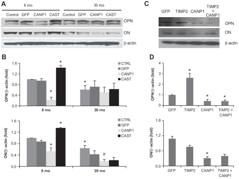
Age-associated central arterial wall stiffness is linked to extracellular matrix remodeling, including fibrosis and vascular calcification. Angiotensin II induces both matrix metalloproteinase 2 (MMP2) and calpain-1 expression and activity in the arterial wall. However, the role of calpain-1 in MMP2 activation and extracellular matrix remodeling remains unknown. Dual histo-immunolabeling demonstrates colocalization of calpain-1 and MMP2 within old rat vascular smooth muscle cells. Overexpression of calpain-1 induces MMP2 transcripts, protein levels, and activity, in part, by increasing the ratio of membrane type 1 MMPs to tissue inhibitor of metalloproteinases 2. These effects of calpain-1 overexpression-induced MMP2 activation are linked to increased collagen I and III production and vascular calcification. In addition, overexpression of calpain-1 also induces transforming growth factor-β1/Smad signaling, elastin degradation, alkaline phosphatase activation, and total calcium content but reduces the expression of calcification inhibitors, osteopontin, and osteonectin, in cultured vascular smooth muscle cells in vitro and in carotid artery rings ex vivo. Furthermore, both calpain-1 and collagen II increase with aging within human aortic intima. Interestingly, in aged human aortic wall, both calpain-1 and collagen II are highly expressed in artherosclerotic plaque areas compared with grossly normal areas. Cross-talk of 2 proteases, calpain-1 and MMP2, leads to secretion of active MMP2, which modulates extracellular matrix remodeling via enhancing collagen production and facilitating vascular calcification. These results establish calpain-1 as a novel molecular candidate to retard age-associated extracellular matrix remodeling and its attendant risk for hypertension and atherosclerosis.


Figures

Figure 1
Figure 1. Calpain-1 induces MMP2 activation in VSMC
A. Photomicrographs of dual labeling for calpain-1 (green) and matrix metalloproteinase-2 (MMP2, red) within old rat VSMC; merged image (right panel). Nuclei are counterstained with DAPI (blue). Magnification: X400. B. Representative Western blot and average MMP2 protein levels in young (8 mo) and old (30 mo) rat VSMC infected with GFP or CANP1 for 48 hrs. C. Representative gelatin zymograph and average MMP2 activity from young (8 mo) and old (30 mo) rat VSMC infected with GFP or CANP1 for 48 hrs. D. Representative gelatin zymograph and average MMP2 activity from young (8 mo) and old (30 mo) rat VSMC infected with control or calpastatin (CAST) adenoviruses. *: p<0.05 compared to GFP infected young VSMC, n=3. †: p<0.05 compared to young blank VSMC, n=3. # p<0.05 compared to GFP infected old VSMC, n=3.
Figure 2
Figure 2. Calpain-1 induces MT1MMP/TIMP2 ratio in VSMC
Representative western blot (A) and average protein levels of TIMP2 and MT1MMP from VSMC infected by GFP or CANP1 adenoviruses for 48 hrs. *: p<0.05 compared to GFP infected 8-mo VSMC, n=3. # p<0.05 compared to GFP infected old VSMC, n=3.
Figure 3
Figure 3. Over-expression of calpain-1 induces collagen type I, II and III, and reduces elastin expression levels in VSMC
A. Representative western blots of collagen I and collagen III in VSMC after infected by GFP or CANP1 adenoviruses for 48 hrs. B. Photomicrographs (X400) of collagen II protein staining (brown color) from young (8-mo) and old (30-mo) rat aortic wall. Nuclei are counterstained with hematoxylin (blue color). L = lumen; and M = media. C and D. Representative Western blots and average protein levels collagen II (C) and elastin (D) in young VSMC infected with GFP or CANP1 for 48 hours. *: p<0.05 compared to GFP infected 8-mo VSMC, n=3.
Figure 4
Figure 4. Over-expression of calpain-1 induces VSMC calcification
A. Representative Alizarin Red S Staining showing that compared to GFP control (top left panel) over-expression of calpain-1 induces local calcification in young (8-mo) VSMC (middle left panel) in high phosphate (2.0 mmol/L) calcification medium. Resultant levels in these treated young VSMC are comparable to the levels of old GFP control (30-mo, lower left panel). Calpain-1 induced calcification is inhibited by TIMP2 (lower right panel). Red deposits indicate deposits of phosphate-containing mineral. GFP infected VSMC without phosphate (lower right panel) is a negative control. Representative Western blots and average protein levels of TGF-β1 (B), and Smad 2/3 (C) in young (8-mo) and old (30-mo) VSMC infected with GFP or CANP1 for 48 hours. *: p<0.05 compared to GFP infected 8-mo VSMC, n=3.
Figure 5
Figure 5. Over-expression of Calpain-1 down-regulates osteoponin (OPN) and osteonectin (ON) levels in VSMC, while over-expression of calpastatin, a specific inhibitor of calpain-1 has the opposite effects
Representative western blot (A, C) and average protein levels (B, D) of OPN and ON in VSMC infected by GFP or CANP1 or CAST or TIMP2 adenoviruses for 48 hrs. *: p<0.05 compared to GFP infected 8-mo VSMC, n=3. # p<0.05 compared to GFP infected old VSMC, n=3.
Figure 6
Figure 6. Over-expression of calpain-1 regulates ECM remodeling, particularly fibrosis and vascular calcification in cultured carotid artery rings
Representative western blot of TIMP2, calpain-1, elastin, TGFβ1, (Figure 6A), Smad 2/3, Collagen I, II, III, (Figure 6B) and OPN and ON (Figure 6C) in cultured carotid artery rings isolated from 8-mo old rat after infected by GFP or CANP1 or TIMP2 adenoviruses for 48 hrs.
Figure 7
Figure 7. Both calpain-1 and collagen II are increased in human aortic intima and arthrosclerosis area
A. Representative western blot and average protein levels of calpain-1 and collagen II in human aortic intima. B. Photomicrographs (X400) of calpain-1, collagen II protein staining (brown color) within arthrosclerosis area from old human aortic wall. Nuclei were counterstained with hematoxylin (blue color). Representative Alizarin Red S staining showing local calcification within the same atherosclerotic area. C. calpain-1 activity is significantly increased in the atherosclerotic area compared to normal area in human aortic wall. *: p<0.05 compared to young ones (A) or normal area (C) in human aortic wall, n=3.

References

    1. Lakatta EG, Levy D. Arterial and cardiac aging: major shareholders in cardiovascular disease enterprises: part I: aging arteries: a “set up” for vascular disease. Circulation. 2003;107:139–146. - PubMed
    1. Lakatta EG. Arterial and cardiac aging: major shareholders in cardiovascular disease enterprises: part III: cellular and molecular clues to heart and arterial aging. Circulation. 2003;107:490–497. - PubMed
    1. Wang M, Monticone RE, Lakatta EG. Arterial aging: a journey into subclinical arterial disease. Curr Opin Nephrol Hypertens. 2010;19:201–207. - PMC - PubMed
    1. Wang M, Khazan B, Lakatta EG. Central Arterial Aging and Angiotensin II Signaling. Curr Hypertens Rev. 2010;6:266–281. - PMC - PubMed
    1. Lakatta EG, Wang M, Najjar SS. Arterial aging and subclinical arterial disease are fundamentally intertwined at macroscopic and molecular levels. Med Clin North Am. 2009;93:583–604. - PMC - PubMed

Publication types

MeSH terms

LinkOut - more resources