. 2012 Sep 24;2(1):49.
doi: 10.1186/2191-219X-2-49.

Estimation of absorbed dose to the kidneys in patients after treatment with 177Lu-octreotate: comparison between methods based on planar scintigraphy

Affiliations

Estimation of absorbed dose to the kidneys in patients after treatment with 177Lu-octreotate: comparison between methods based on planar scintigraphy

Maria Larsson et al. EJNMMI Res. .

Abstract

Background: Lu-[DOTA0, Tyr3]-octreotate (177Lu-octreotate) is used to treat neuroendocrine tumors with high somatostatin-receptor expression. 177Lu-octreotate is mainly excreted via the kidneys, but to some extent, accumulates in the kidney cortex due to, e.g., tubular reabsorption. Renal toxicity is one of the main limiting factors in 177Lu-octreotate treatment. Further knowledge of the biodistribution and dosimetry of 177Lu-octreotate in individual patients is needed. The aim of this study was to estimate the absorbed dose to the kidneys and compare the results obtained with planar imaging and different dosimetric methods: (1) conjugate-view (CV) method using patient-specific kidney sizes, (2) PA method, based on posterior images only, (3) CV method with reduced number of time points (CVreduced data), and (4) CV method using standard kidney sizes (CVstandard size).

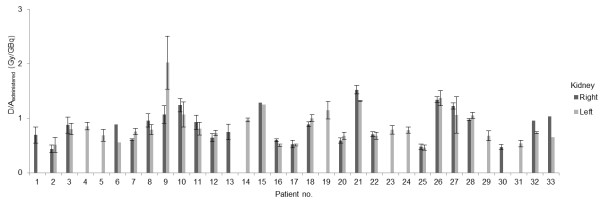
Methods: Totally, 33 patients each received 3.4 to 8.2 GBq of 177Lu-octreotate up to five times, with infusion of lysine and arginine to block the renal uptake. Whole-body planar gamma camera images were acquired on days 0, 1, 2, and 7. The 177Lu concentration in the kidneys was determined by the CV method, and the absorbed dose was estimated with patient-specific organ sizes. Comparison to the CV method was made using posterior images only, together with the influence of the number of time points and with standard organ sizes.

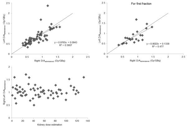
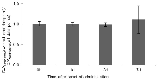
Results: Large interindividual variations were found in the time-activity curve pattern and in the absorbed dose to the kidneys using the CV method: 0.33 to 2.4 Gy/GBq (mean = 0.80 Gy/GBq, SD = 0.30). In the individual patient, the mean deviation of all subsequent kidney doses compared to that of the first administration was 1% (SD = 19%) and 5% (SD = 23%) for the right and left kidneys, respectively. Excluding data for day 7 resulted in large variations in the absorbed dose.

Conclusion: Large interindividual variations in kidney dose were found, demonstrating the need for patient-specific dosimetry and treatment planning.


Figures

Figure 1
Figure 1
The177Lu activity in the right and left kidneys given as percent of injected activity. The 177Lu activity in the right and left kidneys given as percent of injected activity (%IA) for (A) and (B) patient no. 8 (left and right kidneys, treatment cycles 1 to 4) and (C) for patient no. 24 (left kidney, treatment cycles 1 to 4).
Figure 2
Figure 2
The mean absorbed dose per unit administered activity. The mean absorbed dose per unit administered activity, to the right and left kidneys, respectively, for all 33 patients included in the study. Results are given as mean values of all treatment cycles. Patient nos. 6, 15, and 33 only received one treatment cycle, and for patient no. 32 data for the right kidney could only be obtained from one treatment cycle. Patient no. 5 had only one kidney. For patient nos. 1, 4, 13, 14, 19, 23, 24, 29, 30, and 31 only one kidney dose could be evaluated due to high uptake in overlapping and adjacent tissues (see text). Error bars indicate ± 1 SD.
Figure 3
Figure 3
The mean absorbed dose per unit administered activity for each treatment cycle in each patient. The mean absorbed dose per unit administered activity given to (A) right kidney and (B) left kidney for each treatment cycle in each patient. The mean absorbed dose per unit administered activity normalized to the value for the first treatment cycle given to (C) right kidney and (D) left kidney for each patient. Each line corresponds to one patient and the different colors are used to help the reader to follow at least some of the lines to better see different patterns.
Figure 4
Figure 4
Comparison between the mean absorbed dose per unit administered activity to the left and right kidneys. Comparison between the mean absorbed dose per unit administered activity to the left and right kidneys for (A) all treatment cycles and (B) only the first treatment cycle. (C) Ratio between the absorbed dose to the right and left kidneys for all treatment cycles in the studied 22 patients.
Figure 5
Figure 5
Correlation between PA and CV methods. Correlation between the mean absorbed dose per unit administered activity, estimated with the PA and the CV methods for the (A) right and (B) left kidneys.
Figure 6
Figure 6
Mean absorbed dose per unit administered activity, excluding one of the data points (CVreduced datamethod). Mean absorbed dose per unit administered activity to the kidney determined excluding one of the data points (1 h, or 1, 2, or 7 days after administration) divided by the mean absorbed dose per unit administered activity including all data points. Error bars indicate ± 1 SD. Note that the value, when excluding the day 7, data point is underestimated since values of infinity were excluded.

References

    1. Pool SE, Krenning EP, Koning GA, van Eijck CH, Teunissen JJ, Kam B, Valkema R, Kwekkeboom DJ, de Jong M. Preclinical and clinical studies of peptide receptor radionuclide therapy. Semin Nucl Med. 2010;40:209–218. doi: 10.1053/j.semnuclmed.2009.12.001. - DOI - PubMed
    1. Kwekkeboom DJ, Kam BL, van Essen M, Teunissen JJ, van Eijck CH, Valkema R, de Jong M, de Herder WW, Krenning EP. Somatostatin-receptor-based imaging and therapy of gastroenteropancreatic neuroendocrine tumors. Endocr Relat Cancer. 2010;17:R53–R73. doi: 10.1677/ERC-09-0078. - DOI - PubMed
    1. Kwekkeboom DJ, Bakker WH, Kooij PP, Konijnenberg MW, Srinivasan A, Erion JL, Schmidt MA, Bugaj JL, de Jong M, Krenning EP. [177Lu-DOTAOTyr3]octreotate: comparison with [111In-DTPAo]octreotide in patients. Eur J Nucl Med. 2001;28:1319–1325. doi: 10.1007/s002590100574. - DOI - PubMed
    1. Sward C, Bernhardt P, Ahlman H, Wangberg B, Forssell-Aronsson E, Larsson M, Svensson J, Rossi-Norrlund R, Kolby L. [177Lu-DOTA 0-Tyr 3]-octreotate treatment in patients with disseminated gastroenteropancreatic neuroendocrine tumors: the value of measuring absorbed dose to the kidney. World Journal of Surgery. 2010;34:1368–1372. doi: 10.1007/s00268-009-0387-6. - DOI - PubMed
    1. Melis M, Krenning EP, Bernard BF, Barone R, Visser TJ, de Jong M. Localisation and mechanism of renal retention of radiolabelled somatostatin analogues. Eur J Nucl Med Mol Imaging. 2005;32:1136–1143. doi: 10.1007/s00259-005-1793-0. - DOI - PubMed

LinkOut - more resources