Review
. 2013;103(1):10-20.
doi: 10.1159/000342340. Epub 2012 Sep 20.

Paneth cells and necrotizing enterocolitis: a novel hypothesis for disease pathogenesis

Affiliations
Review

Paneth cells and necrotizing enterocolitis: a novel hypothesis for disease pathogenesis

Steven J McElroy et al. Neonatology. 2013.

Abstract

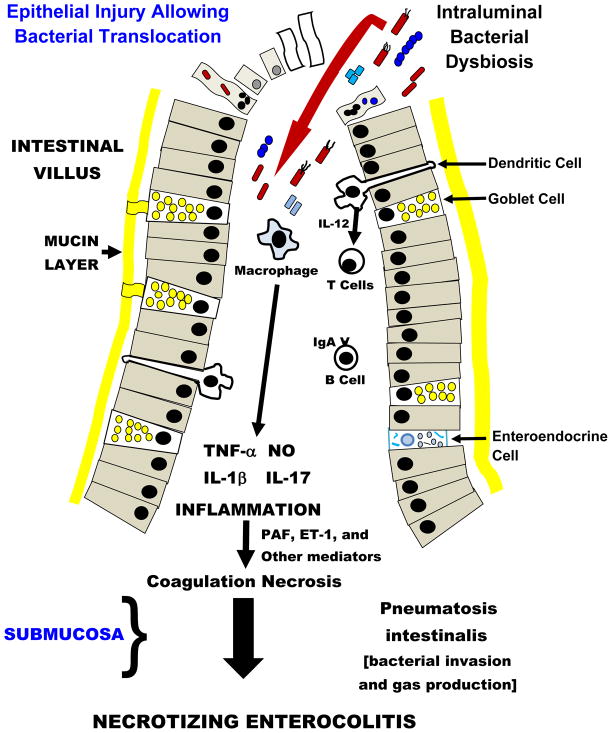
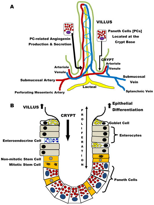
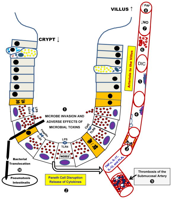
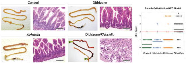
Current models of necrotizing enterocolitis (NEC) propose that intraluminal microbes destroy intestinal mucosa and activate an inflammatory cascade that ends in necrosis. We suggest an alternate hypothesis wherein NEC is caused by injury to Paneth cells (PCs) in the intestinal crypts. PCs are specialized epithelia that protect intestinal stem cells from pathogens, stimulate stem cell differentiation, shape the intestinal microbiota, and assist in repairing the gut. Our novel model of NEC uses neonatal mice and ablates PCs followed by enteral infection. We contrast this model with other animal examples of NEC and the clinical disease. Selective destruction of PCs using dithizone likely releases tumor necrosis factor-α and other inflammatory mediators. We propose that this event produces inflammation in the submucosa, generates platelet-activating factor, and induces a coagulopathy. The role of PCs in NEC is consistent with the onset of disease in preterm infants after a period of PC-related maturation, the central role of PCs in crypt-related homeostasis, the anatomic location of pneumatosis intestinalis close to the crypts, and the proximity of PCs to occluded blood vessels that cause coagulation necrosis of the intestinal villi. We offer this hypothesis to promote new thoughts about how NEC occurs and its potential prevention.


Conflict of interest statement

Disclosure Statement

Drs. McElroy, Underwood and Sherman have no financial conflicts of interest to report.

Figures

Fig 1
Fig 1. Traditional “Top Down” Hypothesis of Necrotizing Enterocolitis
Intraluminal bacterial dysbiosis causes activation of epithelial cell pathogen-associated molecular pattern receptors, especially Toll-like Receptor 4, and may induce either apoptosis (identified as small nucleated cells) or necrosis (identified as ghost cells). Bacterial invasion of epithelia can also initiate anoikis (apoptosis and cell detachment from the basement membrane – cells with bacteria and micronuclei). Failure of the epithelial barrier allows bacterial translocation and results in a vigorous inflammatory response and causes coagulation necrosis, a constant submucosal finding in necrotizing enterocolitis (NEC). The invading bacteria also gain access to the submucosa, where they ferment sugar and make gas-filled bullae called pneumatosis intestinalis. Please note there is no mention of Paneth cells in this conceptual pathophysiology for NEC.
Figure 2
Figure 2. Paneth Cells (PCs) – Normal Anatomy and Function
In addition to their antimicrobial functions, Paneth cells play a vital role regulating the intestinal architecture. A) PCs are located near the vasculature entering and leaving the villi and they regulate blood vessel growth through the secretion of angiogenins and B) PCs are found intertwined with intestinal stem cells in the crypts. PCs are critical to maintaining stem cell homeostasis through Wnt signaling pathways. Cooperation between PCs and stem cells allows orderly regeneration of intestinal surface epithelia on a continuous basis.
Fig 3
Fig 3. Paneth Cells -- The “Bottom Up” Hypothesis of Necrotizing Enterocolitis
In this figure, the sequence of events associated with the ‘Bottom Up’ hypothesis is identified by bold numbers. ➊ Microbial toxins and invasion cause disruption of Paneth cells (PCs). On the luminal surface of PCs, lipopolysaccharide (LPS) receptors CD-14 and Toll-like receptor-4 (TLR-4) are identified. Additionally, the TLR-5 receptor that binds flagellin and TLR-9 receptor that binds CpG are shown. ➋ Disruption of PCs releases their granules into the crypt space, while cytokines likely undergo basolateral discharge into the lamina propria and its adjacent microvasculature. ➌ Released cytokines (tumor necrosis factor-alpha, TNF-α; interleukin-17, IL-17; interleukin-1 beta, IL-1β) and other mediators (phospholipase A2, PLA2) activate inflammation and injure endothelium. ➍ Altered endothelia enhance neutrophil and platelet attachment to their surface. ➎ Intravascular inflammation initiates fibrin aggregation to endothelium, adherent platelets and adherent neutrophils. ➏ A disseminated intravascular coagulopathy ensues with damaging effects to erythrocytes. ➐ Endothelial damage is magnified and markedly decreases production of nitric oxide (NO) which additionally hinders perfusion of the intestinal villi. ➑ This phenomenon also generates platelet activating factor (PAF) and other mediators that cause downstream vasoconstriction in the intestinal villi. ➒ The low flow state and intravascular coagulopathy extends to the submucosal vessels and thrombotic occlusion takes place. This vascular obstruction causes coagulation necrosis of the intestinal mucosa, a common histologic finding in necrotizing enterocolitis (NEC). ➓ Finally, microbes that have invaded the disrupted crypts, move to the submucosa, ferment sugars and produce gas, and thus are the origins of pneumatosis intestinalis, the pathognomonic sign of NEC.
Fig 4
Fig 4. Selective ablation of Paneth cells followed by enteric infection with Klebsiella induces a NEC-like injury in 14- to 16-day-old mice
P14 – P16 CD-1 mice were divided into four groups (control n = 31, dithizone only n = 42, Klebsiella only n = 13 and dithizone/Klebsiella {designated Dith/Kleb}, n = 30). Mice were given either an intraperitoneal of dithizone (75 mg/kg) or an equivalent volume of Li2CO3 diluent. At 6 hours after the injection, mice were given either a gavage feeding of 1 x 10¹¹ CFU Klebsiella pneumoniae per kg body weight suspended in nutrient broth or nutrient broth only. Mice were monitored for 10 hours following gavage and then underwent euthanasia for harvesting of the small intestine. A blinded investigator applied a Lickert system that evaluated villus integrity on a scale of 0 to 4. Mice treated with Dith/Kleb were significantly different from the other groups and their average score was >2 (see red line on scoring graphic) and was considered significant for NEC-like damage (* P<0.001 vs. other groups). The loss of intestinal villi on histologic analyses of the Dith/Kleb-treated pups is remarkable compared to the control groups. Horizontal lines for each group indicate the mean histologic score. In the Dith/Kleb group, the terminal ileum showed transmural necrosis and is displayed between the black and red arrows; intestinal damage is also seen proximally. Internal size scale: bars are 0.1 mm. Permission granted for use of this illustration from Diseases Models and Mechanisms.

References

    1. Kliegman RM, Fanaroff AA. Necrotizing enterocolitis. N Engl J Med. 1984;310:1093–1103. - PubMed
    1. Neu J, Walker WA. Necrotizing enterocolitis. N Engl J Med. 2011;364:255–264. - PMC - PubMed
    1. Hsueh W, De Plaen IG, Caplan MS, Qu XW, Tan XD, Gonzalez-Crussi F. Neonatal necrotizing enterocolitis: clinical aspects, experimental models and pathogenesis. World J Pediatr. 2007;3:17–29.
    1. Grave GD, Nelson SA, Walker WA, Moss RL, Dvorak B, Hamilton FA, Higgins R, Raju TN. New therapies and preventive approaches for necrotizing enterocolitis: report of a research planning workshop. Pediatr Res. 2007;62:510–514. - PubMed
    1. Afrazi A, Sodhi CP, Richardson W, Neal M, Good M, Siggers R, Hackam DJ. New insights into the pathogenesis and treatment of necrotizing enterocolitis: Toll-like receptors and beyond. Pediatr Res. 2011;69:183–188. - PMC - PubMed

Publication types