. 2012 Dec;92(12):1712-25.
doi: 10.1038/labinvest.2012.121. Epub 2012 Sep 24.

Lumican, an extracellular matrix proteoglycan, is a novel requisite for hepatic fibrosis

Affiliations

Lumican, an extracellular matrix proteoglycan, is a novel requisite for hepatic fibrosis

Anuradha Krishnan et al. Lab Invest. 2012 Dec.

Abstract

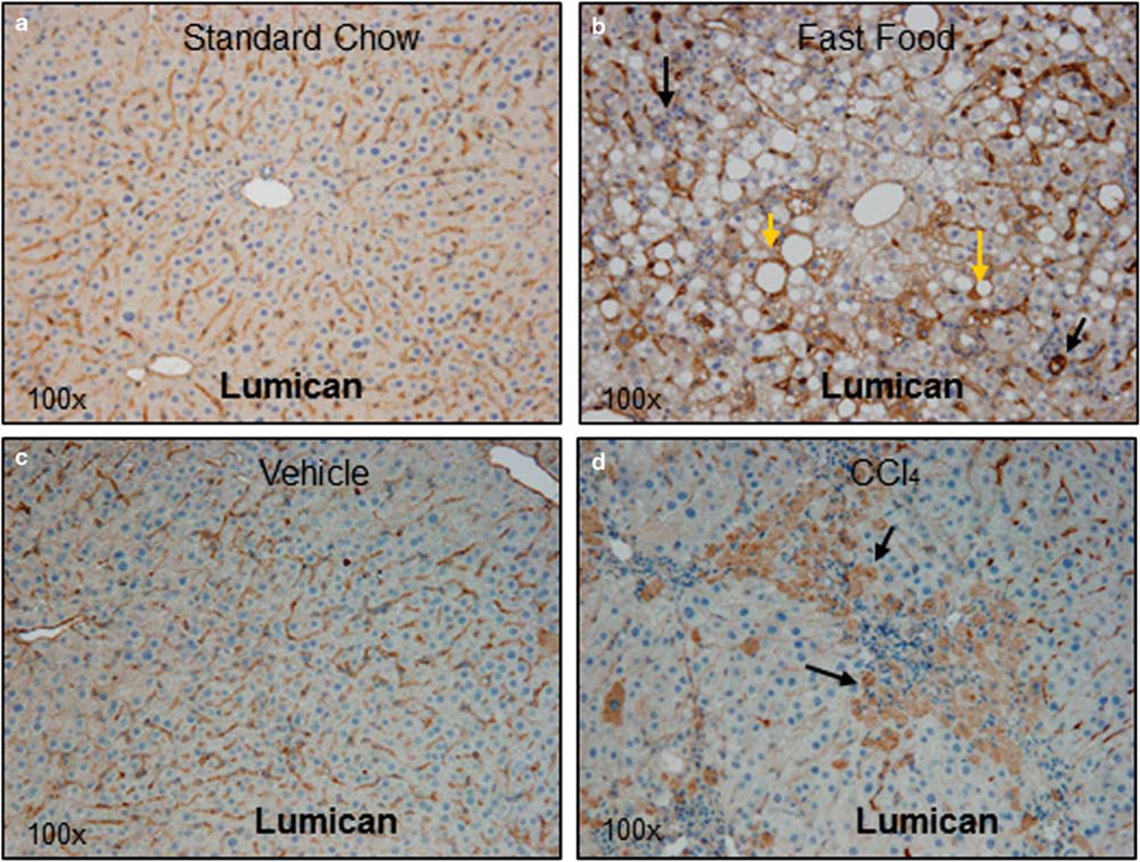
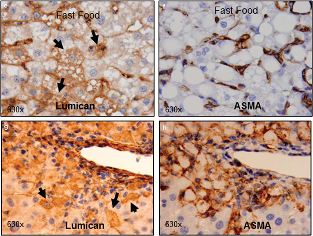
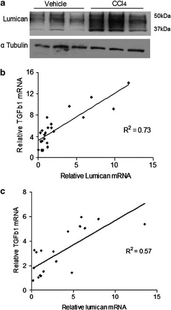
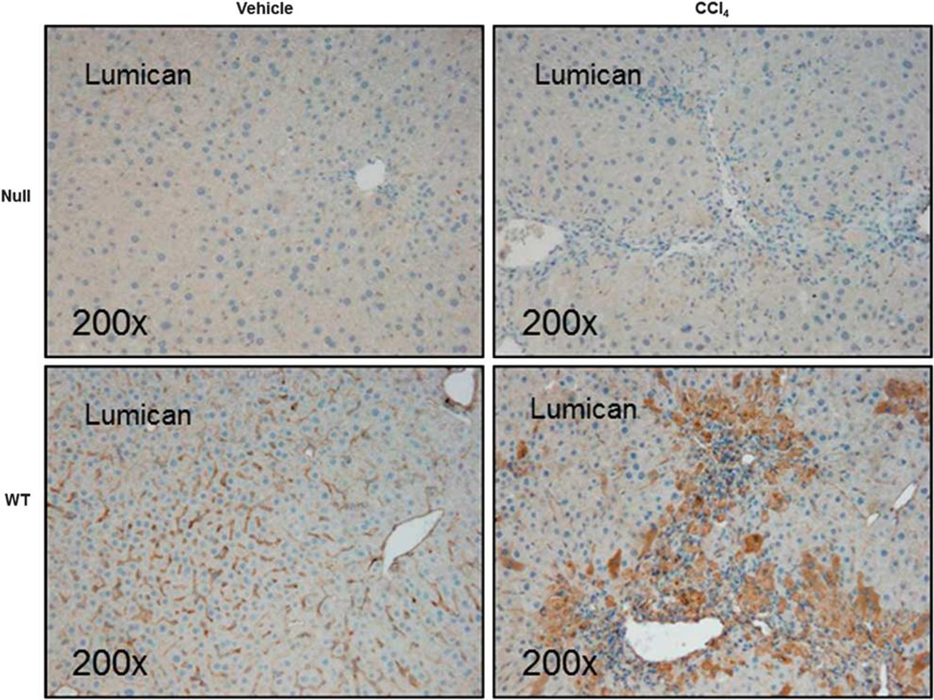
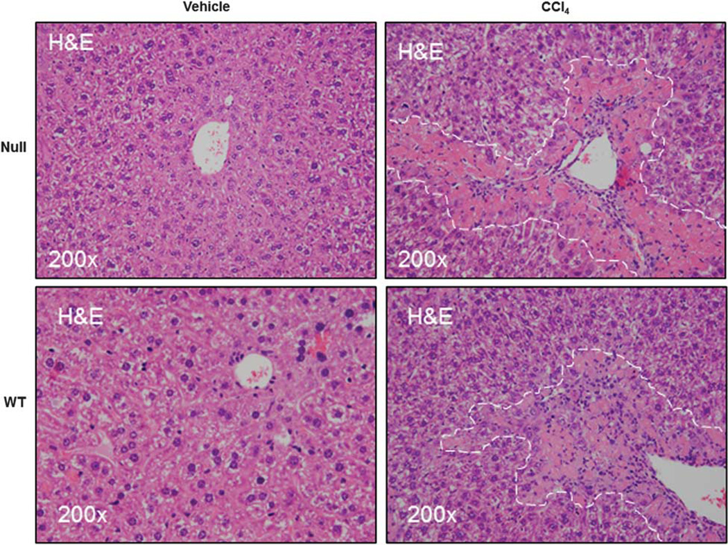
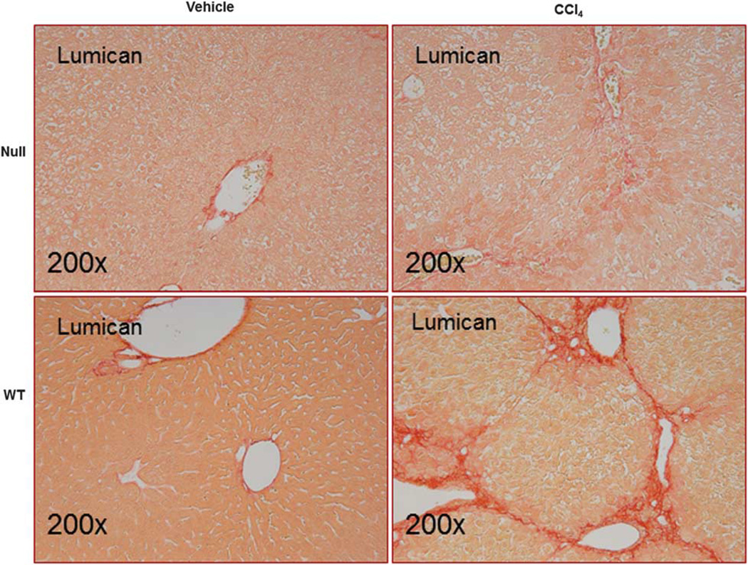
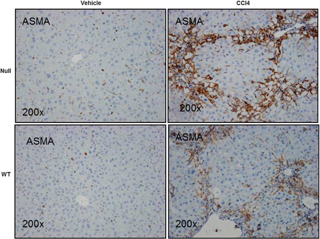
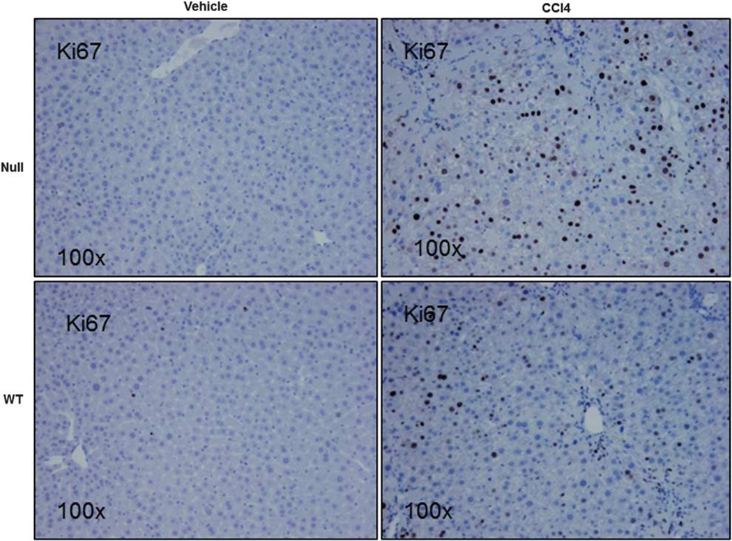
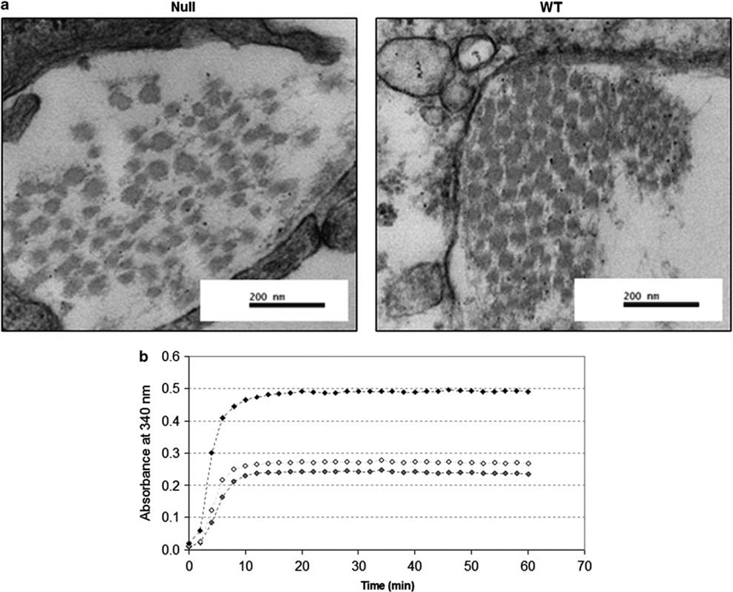
Lumican, an extracellular matrix proteoglycan was previously shown to be upregulated with increasing severity of nonalcoholic steatohepatitis (NASH). Although lumican is involved in collagen fibrillogenesis in extra-hepatic tissues, little is known about the role of lumican in hepatic disease. We therefore determined lumican expression in etiologies other than clinical NASH. Our results indicated that lumican is upregulated in clinical samples of hepatitis C virus infection, in experimental rodent models of chronic and acute liver injury and could additionally be induced in vitro in response to the pro-fibrotic cytokine transforming growth factor β1 (TGFβ1) and to lipotoxic palmitic acid. Together, these results suggested a role for lumican in hepatic fibrosis. To investigate the functional role of lumican in hepatic fibrosis, lumican null (Null) and wild-type (WT) littermates were administered carbon tetrachloride intra-peritoneally. Serum and liver tissue were analyzed for indices of liver injury, fibrosis, matrix turnover, and proliferation. Hepatic fibrosis was greatly reduced in null animals (P<0.05). Paradoxically, gene expression of fibrosis-related genes such as TGFβ1 and collagen 1 was numerically higher in null animals though statistically insignificant from WT animals. On the other hand, α smooth muscle actin expression (α-SMA), a marker for activated fibroblasts, the main contributors of collagen production was significantly higher (P<0.05) in null animals as compared with WT littermates. Among the matrix metalloproteases (MMP), MMP13 was significantly increased (P<0.05) in null animals. Ultra-structural imaging indicated differences in the organization and spatial distribution of hepatic collagen fibrils of null and WT mice. Cell proliferation was significantly increased (P<0.05) in null animals. We conclude that lumican is a prerequisite for hepatic fibrosis. The protective effect of lumican deficiency in hepatic fibrosis appears to be downstream of collagen production and mediated through the combined effects of impaired collagen fibrillogenesis, increased matrix turnover, and an enhanced proliferative response.


Figures

Figure 1
Figure 1
Lumican is upregulated in a rodent model of diet-induced steatohepatitis and in acute hepato-toxin induced liver injury. C57Bl/6 mice were fed for 6 months either standard rodent chow or ‘FF’ (a combination of high fat, cholesterol and sugars). Liver sections were stained for lumican, an ECM proteoglycan. (a) In mice reared on SC, lumican is seen primarily localized to the hepatic sinusoids. (b) In mice reared on FF, cytoplasmic staining of hepatocytes is intense around areas of fat vacuoles (yellow arrows) and around inflammation (black arrows). In a second experiment, J129/B6 mice were administered intra-peritoneal injections of either vehicle (corn oil) or CCl4 @ 1 ml/kg. (c) Lumican is seen primarily localized to the hepatic sinusoids in mice administered vehicle alone whereas (d) in animals administered CCl4, vacuolated hepatocytes clustered around areas of injury (black arrows) stain intensely for lumican. Magnification (ad): ×100. (eh) Upon injury or stress, hepatocytes contribute to lumican production. (eh) Serial sections of liver of mice reared on FF or treated with CCl4 @ 1 ml/kg were stained for lumican and ASMA to compare their in situ localization. (e) Hepatocytes containing lipid vacuoles or (g) injured, vacuolated hepatocytes stain densely for lumican; (f, h) ASMA is restricted to the sinusoids alone. Magnification: ×630.
Figure 1
Figure 1
Lumican is upregulated in a rodent model of diet-induced steatohepatitis and in acute hepato-toxin induced liver injury. C57Bl/6 mice were fed for 6 months either standard rodent chow or ‘FF’ (a combination of high fat, cholesterol and sugars). Liver sections were stained for lumican, an ECM proteoglycan. (a) In mice reared on SC, lumican is seen primarily localized to the hepatic sinusoids. (b) In mice reared on FF, cytoplasmic staining of hepatocytes is intense around areas of fat vacuoles (yellow arrows) and around inflammation (black arrows). In a second experiment, J129/B6 mice were administered intra-peritoneal injections of either vehicle (corn oil) or CCl4 @ 1 ml/kg. (c) Lumican is seen primarily localized to the hepatic sinusoids in mice administered vehicle alone whereas (d) in animals administered CCl4, vacuolated hepatocytes clustered around areas of injury (black arrows) stain intensely for lumican. Magnification (ad): ×100. (eh) Upon injury or stress, hepatocytes contribute to lumican production. (eh) Serial sections of liver of mice reared on FF or treated with CCl4 @ 1 ml/kg were stained for lumican and ASMA to compare their in situ localization. (e) Hepatocytes containing lipid vacuoles or (g) injured, vacuolated hepatocytes stain densely for lumican; (f, h) ASMA is restricted to the sinusoids alone. Magnification: ×630.
Figure 2
Figure 2
Lumican protein is increased in mice treated with CCl4 and correlates with TGFβ1 expression. (a) Western blot analysis of liver lysates of mice treated with CCl4 demonstrate increased expression of lumican as compared with control animals. Glycosylation of lumican protein is indicated by the extensive band that stretches from 37 to 50 kDa. (b, c) Lumican gene expression correlates positively with TGFβ1. Relative gene expression of lumican is plotted against relative gene expression of TGFβ1 (b) in all animals that were fed SC, and FF and (c) in all animals that were administered CCl4 or vehicle alone.
Figure 3
Figure 3
Lumican is over-expressed with CCl4 administration. Lumican null or WT animals were administered CCl4 (n=6) or vehicle (n=4) twice weekly for 1 month @ 1 ml/kg. Lumican immunostaining is minimal in null animals. By contrast, in WT animals, lumican staining closely approximates the bridging lesions seen typically in hepato-toxin induced chronic liver injury. Hepatocytes clustered around areas of tissue injury (bridging vein to vein) and inflammation stain intensely for lumican in addition to the hepatic sinusoids. Magnification: ×200.
Figure 4
Figure 4
Lumican null mice are not protected against hepatic injury. Lumican null or WT animals were administered CCl4 (n=6) or vehicle (n=4) twice weekly for 1 month @ 1 ml/kg. H&E-stained sections of liver show necro-inflammation and hepatocellular vacuolization that appear more severe in Null animals. No injury is seen in liver sections of mice administered vehicle alone.
Figure 5
Figure 5
Lumican null animals show decreased hepatic fibrosis. Liver sections of Null and WT animals administered CCl4 or vehicle twice weekly for 1 month at 1 ml/kg were stained with picrosirius red. There was a twofold decrease in stained area in null animals as compared with the WT animals administered CCl4. Injured and vacuolated hepatocytes are visible in null animals. Magnification: ×200.
Figure 6
Figure 6
Null animals are characterized by increased ASMA staining. Liver sections of Null and WT animals administered CCl4 or vehicle twice weekly for 1 month at 1 ml/kg were stained for ASMA, a marker for myofibroblast and stellate cell activation, the major contributors to collagen production. ASMA was increased (1.3-fold) in null animals as compared with their WT littermates administered CCl4. Magnification: ×200.
Figure 7
Figure 7
Null animals are characterized by increased cell proliferation. Liver sections of Null and WT animals administered CCl4 or vehicle twice weekly for 1 month at 1 ml/kg were stained for Ki67, a marker for proliferating cells. Ki67-positive cells were twofold significantly increased in Null animals as compared with their WT counterparts. Magnification: ×100.
Figure 8
Figure 8
(a) Ultra-structural micrographs of healthy, age-matched Null and WT animals. In null animals, collagen fibrils are irregularly distributed and have unequal diameters. By contrast, in WT animals, collagen fibrils are of even diameter and are packed uniformly. Magnification: ×80 000 (b). Collagen fibril formation was assayed spectrophotometrically with the addition of recombinant lumican protein. Collagen fibril was quantitatively enhanced and hastened by the addition of recombinant lumican and was dose dependent.

References

    1. Kmiec Z. Cooperation of liver cells in health and disease. Adv Anat Embryol Cell Biol. 2001;161:III–XIII. 1–151. - PubMed
    1. Reeves HL, Friedman SL. Activation of hepatic stellate cells–a key issue in liver fibrosis. Front Biosci. 2002;7:d808–d826. - PubMed
    1. Charlton M, Viker K, Krishnan A, et al. Differential expression of lumican and fatty acid binding protein-1: new insights into the histologic spectrum of nonalcoholic fatty liver disease. Hepatology. 2009;49:1375–1384. - PMC - PubMed
    1. Kao WW, Funderburgh JL, Xia Y, et al. Focus on molecules: lumican. Exp Eye Res. 2006;82:3–4. - PMC - PubMed
    1. Chakravarti S, Stallings RL, SundarRaj N, et al. Primary structure of human lumican (keratan sulfate proteoglycan) and localization of the gene (LUM) to chromosome 12q21.3-q22. Genomics. 1995;27:481–488. - PubMed

Publication types

MeSH terms