. 2012 Oct 1;199(1):137-50.
doi: 10.1083/jcb.201206040. Epub 2012 Sep 24.

The Cbp3-Cbp6 complex coordinates cytochrome b synthesis with bc(1) complex assembly in yeast mitochondria

Affiliations

The Cbp3-Cbp6 complex coordinates cytochrome b synthesis with bc(1) complex assembly in yeast mitochondria

Steffi Gruschke et al. J Cell Biol. .

Abstract

Respiratory chain complexes in mitochondria are assembled from subunits derived from two genetic systems. For example, the bc(1) complex consists of nine nuclear encoded subunits and the mitochondrially encoded subunit cytochrome b. We recently showed that the Cbp3-Cbp6 complex has a dual function for biogenesis of cytochrome b: it is both required for efficient synthesis of cytochrome b and for protection of the newly synthesized protein from proteolysis. Here, we report that Cbp3-Cbp6 also coordinates cytochrome b synthesis with bc(1) complex assembly. We show that newly synthesized cytochrome b assembled through a series of four assembly intermediates. Blocking assembly at early and intermediate steps resulted in sequestration of Cbp3-Cbp6 in a cytochrome b-containing complex, thereby making Cbp3-Cbp6 unavailable for cytochrome b synthesis and thus reducing overall cytochrome b levels. This feedback loop regulates protein synthesis at the inner mitochondrial membrane by directly monitoring the efficiency of bc(1) complex assembly.


Figures
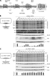
Figure 1.
Figure 1.
Blockage of bc1 complex assembly perturbs cytochrome b expression. (A) Schematic assembly line of the yeast bc1 complex. Assembly factors are shown in bold. (B) Efficiency of mitochondrial translation and synthesis of cytochrome b is perturbed upon bc1 complex assembly inhibition. Mitochondrial translation products of cells carrying a wild-type mitochondrial genome (left) and deletions of the indicated genes were labeled with [35S]methionine for 15 min in vivo. Proteins were extracted, separated on SDS-PAGE, and analyzed by autoradiography and Western blotting. Membranes were probed with antibodies against Mrpl40 to check for equal loading and with antibodies against Cox2 to estimate expression of another mitochondrially encoded protein. Cells were streaked on plates containing either glucose (YPD) or glycerol (YPG) to score for respiratory growth. The steady-state levels of cytochrome b and Cox2 were measured densitometrically, and the Cytb/Cox2 ratio is shown in the diagram. The wild-type ratio was set to 1 and the broken line is drawn at a 50% threshold to qualify as a mutant showing a decreased cytochrome b level. Error bars (mean ± SD) are depicted from n = 3. a.u., arbitrary units. (C) In the absence of cytochrome b, only deletion of CBP3 or CBP6 abolishes expression of mRNAs containing the 5′ UTR of COB. Mitochondrial translation products of cells harboring the cob::ARG8m mitochondrial genome (left) were labeled radioactively and analyzed like described in B. Cox1 is generally only poorly produced when cells are respiratory deficient. The asterisk indicates a degradation product of Arg8. The Arg8/Cox2 ratio was calculated and graphically depicted as in B. n = 3.
Figure 2.
Figure 2.
Ectopically expressed cytochrome b depends on Cbp3–Cbp6 but fails to accumulate to wild-type levels. (A) Scheme of the cox2::COB cob::ARG8m mitochondrial genome. The coding sequence of cytochrome b was inserted by biolistic transformation and homologous recombination into a nonessential region upstream of COX2 in the cob::ARG8m mitochondrial genome. (B) Ectopically expressed cytochrome b can support respiratory growth to a wild-type level. Cells were streaked on plates containing either glucose (YPD) or glycerol (YPG) to score for respiratory growth. Growth on SD-Arg was used to test for expression of Arg8. (C) Ectopically expressed cytochrome b is synthesized efficiently but accumulates poorly. Mitochondrial translation products of cells containing the indicated mitochondrial genomes were labeled with [35S]methionine for 15 min in vivo. Proteins were extracted, separated on SDS-PAGE, and analyzed by autoradiography and Western blotting. (D) Supercomplexes are less abundant when cytochrome b is expressed ectopically. Mitochondria from cells harboring the indicated mitochondrial genomes were lysed in digitonin and subjected to 1D BN PAGE. The gel was stained with Coomassie brilliant blue. Vi, ATP synthase monomer. Vii, ATP synthase dimer. (E) Ectopically expressed cytochrome b requires Cbp3 for stability. Mitochondrial translation products of the indicated cells were pulse-labeled with [35S]methionine for up to 15 min. The fate of newly synthesized proteins was followed for 150 min. Proteins were extracted, separated on SDS-PAGE, and analyzed by autoradiography and Western blotting. The levels of radiolabeled cytochrome b, Arg8, and Cox2 during the pulse were measured densitometrically and are shown in the top diagrams. The 15-min signal of the wild type was set to 1. n = 3. The bottom graphs depict stability of cytochrome b and Arg8 in the strains (calculated as ratio of 150 min/0 min signal) as well as the relative expression level of Arg8 calculated as the Arg8/Cox2 ratio at 150 min (wild-type ratio set to 1). n = 3. For the labeling of Δcbp6 cells, see Fig. S1. (F) Wild-type and Δcbp3 cells containing the cox2::COB cob::ARG8m mitochondrial genome were grown on media containing either glucose (YPD and SD-Arg) or glycerol (YPG) as a carbon source. Error bars indicate mean ± SD. For the labeling of Δcbp6 cells, see Fig. S1.
Figure 3.
Figure 3.
Expression of cob::ARG8m is modulated by the efficiency of bc1 complex assembly when cytochrome b is present. Mitochondrial translation products of cells with the indicated mutations containing the cox2::COB cob::ARG8m mitochondrial genome were labeled with [35S]methionine for 15 min. Proteins were extracted, separated on SDS-PAGE, and analyzed by autoradiography and Western blotting. The asterisk indicates a degradation product of Arg8. The Arg8/Cox2 ratio was calculated and graphically depicted. The wild-type ratio was set to 1. A threshold of 50% of the wild-type signal (broken line) was set to define mutants affected in cob::ARG8m expression. n = 3. Error bars indicate mean ± SD.
Figure 4.
Figure 4.
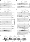
Distinct early assembly intermediates accumulate in cells where expression of the cob::ARG8m reporter is decreased and cytochrome b cannot assemble. (A) Mitochondria of strains carrying the cox2::COB cob::ARG8m or the cob::ARG8m mitochondrial genome were lysed in digitonin, separated on 2D BN/SDS-PAGE, and analyzed by Western blotting with antibodies against cytochrome b, Cbp3, Cbp6, or Cox2. Black arrows indicate the complexes in which cytochrome b is present. White arrows indicate the cytochrome b–free form of Cbp3: one representing the Cbp3–Cbp6 complex (left) and one representing the monomeric Cbp3 (right). T25%, 25% of the material used for 2D BN/SDS PAGE. The top two panels of data (Cytb and Cbp3 for cox::COB cob::ARG8m) in A are presented again in D of this figure and labeled “wild type.” (B) Mitochondria carrying the cox2::COB cob::ARG8m mtDNA were lysed in digitonin, an immunoprecipitation (IP) using an antibody against Cbp3 was performed, and total (top panels) and unbound (bottom panels) fractions were analyzed by 2D BN/SDS-PAGE and Western blotting. For a control of bound proteins, the SDS-eluted fraction (E100%) after the IP was loaded onto the 2D SDS-PAGE and analyzed by Western blotting. T25%, 25% of the mitochondrial lysate before the IP that was used for 2D BN/SDS-PAGE. NB25%, 25% of the unbound material after the IP that was used for 2D BN/SDS-PAGE. These data are presented again in Fig. S2. (C) Mitochondria of the indicated mutants that harbored the cox2::COB cob::ARG8m mitochondrial genome were analyzed as in A. The data for strains Δqcr8, Δcyt1, and Δrip1 in C are presented again in D of this figure. (D) Mitochondria from wild type, Δqcr8, Δcyt1, and Δrip1 were analyzed by 2D BN/SDS-PAGE and Western blotting with the indicated antibodies. The strains correspond to the ones in A and C. The Cytb and Cbp3 data for strains Δqcr8, Δcyt1, and Δrip1 are the same data presented in C, and the Cytb and Cbp3 data for wild-type are the same data presented in A. The gray arrow in the Δcyt1 strain points to the Qcr8 signal as the protein is hardly detectable in this mutant. The asterisk at the lower signal in the Cbp4 blot of the Δrip1 strain corresponds to residual signal of the Qcr7 antibody, with which the membrane was probed before. (E) Schematic drawing of intermediates formed during cytochrome b assembly into the bc1 complex. The exact composition of intermediate III (present in cells lacking cytochrome c1 or Cor2) is at present not known.
Figure 5.
Figure 5.
Newly synthesized cytochrome b assembles in a step-wise fashion through intermediates I, II, and III. (A) Cytochrome b produced from its authentic mRNA forms the same assembly intermediates as ectopically expressed cytochrome b. Mitochondria of the indicated strains with wild-type mtDNA were lysed in digitonin, separated on 2D BN/SDS-PAGE, and analyzed by Western blotting. T25%, 25% of the material used for 2D BN/SDS-PAGE. (B) Assembly pathway of newly synthesized cytochrome b. Wild-type cells were grown in the presence of 4 mg/ml chloramphenicol to deplete mitochondrial translation products and to increase the pool of nuclear encoded subunits, thereby allowing efficient assembly of cytochrome b. Mitochondria were isolated, mitochondrial translation products were radiolabeled, and complexes of cytochrome b were analyzed after the indicated time points by 2D BN/SDS-PAGE followed by autoradiography (for all time points) and Western blotting (for time point 0 min). The nomenclature of the intermediates is identical to Fig. 4 E.
Figure 6.
Figure 6.
Overexpression of Cbp3–Cbp6 allows regaining expression of cob::ARG8m in cells where cytochrome b assembly is disturbed at early or intermediate steps. (A) Less Cbp3–Cbp6 is ribosome-bound in Δqcr8 and Δcyt1 cells. Indicated mitochondria harboring the cox2::COB cob::ARG8m mitochondrial genome were lysed in digitonin and fractionated by centrifugation through a high-density sucrose cushion. The fractions were analyzed by Western blotting and quantified using densitometry (B). n = 3, error bars are depicted. T, 100% total before centrifugation. S, supernatant after centrifugation. P, ribosome-containing pellet after centrifugation. Error bars indicate mean ± SD. (C) Simultaneous overexpression of CBP3 and CBP6 restores expression of the ARG8m reporter in Δqcr8 and Δcyt1 cells. The indicated cells harboring the cox2::COB cob::ARG8m mitochondrial genome were transformed with either empty plasmids or plasmids allowing the overexpression of CBP3 and CBP6 from single copy (CEN) plasmids with the strong TPI promoter. Mitochondrial translation products of these strains were radiolabeled and analyzed by autoradiography and Western blotting. The asterisk indicates a degradation product of Arg8. (D) Overexpression of CBP3 and CBP6 increases the cytochrome b–free pool of the Cbp3–Cbp6 complex. Mitochondria from the strains used in C overexpressing CBP3 and CBP6 were isolated and analyzed as in Fig. 4 A. The nomenclature of the intermediates is identical to Fig. 4 E.
Figure 7.
Figure 7.
Model of assembly of cytochrome b and the feedback loop modulating COB expression. Biogenesis of cytochrome b can be divided into several steps. First, cytochrome b is synthesized on membrane-associated ribosomes that have the Cbp3–Cbp6 complex bound to the tunnel exit. For this step, the other translational activators Cbs1 and Cbs2 are also required. Next, the fully synthesized cytochrome b interacts with Cbp3–Cbp6 and is released from the ribosome. Cbp4 is then recruited to this complex, giving rise to assembly intermediate I. Qcr7 and Qcr8 are added to cytochrome b to form intermediate II, which provokes release of Cbp3–Cbp6 from cytochrome b. Next, the intermediate assembling subunits Cor1, Cor2, cytochrome c1, and Qcr6 are added to form intermediate III and IV. The addition of Qcr9, Qcr10, and Rip1 completes assembly of the bc1 complex. When formation of intermediate IV is compromised, intermediates I, II, and III pile up. Through this process, Cbp3–Cbp6 is sequestered in intermediate I and is no longer available to activate further rounds of COB expression.

References

    1. Atkinson A., Smith P., Fox J.L., Cui T.Z., Khalimonchuk O., Winge D.R. 2011. The LYR protein Mzm1 functions in the insertion of the Rieske Fe/S protein in yeast mitochondria. Mol. Cell. Biol. 31:3988–3996 10.1128/MCB.05673-11 - DOI - PMC - PubMed
    1. Barrientos A., Korr D., Tzagoloff A. 2002. Shy1p is necessary for full expression of mitochondrial COX1 in the yeast model of Leigh’s syndrome. EMBO J. 21:43–52 10.1093/emboj/21.1.43 - DOI - PMC - PubMed
    1. Barrientos A., Zambrano A., Tzagoloff A. 2004. Mss51p and Cox14p jointly regulate mitochondrial Cox1p expression in Saccharomyces cerevisiae. EMBO J. 23:3472–3482 10.1038/sj.emboj.7600358 - DOI - PMC - PubMed
    1. Bonnefoy N., Fox T.D. 2000. In vivo analysis of mutated initiation codons in the mitochondrial COX2 gene of Saccharomyces cerevisiae fused to the reporter gene ARG8m reveals lack of downstream reinitiation. Mol. Gen. Genet. 262:1036–1046 10.1007/PL00008646 - DOI - PubMed
    1. Bonnefoy N., Fox T.D. 2007. Directed alteration of Saccharomyces cerevisiae mitochondrial DNA by biolistic transformation and homologous recombination. Methods Mol. Biol. 372:153–166 10.1007/978-1-59745-365-3_11 - DOI - PMC - PubMed

Publication types

MeSH terms