Primary osseous tumors of the hindfoot: why the delay in diagnosis and should we be concerned?
- PMID: 23008022
- PMCID: PMC3563817
- DOI: 10.1007/s11999-012-2570-6
Primary osseous tumors of the hindfoot: why the delay in diagnosis and should we be concerned?
Abstract
Background: Bony tumors of the foot account for approximately 3% of all osseous tumors. Diagnosis is frequently delayed as a result of lack of clinician familiarity and as a result of their rarity. The reasons for the delays, however, are unclear.
Questions/purposes: We therefore determined (1) how hindfoot tumors present and the specific reasons for delay in diagnosis; (2) whether the spectrum of disease varies between the talus and calcaneus; and (3) how these patients were treated.
Methods: We retrospectively reviewed the medical notes and imaging for all patients with 34 calcaneal and 23 talar tumors recorded in the Scottish Bone Tumour Registry. Demographics, presentation, investigation, histology, management, recurrence, and mortality were recorded.
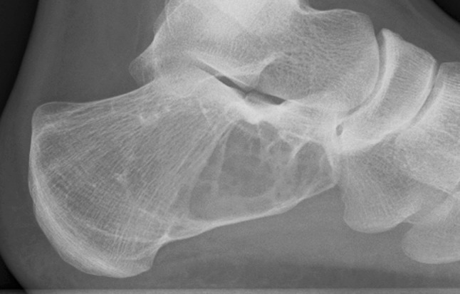
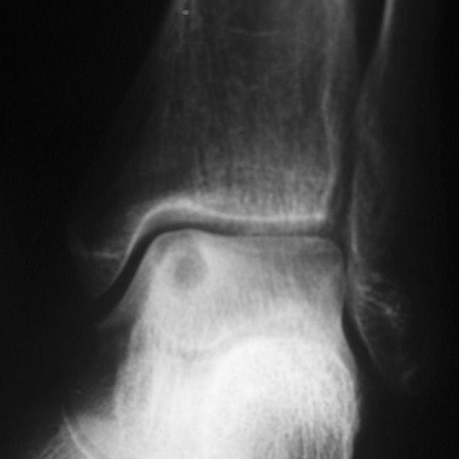
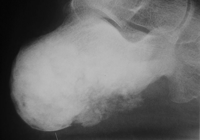
Results: Hindfoot tumors present with pain and often swelling around the heel (calcaneus) or ankle (talus), most often misdiagnosed as soft tissue injury. Calcaneal lesions were more likely to be malignant than talar lesions: 13 of 34 versus three of 23.
Conclusions: Clinicians should be aware that hindfoot tumors can be initially misdiagnosed as soft tissue injuries and suspicion of a tumor should be raised in the absence of trauma or persistent symptoms. Lesions affecting the calcaneus are more likely to be malignant. Early diagnosis and adjuvant therapy are important.
Level of evidence: Level IV, therapeutic study. See Guidelines for Authors for a complete description of levels of evidence.
Figures


Similar articles
-
Massive Chondroblastoma of the Talus: Treatment With En Bloc Talectomy and Tibiocalcaneal Arthrodesis: Long-Term Follow-up of a Case.Foot Ankle Spec. 2017 Jun;10(3):274-277. doi: 10.1177/1938640016676339. Epub 2016 Oct 25. Foot Ankle Spec. 2017. PMID: 27798068
-
Surgical Reconstruction for Talocalcaneal Coalitions With Severe Hindfoot Valgus Deformity.J Pediatr Orthop. 2017 Jun;37(4):293-297. doi: 10.1097/BPO.0000000000000642. J Pediatr Orthop. 2017. PMID: 26371942
-
The presence of calcaneal fibular remodeling associated with middle facet talocalcaneal coalition: a retrospective CT review of 35 feet. Investigations involving middle facet coalitions--Part II.J Foot Ankle Surg. 2008 Jul-Aug;47(4):288-94. doi: 10.1053/j.jfas.2008.04.004. J Foot Ankle Surg. 2008. PMID: 18590890
-
Hindfoot Fractures: Injury Patterns and Relevant Imaging Findings.Radiographics. 2022 May-Jun;42(3):661-682. doi: 10.1148/rg.210167. Epub 2022 Mar 11. Radiographics. 2022. PMID: 35275783 Review.
-
Metastatic endometrial carcinoma of the foot. A case report.J Am Podiatr Med Assoc. 1996 Jul;86(7):331-3. doi: 10.7547/87507315-86-7-331. J Am Podiatr Med Assoc. 1996. PMID: 8757485 Review. No abstract available.
Cited by
-
Primary tumours of the calcaneus.Oncol Lett. 2018 Jun;15(6):8901-8914. doi: 10.3892/ol.2018.8487. Epub 2018 Apr 13. Oncol Lett. 2018. PMID: 29928329 Free PMC article. Review.
-
Delay in Diagnosis and Treatment of Bone Sarcoma-Systematic Review.Cancers (Basel). 2025 Mar 14;17(6):981. doi: 10.3390/cancers17060981. Cancers (Basel). 2025. PMID: 40149315 Free PMC article. Review.
-
Tumours of the talus - A pictorial review.J Clin Orthop Trauma. 2020 May-Jun;11(3):410-416. doi: 10.1016/j.jcot.2020.03.021. Epub 2020 Apr 11. J Clin Orthop Trauma. 2020. PMID: 32405200 Free PMC article.
-
Osteolytic lesions of the calcaneus: results from a multicentre study.Int Orthop. 2013 Sep;37(9):1851-6. doi: 10.1007/s00264-013-2042-y. Epub 2013 Aug 22. Int Orthop. 2013. PMID: 23974835 Free PMC article.
-
Reconstruction after Talar Tumor Resection: A Systematic Review.Curr Oncol. 2022 Dec 12;29(12):9788-9800. doi: 10.3390/curroncol29120769. Curr Oncol. 2022. PMID: 36547183 Free PMC article.
References
Publication types
MeSH terms
LinkOut - more resources
Full Text Sources
Other Literature Sources
Medical
Research Materials