

Widespread osteonecrosis in children with leukemia revealed by whole-body MRI
- PMID: 23008023
- PMCID: PMC3492614
- DOI: 10.1007/s11999-012-2579-x
Widespread osteonecrosis in children with leukemia revealed by whole-body MRI
Abstract
Background: Confirmation of early long-bone epiphyseal osteonecrosis in pediatric patients with leukemia allows for medical and surgical intervention before articular surface collapse. MRI detects early osteonecrosis, but multiple focused MR images are required to capture all lesions.
Questions/purposes: We determined whether whole-body MRI (WB-MRI) could (1) assist in diagnosing long-bone epiphyseal and other osteonecroses, (2) characterize articular surface involvement, and (3) detect preferential sites for osteonecrosis.
Patients and methods: We retrospectively reviewed prospectively collected data on all 11 pediatric patients newly diagnosed with leukemia who had musculoskeletal pain develop that persisted 4 weeks or more during leukemia treatment. All were screened for osteonecrosis by WB-MRI, which consisted of a one-time scan of the entire body. Osteonecrosis was defined as circumscribed lesions with a distinct rim of low signal intensity in the normally high-intensity marrow on T1-weighted images and high signal intensity in the normally low-intensity marrow on short-tau inversion recovery images.
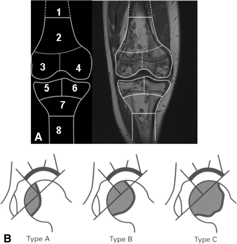
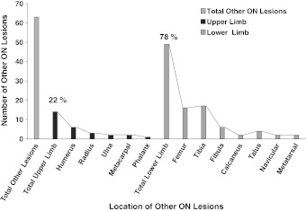
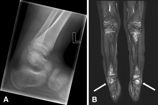
Results: WB-MRI confirmed osteonecrosis in nine of 11 patients. All patients had multisite lesions; eight had long-bone epiphyseal lesions, which comprised 66 of 129 (51%) of all lesions. Osteonecrosis involving greater than 50% of the epiphyseal surface was present in 57% of distal femoral and proximal tibial lesions. All humeral and femoral head lesions involved more than 1/3 of the medial surface volume but were asymptomatic. No articular collapse was present. All osteonecrotic lesions were more common in the lower extremities.
Conclusions: WB-MRI confirmed early epiphyseal osteonecrosis, with quantification of articular surface involvement. Lower limbs were preferentially affected, but asymptomatic humeral head osteonecrosis was present in five of nine patients.
Level of evidence: Level IV, diagnostic study. See Instructions for Authors for a complete description of levels of evidence.
Figures
Similar articles
-
[Whole-body MR imaging in children with suspected osteonecrosis after intensive chemotherapy: preliminary results].Rofo. 2008 Mar;180(3):238-45. doi: 10.1055/s-2008-1027185. Rofo. 2008. PMID: 18278731 German.
-
Unique MRI findings as an early predictor of osteonecrosis in pediatric acute lymphoblastic leukemia.AJR Am J Roentgenol. 2012 May;198(5):W432-9. doi: 10.2214/AJR.11.7367. AJR Am J Roentgenol. 2012. PMID: 22528924 Free PMC article.
-
How does osteonecrosis about the knee progress in young patients with leukemia?: a 2- to 7-year study.Clin Orthop Relat Res. 2010 Sep;468(9):2454-9. doi: 10.1007/s11999-010-1358-9. Epub 2010 Jun 26. Clin Orthop Relat Res. 2010. PMID: 20582497 Free PMC article.
-
MR imaging of epiphyseal lesions of the knee: current concepts, challenges, and controversies.Radiol Clin North Am. 2005 Jul;43(4):655-72, vii-viii. doi: 10.1016/j.rcl.2005.02.002. Radiol Clin North Am. 2005. PMID: 15893529 Review.
-
Magnetic resonance imaging and differential diagnosis of epiphyseal osteonecrosis.Semin Musculoskelet Radiol. 2001;5(1):57-67. doi: 10.1055/s-2001-12924. Semin Musculoskelet Radiol. 2001. PMID: 11371336 Review.
Cited by
-
Systemic Sarcoidosis With Neurosarcoidosis Features as a Risk Factor for Multifocal Osteonecrosis.Cureus. 2024 Aug 13;16(8):e66791. doi: 10.7759/cureus.66791. eCollection 2024 Aug. Cureus. 2024. PMID: 39268259 Free PMC article.
-
Value of whole-body magnetic resonance imaging for screening multifocal osteonecrosis in patients with polymyositis/dermatomyositis.Br J Radiol. 2017 May;90(1073):20160780. doi: 10.1259/bjr.20160780. Epub 2017 Mar 29. Br J Radiol. 2017. PMID: 28355130 Free PMC article.
-
How PET/MR Can Add Value For Children With Cancer.Curr Radiol Rep. 2017 Mar;5(3):15. doi: 10.1007/s40134-017-0207-y. Epub 2017 Feb 21. Curr Radiol Rep. 2017. PMID: 28695063 Free PMC article.
-
Nonbacterial and bacterial osteomyelitis in children: a case-control retrospective study.Front Pediatr. 2023 May 3;11:1067206. doi: 10.3389/fped.2023.1067206. eCollection 2023. Front Pediatr. 2023. PMID: 37206973 Free PMC article.
-
Osteonecrosis of the Shoulders in Pediatric Patients Treated for Leukemia or Lymphoma: Single-Institutional Experience.J Pediatr Orthop. 2019 Feb;39(2):104-110. doi: 10.1097/BPO.0000000000000900. J Pediatr Orthop. 2019. PMID: 28452860 Free PMC article.
References
MeSH terms
Substances
LinkOut - more resources
Full Text Sources
Medical
Research Materials
