. 2015 Jan;9(1):1-12.
doi: 10.1002/term.1602. Epub 2012 Sep 24.

Inducing endoderm differentiation by modulating mechanical properties of soft substrates

Affiliations

Inducing endoderm differentiation by modulating mechanical properties of soft substrates

Maria Jaramillo et al. J Tissue Eng Regen Med. 2015 Jan.

Abstract

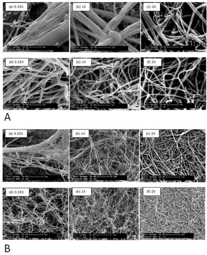
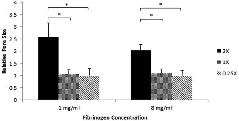
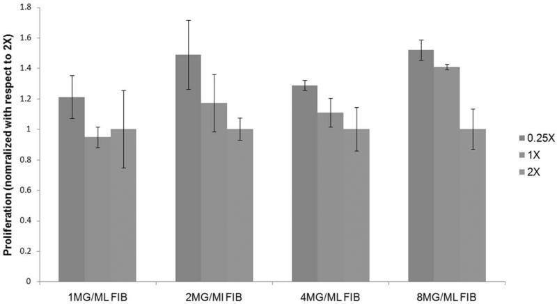
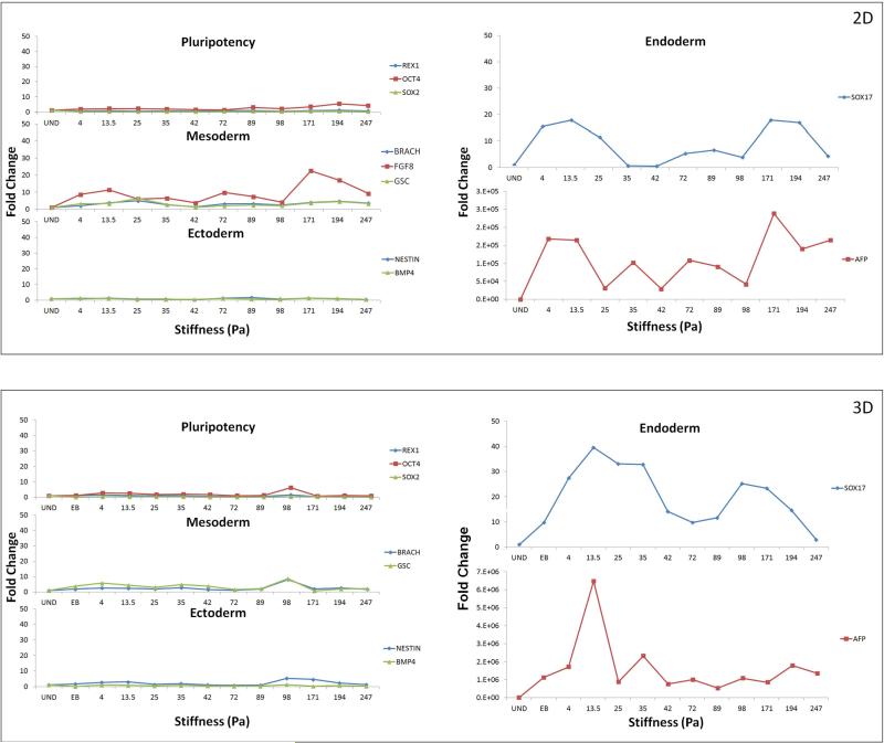
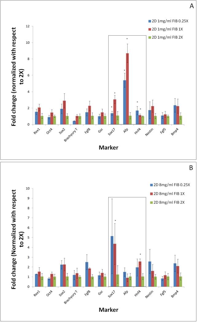
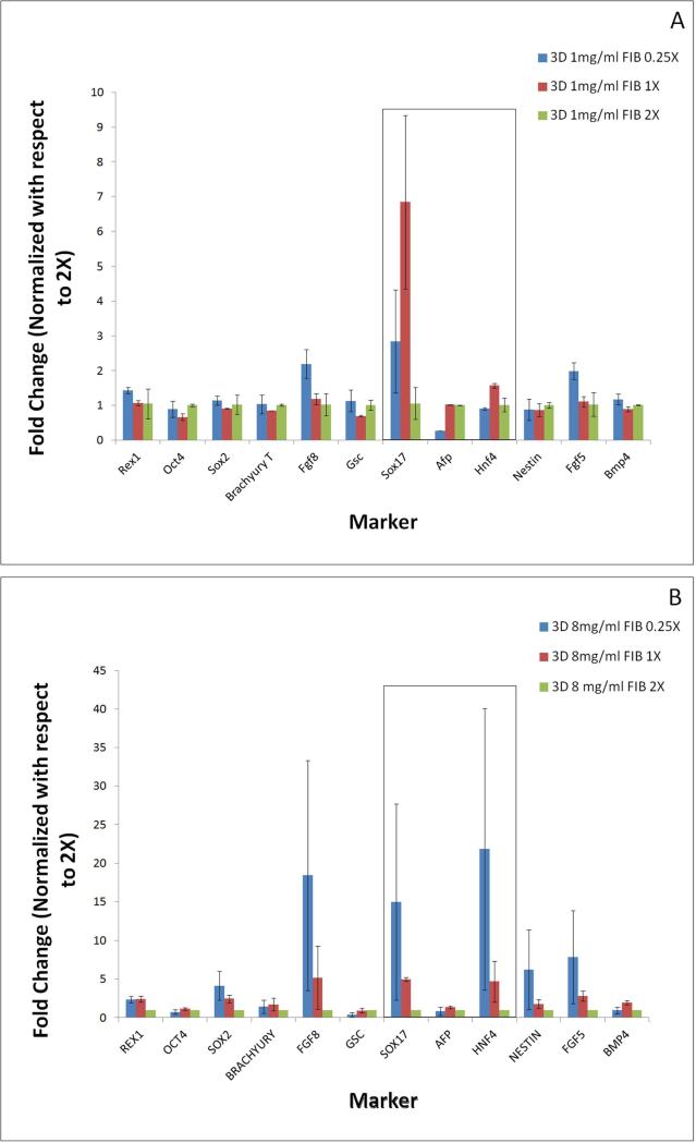
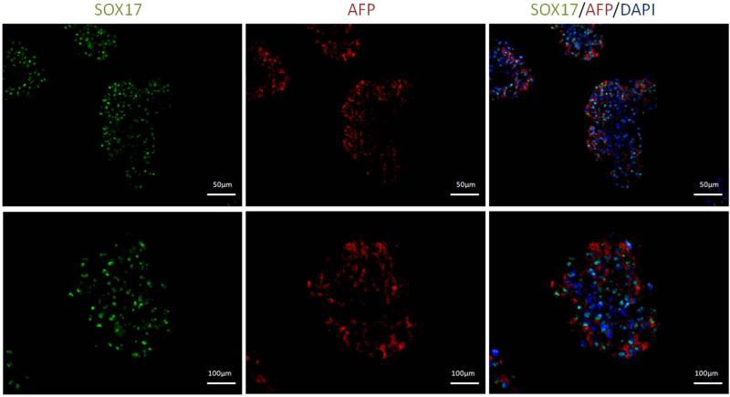
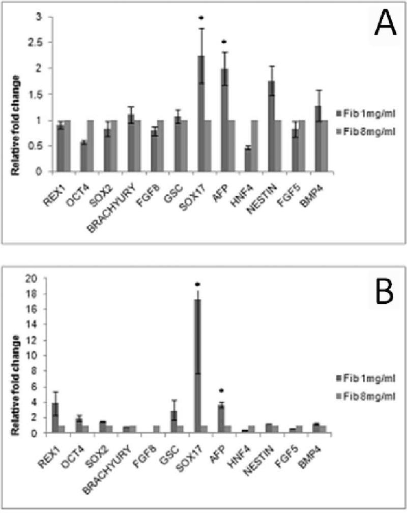
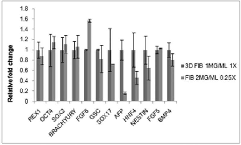
Early embryonic stem cell (ESC) differentiation is marked by the formation of three germ layers from which all tissues types arise. Conventionally, ESCs are differentiated by altering their chemical microenvironment. Recently however, it was established that a mechanical microenvironment can also contribute towards cellular phenotype commitment. In this study, we report how the cellular mechanical microenvironment of soft substrates affects the differentiation and phenotypic commitment of ESCs. Mouse ESCs were cultured in a fibrin hydrogel matrix in 2D and 3D cultures. The gelation characteristics of the substrates were modulated by systematically altering the fibrinogen concentration and the fibrinogen-thrombin crosslinking ratio. Analysis of the ESCs cultured on different substrate conditions clearly illustrated the strong influence that substrate physical characteristics assert on cellular behaviours. Specifically, it was found that ESCs had a higher proliferation rate in gels of lower stiffness. Early differentiation events were studied by analyzing the gene and protein expression levels of early germ layer markers. Our results revealed that lower substrate stiffness elicited stronger upregulation of endoderm related genes Sox17, Afp and Hnf4 compared to stiffer substrates. While both 2D and 3D cultures showed a similar response, the effects were much stronger in 3D culture. These results suggest that physical cues can be used to modulate ESC differentiation into clinically relevant tissues such as liver and pancreas.

Keywords: differentiation; embryonic stem cells; endoderm; fibrin hydrogel; mechanical properties; soft substrates.


Figures

Figure 1
Figure 1
(A)Effect of varying the fibrinogen concentration as well as the fibrinogen to thrombin ratios on the microstructure of the gels imaged at a high magnification of 20KX. In (a)-(c) 1 mg/ml of fibrinogen at fibrinogen to thrombin ratios of 0.25X, 1X, and 2X respectively. In (d)-(f) 8 mg/ml of fibrinogen at fibrinogen to thrombin ratios of 0.25X, 1X, and 2X respectively. (B) Effect of varying the fibrinogen concentration as well as the fibrinogen to thrombin ratios on the microstructure of the gels observed at a low magnification of 5KX. In (a)-(c) 1 mg/ml of fibrinogen at fibrinogen to thrombin ratios of 0.25X, 1X, and 2X, respectively. In (d)-(f) 8 mg/ml of fibrinogen at fibrinogen to thrombin ratios of 0.25X, 1X, and 2X, respectively.
Figure 2
Figure 2
The average pore size calculated using ImageJ for 1 mg/ml and 8 mg/ml fibrinogen concentrations and all three fibrinogen to thrombin ratios normalized to 2X for both fibrinogen concentrations. * p<0.05 compared to highest stiffness group.
Figure 3
Figure 3
Representative images of cells plated on gels of identical fibrinogen concentration, but varying cross-linking fibrinogen/thrombin ratio: 0.25X (Left) and 2X (Right). Cells attach, but do not spread out, instead, they form cell clusters. Cell cluster size greater in gels with lower cross-linking ratio.
Figure 4
Figure 4
Comparison of the proliferation of mouse ES cells plated on gels of different fibrinogen concentration and varying cross-linking ratio after 24 hours of plating. Y-Axis represents alamar blue fluorescence. All results normalized with respect to the lowest cross-linking ratio for each group. At all fibrinogen concentrations proliferation was found to decrease as the cross-linking increased. * p<0.05 compared to highest stiffness group.
Figure 5
Figure 5
The effect of changing substrate stiffness on the embryonic stem cells pluripotency and germ layer commitment in 2-dimensional (a) and 3-dimensional (b) configurations. Results are normalized with respect to undifferentiated cells. Most significant effect was observed in the up-regulation of endoderm markers SOX17 and AFP in lower range stiffness gels. Effect was more prominent in 3-dimensional configurations.
Figure 6
Figure 6
The effect of changing substrate cross-linking ratios on the embryonic stem cell pluripotency and germ layer commitment, analyzed at extreme fibriniogen concentrations of 1mg/ml (a) and 8mg/ml 1mg/ml (b) in 2-dimensional configuration. Results are normalized with respect to highest cross-linking ratio of 2X. Most significant effect was observed in the up-regulation of endoderm markers when compared between cells differentiated on gels of lower cross-linking (0.25X, 1X) and higher cross-linking (2X) ratio. * p<0.05 compared to highest stiffness group.
Figure 7
Figure 7
The effect of changing substrate cross-linking ratios on the embryonic stem cell pluripotency and germ layer commitment, analyzed at extreme fibriniogen concentrations of 1mg/ml (a) and 8mg/ml 1mg/ml (b) in 3-dimensional configuration. Results are normalized with respect to highest cross-linking ratio of 2X. Most significant effect was observed in the up-regulation of endoderm markers when compared between cells differentiated on gels of lower cross-linking (0.25X, 1X) and higher cross-linking (2X) ratio. * p<0.05 compared to highest stiffness group.
Figure 8
Figure 8
Immunocytochemistry images of cells plated at 1mg/ml and 1X. High expression of endoderm markers AFP (Red) and Sox17 (Green) was observed in the cell clusters which were also found to be highly co-expressed.
Figure 9
Figure 9
Pluripotency and germ layer markers of ES cells differentiated in substrates of varying and extreme fibrinogen concentration but same cross-linking ratio of 1X, in (A) 2-dimensional and (B) 3-dimensional culture. Significant upregulation of endoderm markers was found when cells were differentiated on gels obtained at lower fibrinogen concentration. * p<0.05 compared to highest stiffness group.
Figure 10
Figure 10
Marker expression comparison between 1mg/ml fibrinogen obtained at fibrinogen/thrombin cross-linking ratio of 1X and fibrinogen concentration of 2mg/ml obtained atfibrinogen/thrombin cross-linking ratio of 0.25X. Figure shows expression of most markers to be equivalent in both groups except for that of endoderm markers. Results normalized with respect to 1mg/ml Fibrinogen 1X cross-linking ratio.

References

    1. Antoine M, Wirz W, Tag CG, Mavituna M, Emans N, Korff T, et al. Expression pattern of fibroblast growth factors (FGFs), their receptors and antagonists in primary endothelial cells and vascular smooth muscle cells. Growth Factors. 2005;23(2):87–95. - PubMed
    1. Banerjee A, Arha M, Choudhary S, Ashton RS, Bhatia SR, Schaffer DV, et al. The influence of hydrogel modulus on the proliferation and differentiation of encapsulated neural stem cells. Biomaterials. 2009;30(27):4695–4699. - PMC - PubMed
    1. Banerjee I, Sharma N, Yarmush M. Impact of co-culture on pancreatic differentiation of embryonic stem cells. Journal of Tissue Engineering and Regenerative Medicine. 2010 n/a-n/a. - PubMed
    1. Blombäck B, Bark N. Fibrinopeptides and fibrin gel structure. Biophysical Chemistry. 2004;112(2-3):147–151. - PubMed
    1. Blum M, Gaunt SJ, Cho KWY, Steinbeisser H, Blumberg B, Bittner D, et al. Gastrulation in the mouse: The role of the homeobox gene goosecoid. Cell. 1992;69(7):1097–1106. - PubMed

Publication types

MeSH terms

LinkOut - more resources