. 2012 Oct 22;209(11):2113-26.
doi: 10.1084/jem.20120532. Epub 2012 Sep 24.

Induction of tumoricidal function in CD4+ T cells is associated with concomitant memory and terminally differentiated phenotype

Affiliations

Induction of tumoricidal function in CD4+ T cells is associated with concomitant memory and terminally differentiated phenotype

Daniel Hirschhorn-Cymerman et al. J Exp Med. .

Abstract

Harnessing the adaptive immune response to treat malignancy is now a clinical reality. Several strategies are used to treat melanoma; however, very few result in a complete response. CD4(+) T cells are important and potent mediators of anti-tumor immunity and adoptive transfer of specific CD4(+) T cells can promote tumor regression in mice and patients. OX40, a costimulatory molecule expressed primarily on activated CD4(+) T cells, promotes and enhances anti-tumor immunity with limited success on large tumors in mice. We show that OX40 engagement, in the context of chemotherapy-induced lymphopenia, induces a novel CD4(+) T cell population characterized by the expression of the master regulator eomesodermin that leads to both terminal differentiation and central memory phenotype, with concomitant secretion of Th1 and Th2 cytokines. This subpopulation of CD4(+) T cells eradicates very advanced melanomas in mice, and an analogous population of human tumor-specific CD4(+) T cells can kill melanoma in an in vitro system. The potency of the therapy extends to support a bystander killing effect of antigen loss variants. Our results show that these uniquely programmed effector CD4(+) T cells have a distinctive phenotype with increased tumoricidal capability and support the use of immune modulation in reprogramming the phenotype of CD4(+) T cells.


Figures

Figure 1.
Figure 1.
CTX and OX86 synergize with Trp1 CD4 T cells to eradicate large established tumors. (A) C57BL/6 mice (6–10/group) were inoculated intradermally in the flank with B16 cells. After 3 wk, mice were injected with CTX. The next day, mice were injected with OX86 (or IgG as control) with or without Trp1 cells as indicated. Tumors were measured periodically. Top: graphs represent tumor area of individual mice over time for each treatment. Middle: Kaplan-Meir overall survival curves. Bottom: representative photographs of mice treated with the triple combination therapy at day 0 or 21 after treatment. (B) C57BL/6 mice (7–11/group) inoculated intradermally in the flank with B16 cells. After 3 wk, mice were injected with CTX. The next day, Trp1 cells were transferred to all mice and groups were injected with 1 dose of agonists Abs (OX40, GITR, or CD40) or three doses of antagonist Abs (CTLA-4 or PD-L1) given 3 d apart. Tumor size was measured periodically. Graphs represent tumor area of individual mice over time for each treatment. (C) C57BL/6 mice (7–11/group) were inoculated intradermally in the flank with B16 cells. After 3 wk, mice were injected with CTX. The next day, Trp1 cells or Pmel-1 CD8+ T cells were injected and mice received OX86 (or IgG as control). Tumor size was measured periodically. Graphs represent tumor area of individual mice over time for each treatment. (D) C57BL/6 mice (10/group) were inoculated intradermally in the flank with Hep-55.1C or Hep-55.1C-Trp1 cells. On day 10, mice were injected with CTX. On day 11, mice received Trp1 cells and OX86 or IgG. Plots represent tumor area over time for each individual mouse. **, P < 0.005. All experiments were repeated at least twice with similar results.
Figure 2.
Figure 2.
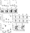
Combination therapy increases Trp1 T eff to T reg cell ratio by expanding T eff cells and reducing T reg cells via activation-induced cell death. C57BL/6 mice (4–5/group) were injected subcutaneously in the flank with B16 cells in Matrigel. 3 wk after tumor challenge, CTX or PBS was injected. The next day, Trp1 cells were transferred followed by injection of OX86 (or IgG as control). On day 14 after treatment, single cell suspensions were prepared from TDLNs and tumors. Cells were stained and analyzed by flow cytometry. (A) Graph represents T eff/T reg cell ratio ± SEM in tumors and TDLNs. *, P < 0.05, ^, P = 0.0599. (B) Representative plots showing CD45.1 (Trp-1 cells) versus CD4 for each group pregated on ViD Foxp3. ViD is a viability dye (ViD: live cells). (C) Graph represents Ki67 MFIs ± SEM of CD45.1+ CD4+ population in TDLNs. **, P < 0.01. (D) Representative plots of Foxp3 versus CD4 depicting the percentage of Trp1+ Foxp3+ in tumors and TDLNs. Events were pregated on ViD CD45.1+. (E) The graph represents the percentage of dead Trp1 T reg cells (ViD+ in the Foxp3+ CD45.1+ gate) in tumors and TDLNs. *, P < 0.05; **, P < 0.01. Error bars represent SEM. (F) Representative plots of activated dead Trp1 T reg cells ViD+ versus Ki67. Events were pregated on CD45.1+ Foxp3+. The experiments were repeated at least five times with equivalent results.
Figure 3.
Figure 3.
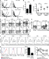
Direct cytotoxicity by Trp1 cells as a result of OX40 engagement during CTX-induced lymphopenia is Eomes dependent. (A) C57BL/6 wild-type or Rag1−/− mice (6–10/group) were inoculated intradermally in the flank with B16 cells. After 3 wk, mice were injected with CTX, followed the next day by OX86 (or IgG as control) and Trp1 cells. Tumors were measured periodically. Graphs represent tumor area of individual mice over time for each treatment. (B) C57BL/6 mice (10 mice/group) were injected subcutaneously with B16 in Matrigel. 3 wk after tumor challenge, CTX was injected. The next day, mice were injected with Trp1 cells and OX86 (or IgG). On day 14 after treatment, Trp1 cells were purified by FACS from splenocytes and were used for in vitro killing assays using B16 cells as targets at a 10:1 effector to target ratio. Means of three individual wells and SEM are shown. *, P < 0.05. (C) Single cell suspensions of TDLNs and spleens from mice that were treated, as in B (8 mice/group), were stimulated overnight with Trp1 peptide in the presence of monensin and anti–CD107a-FITC. The next day, the samples were stained for CD45.1 (Trp1 cells) and other phenotypic markers and analyzed by flow cytometry. Plots show CD107a MFIs of individual mice gated on ViD CD45.1+ CD4+ Foxp3. *, P < 0.005; **, P < 0.05. Error bars represent SEM. (D) Single cell suspensions of TDLNs and spleens from mice that were treated as in B (4–5 mice/group) were stained and analyzed by flow cytometry for GrzB and other phenotypic markers on day 14 after treatment. Representative plots are shown for GrzB versus CD45.1. Events were pregated on ViD, CD4+, and Foxp3. (E) C57BL/6 mice (7–10/group) were treated as in B and bled on day 14 after treatment. PBMCs were stained for GrzB and Eomes and analyzed by flow cytometry. Events were pregated on ViD, CD45.1+, CD4+, and Foxp3. Representative plots are shown of GrzB versus Eomes. (F) C57BL/6 mice (4–5/group) were injected subcutaneously with B16 in Matrigel. 3 wk after tumor challenge, CTX was injected. The next day, mice were injected with Trp1 cells and OX86, DTA-1, FGK45, 9D9, 10F.9G2, or IgG. Two additional doses of 9D9 and 10F.9G2 were given every 3 d. On day 14 after treatment, single cell suspensions were stained for Eomes and other phenotypic markers and analyzed by flow cytometry. Events were pregated on ViD, CD45.1+, CD4+, and Foxp3. Representative plots are shown of GrzB versus Eomes. (G) In vitro activated Trp1 cells were transduced with Eomes shRNA-GFP retrovirus or GFP retrovirus as control. Transduced cells were FACS sorted on GFPhigh gate 5 d later. Cells were stained intracellularly for Eomes, GrzB, and T-bet and analyzed by flow cytometry. Representative histograms are shown. (H) In vitro cytotoxicity assay with transduced Trp1 cells sorted on GFPhigh gate. B16 cells were used as targets at a 10:1 effector to target ratio. Bars represent the means of duplicate wells and the SEM is shown. (I) C57BL/6 Rag1−/− mice (6/group) were inoculated intradermally in the flank with B16 cells. 3 wk later, CTX was injected followed the next day by the transfer of 70,000 transduced Trp1 cells (sorted on GFPhigh) and injection of OX86. Tumor area was periodically monitored. Each point represents the mean tumor area and error bars are SEM. *, P < 0.05. All experiments were repeated at least twice with similar results.
Figure 4.
Figure 4.
CTX and OX86 promotes Trp1 cells to acquire terminal differentiation and memory phenotype with mixed Th1/Th2 cytokine secretion. (A) C57BL/6 mice (4–5 mice/group) were injected subcutaneously in the flank with B16 in Matrigel. 3 wk after tumor challenge, CTX was injected. The next day, Trp1 cells were transferred followed by injection of OX86 or IgG. On day 14 after treatments, single cell suspensions from TDLNs were stained and analyzed by flow cytometry for Eomes, Klrg1, Bcl-6, T-bet, PD-1, CD127, and CD62L. Events were pregated on ViD CD4+ CD45.1+ Foxp3. *, P < 0.05; **, P < 0.005; ***, P < 0.0005; ^, P = 0.0632. Experiment was repeated at least three times with similar results. Error bars represent SEM. (B) C57BL/6 mice (10 mice/group) were injected subcutaneously in the flank with B16 in Matrigel. 3 wk after tumor challenge, CTX was injected. The next day, Trp1 cells were transferred followed by injection of OX86 or IgG. On day 14 after treatments, tumors from each group were pooled and single cell suspensions were prepared. Trp1 cells were purified by MACS and co-cultured with irradiated APCs (pulsed with Trp1 peptide). After 4 d, cytokines levels were measured in supernatants by cytokine bead arrays. Graphs show the means ± SEM of three individual wells per condition. *, P < 0.05; **, P < 0.01; ***, P < 0.001. (C–H) C57BL/6 mice (4–5 mice/group) were injected subcutaneously in the flank with B16 in Matrigel. 3 wk after tumor challenge, CTX was injected. The next day, Trp1 cells were transferred and injected with OX86 (or IgG). On day 14 after treatment, single cell suspensions were prepared from tumors and incubated for 8 h in the presence of APCs pulsed with Trp1 peptide. The cells were stained and analyzed by intracellular flow cytometric analysis. (C) Representative plot of IFN-γ versus CD45.1. Events were pregated on ViD CD4+ Foxp3. (D) Representative plot of TNF versus CD45.1. Events were pregated on ViD CD4+ Foxp3. (E) Representative plot of TNF versus IFN-γ. Events were pregated on ViD CD4+ Foxp3 CD45.1+. (F) Representative plot of IL-4 versus CD45.1. Events were pregated on ViD CD4+ Foxp3. (G) Representative plot of IL-4 versus IFN-γ pregated on ViD CD4+ Foxp3 CD45.1+. (H) Representative plot of IL-17 versus CD45.1 pregated on ViD CD4+ Foxp3. Experiments were repeated at least three times with similar results.
Figure 5.
Figure 5.
Triple combination therapy promotes bystander tumor killing of antigen loss variants. C57BL/6 mice (8/group) were inoculated intradermally in the flank with B16 cells, B78H1, B16, and B78H1 in opposite flanks, or a B16:B78H1 mixture. 3 wk later, CTX was injected followed the next day by treatment with Trp1 cells and OX86 as described in the top diagram. Graphs represent tumor area of individual mice over time for each treatment. Similar results were obtained in two individual experiments.
Figure 6.
Figure 6.
Human and mouse tumor-specific CD4+ T cells showed enhanced anti-tumor lytic activity with OX40 ligation in vitro. (A) Purified Trp1 cells were incubated with APCs and Trp1 peptide and, 48 h after incubation, 10 µg/ml OX86 (or IgG as control) was added to the cultures. After 5 d, the cells were subjected to in vitro cytotoxicity assay using B16 cells as targets at 10:1 effector to target ratio. Graphs show the mean percent killing of duplicate wells. Error bars represent SEM. (B) NY-ESO-1–specific CD4+ T cell lines from three different patients were expanded with plate-bound anti-CD3 and IL-2 in the presence or absence of 10 ng/ml of recombinant human OX40L-Fc. After 10 or 14 d in culture (ptn 1 or 7 and 10, respectively), the CD4+ T cell lines were subjected to an in vitro cytotoxicity assays using patient-matched melanoma cell lines (SKMEL-ptn 1, 7, and 10) pulsed with NY-ESO-1 peptide pools as targets. Bars depict the means of percentage of targets killed of duplicate or triplicate wells/treatment. Errors bars represent SEM. ***, P = 0.0004. Experiments were repeated at least three times with similar results. (C) NY-ESO-1–specific CD4+ T cell lines (patient 7) were expanded with plate-bound anti-CD3/CD28 and IL-2 in the presence or absence of 5 ng/ml of recombinant human OX40L-Fc. After 7 d, RNA was extracted and subjected to RT-PCR for EOMES (Eomes), KLRG1 (KLRG1), TBX21 (T-bet), BCL6 (Bcl-6), PDCD1 (PD-1), IL7R (CD127), and SELL (CD62L). Graphs represent means of triplicate wells/treatment. Error bars represent SEM. *, P < 0.05; ^, P = 0.056. Experiments were repeated at least twice with similar results in cell lines derived from two patients.

References

    1. Al-Shamkhani A., Birkeland M.L., Puklavec M., Brown M.H., James W., Barclay A.N. 1996. OX40 is differentially expressed on activated rat and mouse T cells and is the sole receptor for the OX40 ligand. Eur. J. Immunol. 26:1695–1699 10.1002/eji.1830260805 - DOI - PubMed
    1. Apetoh L., Ghiringhelli F., Tesniere A., Obeid M., Ortiz C., Criollo A., Mignot G., Maiuri M.C., Ullrich E., Saulnier P., et al. 2007. Toll-like receptor 4-dependent contribution of the immune system to anticancer chemotherapy and radiotherapy. Nat. Med. 13:1050–1059 10.1038/nm1622 - DOI - PubMed
    1. Awwad M., North R.J. 1988. Cyclophosphamide (Cy)-facilitated adoptive immunotherapy of a Cy-resistant tumour. Evidence that Cy permits the expression of adoptive T-cell mediated immunity by removing suppressor T cells rather than by reducing tumour burden. Immunology. 65:87–92 - PMC - PubMed
    1. Banerjee A., Gordon S.M., Intlekofer A.M., Paley M.A., Mooney E.C., Lindsten T., Wherry E.J., Reiner S.L. 2010. Cutting edge: The transcription factor eomesodermin enables CD8+ T cells to compete for the memory cell niche. J. Immunol. 185:4988–4992 10.4049/jimmunol.1002042 - DOI - PMC - PubMed
    1. Belz G.T., Kallies A. 2010. Effector and memory CD8+ T cell differentiation: toward a molecular understanding of fate determination. Curr. Opin. Immunol. 22:279–285 10.1016/j.coi.2010.03.008 - DOI - PubMed

Publication types

MeSH terms