. 2012:2012:526236.
doi: 10.1155/2012/526236. Epub 2012 Sep 13.

Influence of bisphosphonate treatment on medullary macrophages and osteoclasts: an experimental study

Affiliations

Influence of bisphosphonate treatment on medullary macrophages and osteoclasts: an experimental study

Natalia Daniela Escudero et al. Bone Marrow Res. 2012.

Abstract

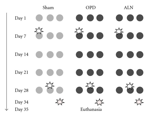
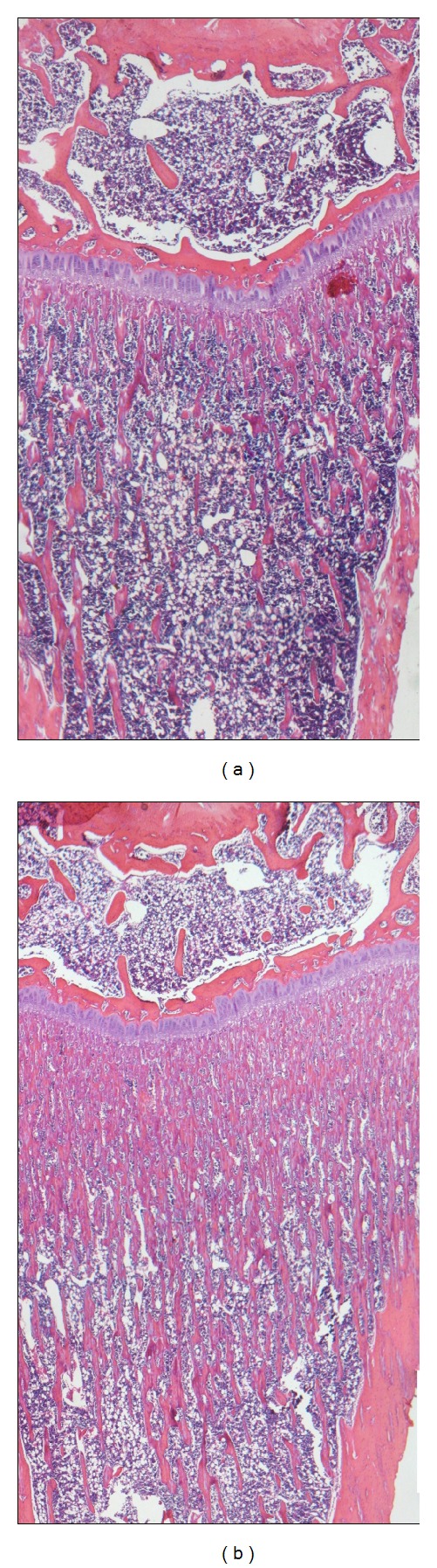
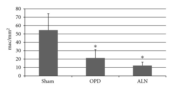
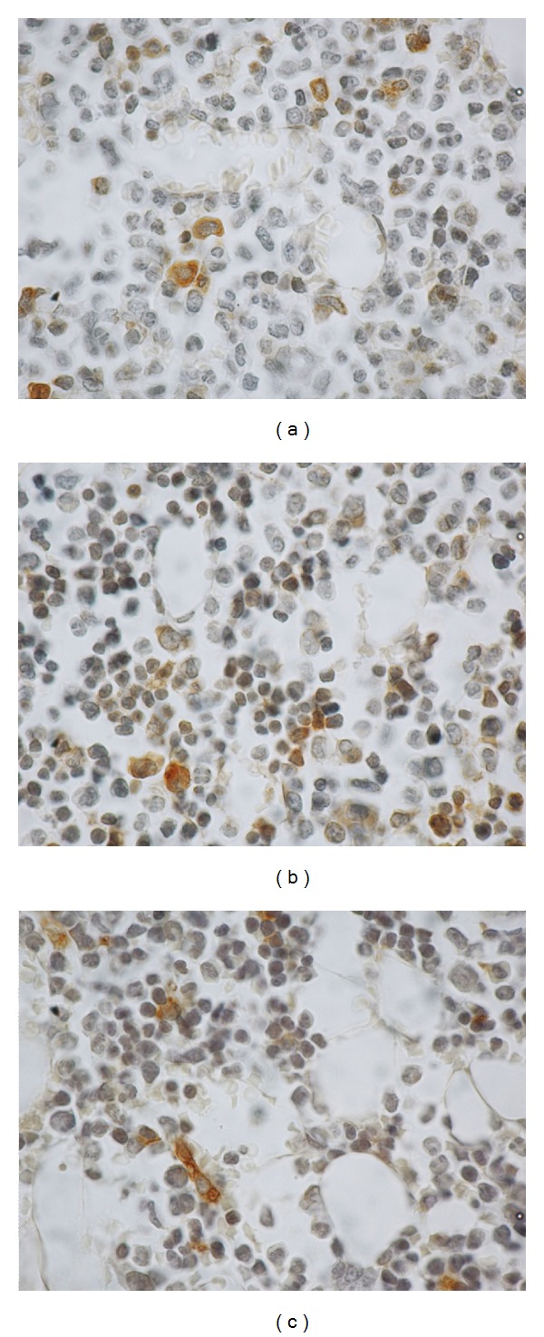
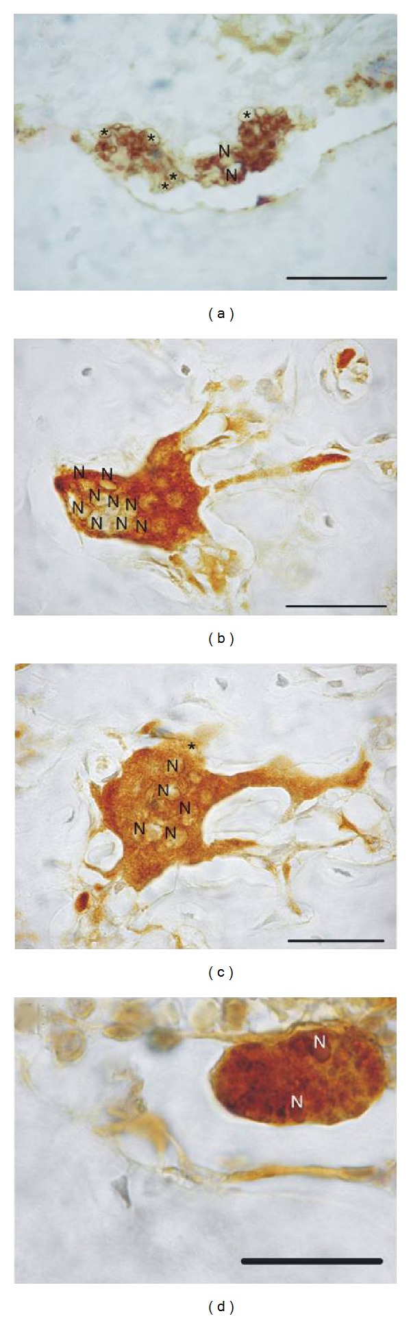
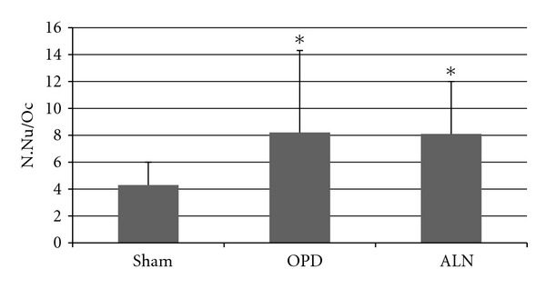
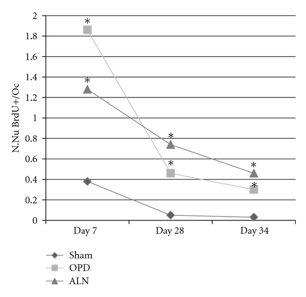
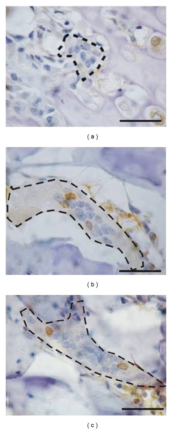
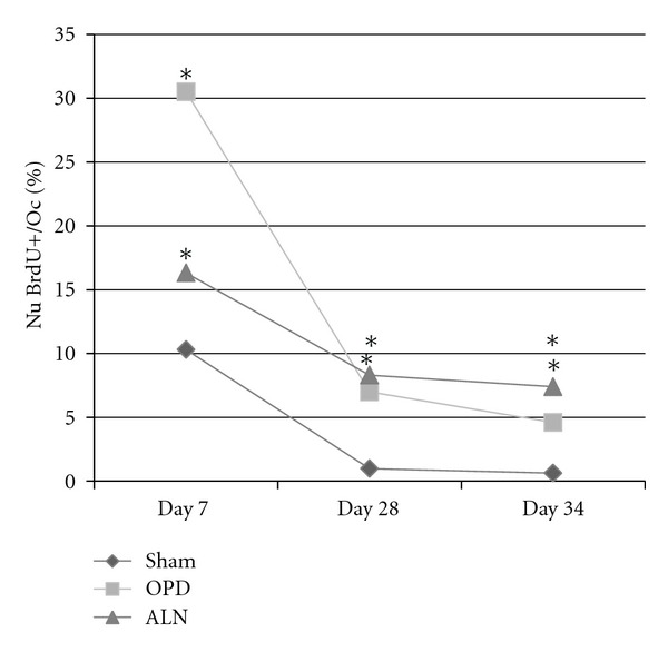
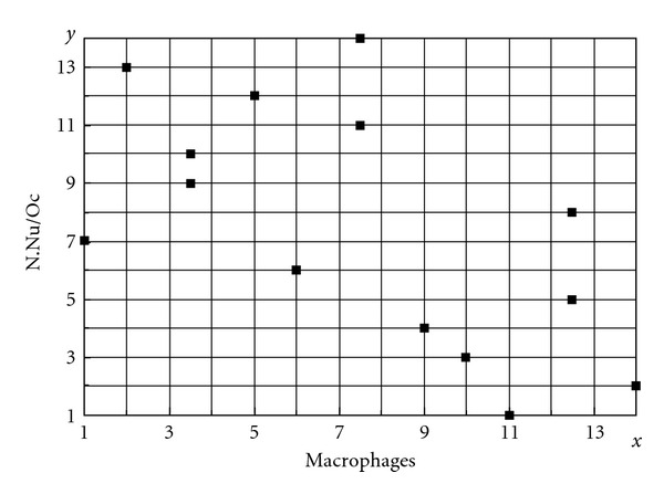
Nitrogen-containing bisphosphonates are widely used for treating diverse bone pathologies. They are anticatabolic drugs that act on osteoclasts inhibiting bone resorption. It remains unknown whether the mechanism of action is by decreasing osteoclast number, impairing osteoclast function, or whether they continue to effectively inhibit bone resorption despite the increase in osteoclast number. There is increasing evidence that bisphosphonates also act on bone marrow cells like macrophages and monocytes. The present work sought to evaluate the dynamics of preosteoclast fusion and possible changes in medullary macrophage number in bisphosphonate-treated animals. Healthy female Wistar rats received olpadronate, alendronate, or vehicle during 5 weeks, and 5-bromo-2-deoxyuridine (BrdU) on day 7, 28, or 34 of the experiment. Histomorphometric studies were performed to study femurs and evaluate: number of nuclei per osteoclast (N.Nu/Oc); number of BrdU-positive nuclei (N.Nu BrdU+/Oc); percentage of BrdU-positive nuclei per osteoclast (%Nu.BrdU+/Oc); medullary macrophage number (mac/mm(2)) and correlation between N.Nu/Oc and mac/mm(2). Results showed bisphosphonate-treated animals exhibited increased N.Nu/Oc, caused by an increase in preosteoclast fusion rate and evidenced by higher N.Nu BrdU+/Oc, and significantly decreased mac/mm(2). Considering the common origin of osteoclasts and macrophages, the increased demand for precursors of the osteoclast lineage may occur at the expense of macrophage lineage precursors.


Figures

Figure 1
Figure 1
Experimental Design. Female Wistar rats were used, divided into three groups: Sham, OPD, and ALN. Each one of the groups was divided into three subgroups and the columns of circles indicate these subsets into which the animals in each group were divided. The rows show weekly administration of vehicle (light gray) or 0.3 mg/kg of the corresponding bisphosphonate olpadronate or alendronate (dark gray). The star indicates the time of administration of the single dose of BrdU, that is, days 7, 28, or 34 of the experiment. All animals were euthanized on day 35 of the experiment.
Figure 2
Figure 2
Microphotographs of hematoxylin-eosin-stained histological sections of distal tibia. Animals treated with BPs showed a larger number of subchondral trabecualae (b) compared to sham (a).
Figure 3
Figure 3
Number of macrophages in diaphyseal bone marrow. The number of macrophages, measured using IHC ED1 detection, was significantly lower in bisphosphonate-treated animals compared to sham (*: Anova p < 0.01 compared to sham).
Figure 4
Figure 4
Macrophages in diaphyseal bone marrow. Microphotographs of histologic sections with ED1 immunohistochemical detection and hematoxylin-counterstain. Brown cells correspond to positive cells (macrophages). (a) shows a microphotograph of a sham animal, (b) an olpadronate-treated animal and (c) and alendronate-treated one. (b) and (c) show a lower number of positive cells.
Figure 5
Figure 5
Microphotographs of osteoclasts stained for IHC detection of ED1. Microphotograph (a) shows the distribution pattern of the marker in the cytoplasm of osteoclasts of sham group sections; the light areas indicate abundant vesicles (*). (b) and (c) show osteoclasts corresponding to olpadronate and alendronate-treated animals, respectively. The cytoplasm exhibits a homogenous distribution pattern of ED1. (d) shows an apoptotic osteoclast staining intensely positive for ED1. N: nucleus. The bar represents 50 microns.
Figure 6
Figure 6
Mean number of nuclei in osteoclasts. The mean number of nuclei per osteoclast was evaluated in BrdU-detection sections, was significantly higher in bisphosphonate-treated animals compared to sham (*: Kruskal Wallis p < 0.01). Both positive and negative nuclei were counted to assess this parameter.
Figure 7
Figure 7
The number in BrdU-positive nuclei per osteoclast was greater in bisphosphonate-treated animals at all three BrdU administration times (*: Kruskal Wallis p < 0.001 for days 7 and 34, p < 0.05 for day 28). Standard deviations are not represented because of high dispersion, which produces lines superposition (see data on text).
Figure 8
Figure 8
Microphotographs of osteoclasts stained immunohistochemically for BrdU. (a): dotted line: osteoclasts corresponding to the sham group showing 8 nuclei, none of which were BrdU-positive. (b): osteoclast corresponding to an olpadronate-treated animal showing at least 15 nuclear profiles, 3 of which were BrdU-positive. (c): osteoclast corresponding to an ALN-treated animal showing 2 BrdU-positive nuclei out of more than 25. The bar represents 50 microns.
Figure 9
Figure 9
Percentage of BrdU positive-nuclei per osteoclast. The percentage of BrdU-positive nuclei per osteoclast was higher in the bisphosphonate-treated groups at all three BrdU administration times (day 7, 28, or 35 of the experiment corresponding to one month, one week, and one day before euthanasia resp.) (*Kruskal Wallis p < 0.05 compared to sham).
Figure 10
Figure 10
Number of nuclei per osteoclast and number of macrophages. These two variables showed a negative correlation (−0.6), as number of nuclei increased in BP-treated animals, the number of macrophages in bone marrow consequently decreased (n sham = 7, n BP-treated = 7). p < 0.05, Spearman's test.

Similar articles

Cited by

References

    1. Hiroi-Furuya E, Kameda T, Hiura K, et al. Etidronate (EHDP) inhibits osteoclastic-bone resorption, promotes apoptosis and disrupts actin rings in isolate-mature osteoclasts. Calcified Tissue International. 1999;64(3):219–223. - PubMed
    1. Benford HL, McGowan NWA, Helfrich MH, Nuttall ME, Rogers MJ. Visualization of bisphosphonate-induced caspase-3 activity in apoptotic osteoclasts in vitro. Bone. 2001;28(5):465–473. - PubMed
    1. Van Beek ER, Löwik CWGM, Papapoulos SE. Bisphosphonates suppress bone resorption by a direct effect on early osteoclast precursors without affecting the osteoclastogenic capacity of osteogenic cells: the role of protein geranylgeranylation in the action of nitrogencontaining bisphosphonates on osteoclast precursors. Bone. 2002;30(1):64–70. - PubMed
    1. Sudhoff H, Jung JY, Ebmeyer J, Faddis BT, Hildmann H, Chole RA. Zoledronic acid inhibits osteoclastogenesis in vitro and in a mouse model of inflammatory osteolysis. Annals of Otology, Rhinology and Laryngology. 2003;112(9):780–786. - PubMed
    1. Kwak HB, Kim JY, Kim KJ, et al. Risedronate directly inhibits osteoclast differentiation and inflammatory bone loss. Biological and Pharmaceutical Bulletin. 2009;32(7):1193–1198. - PubMed