

Adhesion GPCRs are widely expressed throughout the subsections of the gastrointestinal tract
- PMID: 23009096
- PMCID: PMC3526421
- DOI: 10.1186/1471-230X-12-134
Adhesion GPCRs are widely expressed throughout the subsections of the gastrointestinal tract
Abstract
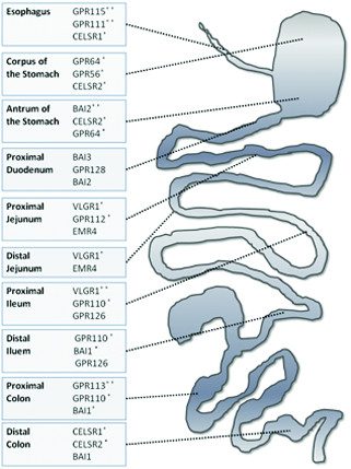
Background: G protein-coupled receptors (GPCRs) represent one of the largest families of transmembrane receptors and the most common drug target. The Adhesion subfamily is the second largest one of GPCRs and its several members are known to mediate neural development and immune system functioning through cell-cell and cell-matrix interactions. The distribution of these receptors has not been characterized in detail in the gastrointestinal (GI) tract. Here we present the first comprehensive anatomical profiling of mRNA expression of all 30 Adhesion GPCRs in the rat GI tract divided into twelve subsegments.
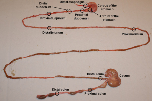
Methods: Using RT-qPCR, we studied the expression of Adhesion GPCRs in the esophagus, the corpus and antrum of the stomach, the proximal and distal parts of the duodenum, ileum, jejunum and colon, and the cecum.
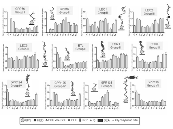
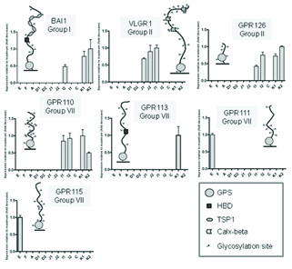
Results: We found that twenty-one Adhesion GPCRs (70%) had a widespread (expressed in five or more segments) or ubiquitous (expressed in eleven or more segments) distribution, seven (23%) were restricted to a few segments of the GI tract and two were not expressed in any segment. Most notably, almost all Group III members were ubiquitously expressed, while the restricted expression was characteristic for the majority of group VII members, hinting at more specific/localized roles for some of these receptors.
Conclusions: Overall, the distribution of Adhesion GPCRs points to their important role in GI tract functioning and defines them as a potentially crucial target for pharmacological interventions.
Figures

Similar articles
-
Comprehensive analysis of localization of 78 solute carrier genes throughout the subsections of the rat gastrointestinal tract.Biochem Biophys Res Commun. 2011 Aug 12;411(4):702-7. doi: 10.1016/j.bbrc.2011.07.005. Epub 2011 Jul 13. Biochem Biophys Res Commun. 2011. PMID: 21781957
-
Anatomical and histological profiling of orphan G-protein-coupled receptor expression in gastrointestinal tract of C57BL/6J mice.Cell Tissue Res. 2009 Nov;338(2):257-69. doi: 10.1007/s00441-009-0859-x. Epub 2009 Sep 10. Cell Tissue Res. 2009. PMID: 19763624
-
Expression profile of the entire family of Adhesion G protein-coupled receptors in mouse and rat.BMC Neurosci. 2008 Apr 29;9:43. doi: 10.1186/1471-2202-9-43. BMC Neurosci. 2008. PMID: 18445277 Free PMC article.
-
The adhesion GPCRs: a unique family of G protein-coupled receptors with important roles in both central and peripheral tissues.Cell Mol Life Sci. 2007 Aug;64(16):2104-19. doi: 10.1007/s00018-007-7067-1. Cell Mol Life Sci. 2007. PMID: 17502995 Free PMC article. Review.
-
Adhesion family of G protein-coupled receptors and cancer.Chang Gung Med J. 2012 Jan-Feb;35(1):15-27. doi: 10.4103/2319-4170.106170. Chang Gung Med J. 2012. PMID: 22483424 Review.
Cited by
-
Self-Nanoemulsifying Drug Delivery Systems for Enhancing Solubility, Permeability, and Bioavailability of Sesamin.Molecules. 2020 Jul 8;25(14):3119. doi: 10.3390/molecules25143119. Molecules. 2020. PMID: 32650503 Free PMC article.
-
The 17β-estradiol induced upregulation of the adhesion G-protein coupled receptor (ADGRG7) is modulated by ESRα and SP1 complex.Biol Open. 2019 Jan 14;8(1):bio037390. doi: 10.1242/bio.037390. Biol Open. 2019. PMID: 30598481 Free PMC article.
-
Deletion of Gpr128 results in weight loss and increased intestinal contraction frequency.World J Gastroenterol. 2014 Jan 14;20(2):498-508. doi: 10.3748/wjg.v20.i2.498. World J Gastroenterol. 2014. PMID: 24574718 Free PMC article.
-
Gene Expression Profiling Reveals Functional Specialization along the Intestinal Tract of a Carnivorous Teleostean Fish (Dicentrarchus labrax).Front Physiol. 2016 Aug 25;7:359. doi: 10.3389/fphys.2016.00359. eCollection 2016. Front Physiol. 2016. PMID: 27610085 Free PMC article.
-
Defining the gene repertoire and spatiotemporal expression profiles of adhesion G protein-coupled receptors in zebrafish.BMC Genomics. 2015 Feb 8;16(1):62. doi: 10.1186/s12864-015-1296-8. BMC Genomics. 2015. PMID: 25715737 Free PMC article.
References
-
- Krasnoperov V, Bittner MA, Holz RW, Chepurny O, Petrenko AG. Structural requirements for alpha-latrotoxin binding and alpha-latrotoxin-stimulated secretion. A study with calcium-independent receptor of alpha-latrotoxin (CIRL) deletion mutants. J Biol Chem. 1999;274(6):3590–3596. doi: 10.1074/jbc.274.6.3590. - DOI - PubMed
Publication types
MeSH terms
Substances
LinkOut - more resources
Full Text Sources
