. 2012 Sep 26;3(1):22.
doi: 10.1186/2042-6410-3-22.

Sex differences in microRNA regulation of gene expression: no smoke, just miRs

Affiliations

Sex differences in microRNA regulation of gene expression: no smoke, just miRs

Christopher P Morgan et al. Biol Sex Differ. .

Abstract

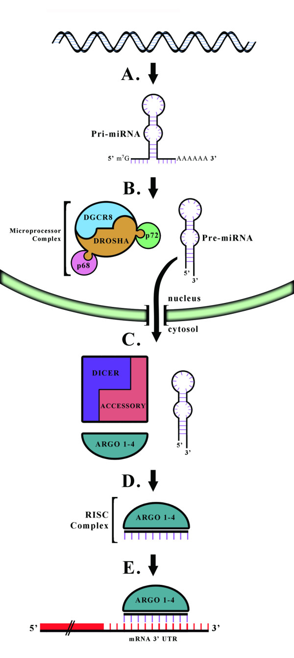
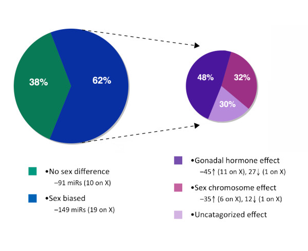
Males and females differ widely in morphology, physiology, and behavior leading to disparities in many health outcomes, including sex biases in the prevalence of many neurodevelopmental disorders. However, with the exception of a relatively small number of genes on the Y chromosome, males and females share a common genome. Therefore, sexual differentiation must in large part be a product of the sex biased expression of this shared genetic substrate. microRNAs (miRs) are small non-coding RNAs involved in the post-transcriptional regulation of up to 70% of protein-coding genes. The ability of miRs to regulate such a vast amount of the genome with a high degree of specificity makes them perfectly poised to play a critical role in programming of the sexually dimorphic brain. This review describes those characteristics of miRs that make them particularly amenable to this task, and examines the influences of both the sex chromosome complement as well as gonadal hormones on their regulation. Exploring miRs in the context of sex differences in disease, particularly in sex-biased neurodevelopmental disorders, may provide novel insight into the pathophysiology and potential therapeutic targets in disease treatment and prevention.


Figures

Figure 1
Figure 1
Schematic representation of miRNA (miR) biogenesis. A, Most miRs are transcribed as polycistronic primary-miRs (pri-miRs) by RNA polymerase II, before undergoing 5' capping and 3' polyadenylation. Portions of the pri-miR folds back on itself to form double-stranded stem-loop structures. B, The Microprocessor complex cleaves these stem-loops from pri-miRs, generating 50–70 bp pre-miRs with a short 3' overhang. Drosha and DGCR8 are the obligate components of the Microprocessor complex, though additional accessory proteins, such as the RNA helicases P68 and P72, can regulate the activity of the complex. C, The short 3' overhang is recognized by components of the nuclear export machinery, leading to active transport of pre-miRs out into the cytosol. In the cytosol, Dicer acts in complex with accessory proteins to process the pre-miRs into double stranded 22 bp duplexes. D, Dicer then assists in loading one strand of this duplex, the guide strand, into the Argonaute-containing RISC complex. E, Mature miRs guide the RISC complex to specific mRNA targets. miRs identify mRNA targets through regions of sequence homology in the mRNA's 3' UTR. The outcome of this interaction can depend on the degree of sequence complementarity and the specific Argonaute present in the RISC complex, but destabilization of the mRNA and subsequent degradation is likely.
Figure 2
Figure 2
The neonatal brain displays extensive sex bias in miR expression, which appears to result from both gonadal hormone and sex chromosomal regulation. In our previous studies, the expression of 240 miRs was assayed in postnatal day 2 whole brains from male and female mice. To determine the role of organizational estradiol in the male brain to program the miR environment, the aromatase inhibitor, formestane, was administered to males at this time and brain tissue compared with that from control males and females. Of these 240 miRs, 149 showed sex-biased expression. These 149 miRs were then further subdivided by: 1) their apparent responsiveness to estradiol, where detected sex differences were ameliorated by formestane treatment making them regulated at some level by gonadal hormones, 2) as likely attributable to X chromosome differences where females showed higher levels (likely due to X gene dosage) and showed no changes in males treated with formestane, or 3) uncategorized effect where the pattern of change did not fit either model of gonadal hormone effect or X-linkage. Of the 149 miRs with a basal sex difference, changes related to estradiol occurred for almost half of these genes (72 miRs), where aromatase inhibition dysmasculinized male expression patterns to look more like that of the females. An effect of sex chromosomes was estimated for 47 miRs, where aromatase inhibition had no affect on male expression. Analysis criteria found that neither regulatory mechanism could be attributed to 30 miRs. (Adapted from Morgan and Bale, 2011 [11]).

References

    1. Yeh S-H, Chen P-J. Gender Disparity of Hepatocellular Carcinoma: The Roles of Sex Hormones. Oncology. 2010;78:172–179. - PubMed
    1. Ober C, Loisel DA, Gilad Y. Sex-specific genetic architecture of human disease. Nature reviews. Genetics. 2008;9:911–922. - PMC - PubMed
    1. Voskuhl R. Sex differences in autoimmune diseases. Biol Sex Differ. 2011;2:1. doi: 10.1186/2042-6410-2-1. - DOI - PMC - PubMed
    1. Mendell JT, Olson EN. MicroRNAs in Stress Signaling and Human Disease. Cell. 2012;148:1172–1187. doi: 10.1016/j.cell.2012.02.005. - DOI - PMC - PubMed
    1. Chang T-C, Mendell JT. microRNAs in vertebrate physiology and human disease. Annu Rev Genomics Hum Genet. 2007;8:215–239. doi: 10.1146/annurev.genom.8.080706.092351. - DOI - PubMed

LinkOut - more resources