Review
. 2013 Jan;21(1):1-26.
doi: 10.3109/1061186X.2012.723213. Epub 2012 Sep 26.

Biological rationale for the design of polymeric anti-cancer nanomedicines

Affiliations
Review

Biological rationale for the design of polymeric anti-cancer nanomedicines

Yan Zhou et al. J Drug Target. 2013 Jan.

Abstract

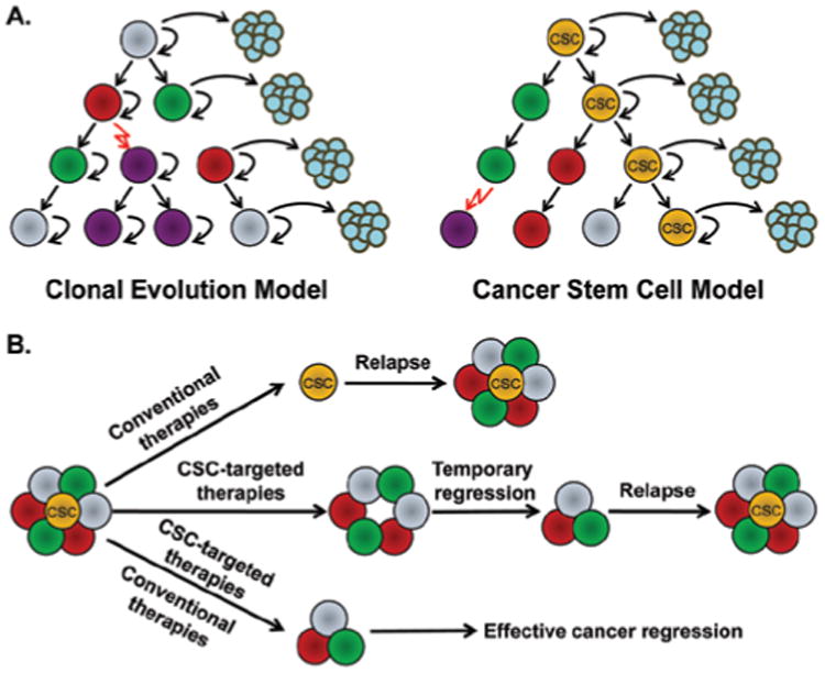
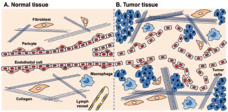
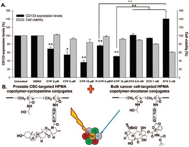
Understanding the biological features of cancer is the basis for designing efficient anti-cancer nanomedicines. On one hand, important therapeutic targets for anti-cancer nanomedicines need to be identified based on cancer biology, to address the unmet medical needs. On the other hand, the unique pathophysiological properties of cancer affect the delivery and interactions of anti-cancer nanomedicines with their therapeutic targets. This review discusses several critical cancer biological properties that challenge the currently available anti-cancer treatments, including cancer heterogeneity and cancer stem cells, the complexcity of tumor microenvironment, and the inevitable cancer metastases. In addition, the biological bases of the enhanced permeability and retention (EPR) effect and tumor-specific active targeting, as well as the physiological barriers for passive and active targeting of anti-cancer nanomedicines are covered in this review. Correspondingly, possible nanomedicine strategies to target cancer heterogeneity, cancer stem cells and metastases, to overcome the challenges related to tumor passive targeting and tumor penetration, and to improve the interactions of therapeutic payloads with the therapeutic targets are discussed. The focus is mainly on the designs of polymeric anti-cancer nanomedicines.


Figures

Figure 1
Figure 1
(A) Two models of tumor heterogeneity. The left cartoon shows the clonal evolution model: cancer cells are phenotypically heterogeneous, but they acquire the tumorigenic ability randomly, with the equal potential to generate new tumors; the right cartoon illustrates the CSC model: only a subset of cancer cells (CSC) have the ability to self-renew and form new tumors, whereas most other cancer cells are depleted of this ability. (B) The rationale for the development of anti-cancer therapeutics based on CSC model. Conventional therapies kill non-CSCs but fail to eliminate CSCs, resulting in relapse; CSC-targeted therapies inhibit CSCs and lead to temporary cancer regression, but may not stop the generation of new CSCs from bulk cancer cells; the combination of conventional and CSC-targeted therapies will effectively target both bulk cancer cells and CSCs, leading to effective cancer regression. Adapted with permission from Reya et al. (2001).
Figure 2
Figure 2
The structural differences between normal and tumor tissue microenvironment. (A) Normal tissues have blood vessels with tightly aligned endothelial cells and pericytes. The extracellular matrix is comprised of a loose network of collagen fibers and a few fibroblasts and macrophages. Lymph vessels are present. (B) Tumor tissues contain leaky and tortuous blood vessels with irregular blood flow. Pericytes covering outside the tumor vasculatures are still present. The tumor extracellular matrix is much denser than normal tissues, with thicker network of collagen fibers, more fibroblasts and macrophages. In addition, tumors usually lack functional lymph vessels. All the above features contribute to the increased interstitial fluid pressure (IFP). Adapted with permission from Heldin et al. (2004).
Figure 3
Figure 3
(A) Summary of changes on CD133+ prostate CSCs and whole cell viabilities following HPMA copolymer-cyclopamine conjugate, free cyclopamine or docetaxel treatments on RC-92a/hTERT prostate cancer cells in vitro (Zhou et al., 2012). Black columns: CD133 expression level (%); gray columns: Cell viability (%). The data are presented as mean ± SD of the experiments done in triplicate. *p < 0.05; **p < 0.01. Vehicle (DMSO) treated and untreated cells were used as controls. (B) The scheme of the combination HPMA copolymer-based macromolecular therapeutics for improving the treatment of prostate cancer, by targeting both bulk cancer cells and prostate CSCs. Adapted with permission from Zhou et al. (2012).
Figure 4
Figure 4
The antitumor and anti-CSC effect of the thermal enhancement with gold nanoshells in combination with radiation in the BCM-2665 breast cancer model. (A) Tumor volume changes following radiation (IR, red triangle) and the combination of radiation and hyperthermia (IR + HT, yellow diamond). (B) The changes in the percentages of ALDH + breast CSCs following IR (red) and IR + HT (yellow) treatments. (C) The changes in mammosphere forming efficiency (MSFE) following IR (red) and IR + HT (yellow) treatments. (D) EdU incorporation following IR (red) and IR + HT (yellow) treatments (*p < 0.05; **p < 0.001). The % changes in B–D were normalized to the mock treatment. Adapted with permission from Atkinson et al. (2010).
Figure 5
Figure 5
The effect of i.p. administered HPMA copolymer-DOX conjugate and free DOX on the growth of both sensitive (left) and resistant (right) human ovarian carcinoma xenografts in mice. Triangles: HPMA copolymer-DOX conjugate; squares: free DOX; circles: control. The data are presented as mean ± SE (Minko et al., 2000). Reprinted with permission from Kopeček & Kopečková (2010).
Figure 6
Figure 6
(A) The structure of Activatable Cell Penetrating Peptide-conjugated Dendrimer (ACPPD). Multiple ACPPs are covalently attached via the polycationic segments (a) to the dendrimer (gray circle). Yellow ovals (d) demonstrated the payloads such as Cy5 and/or Gd. Upon exposure to MMP-2 or -9 in tumor microenvironment, the linkers (b) are cleaved and polyanions (c) are released, leaving the cationic dendrimers for cell entry. (B–E) The enhanced uptake of Gd and Cy5 dually labeled ACPPDs in regions with high MMP activities and infiltrative tumors. (B–C) Axial MR and fluorescence images of a transgenic PyMT mouse before surgery (white arrows: the tumor burdens); (D–E) Axial MR and fluorescence images of the same mouse after surgical removal of tumor under white light. Red arrows: regions of residual hyperintensity on MR and fluorescence imaging; (F) Regions of hyperintensity on MRI were removed under fluorescence imaging and stained with hematoxylin/eosin to verify the presence of tumor. Adapted with permission from Olson et al. (2010).
Figure 7
Figure 7
Inhibition of metastatic 4T1 mammary adenocarcinoma in the tibia by HPMA copolymer-PTX-ALN conjugate. (A) Chemical structure of the conjugate and the release mechanism of PTX and ALN from the conjugate. (B) Fluorescent images of 4T1 mCherry tumors in the tibia following single, combination of free drugs, and HPMA copolymer-PTX-ALN conjugate treatments on day 15. The treatments were administered i.v. every other day. Scale bar represents 15 mm. (C) Antitumor efficacies of HPMA copolymer-PTX-ALN conjugate (closed squares), free PTX plus ALN (open squares), free PTX (closed circles), and free ALN (open triangles) as compared to vehicle (open circles) and saline (closed triangles) controls. Y-axis represents the fluorescent intensities of the tumors as measured quantitatively by intravital noninvasive fluorescence imaging. The data are presented as mean ± SE. PTX, paclitaxel; ALN, alendronate. Adapted with permission from Miller et al. (2011).
Figure 8
Figure 8
(A) The chemical structure of Tyr-HPMA-copolymer. (B) The relationship between the molecular size of i.v. administered 125I-labeled Tyr-HPMA-copolymers and the plasma concentration (AUC), tumor accumulation and renal clearance (CL) in sarcoma-180 tumor-bearing mice (Seymour et al., 1995; Fang et al., 2011). (C) The chemical structure of the branched HPMA copolymer-DOX conjugate. (D) Tumor accumulation of DOX in OVCAR-3 carcinoma bearing nu/nu mice after i.v. bolus of free DOX or HPMA copolymer-DOX conjugate with different molecular weights (Shiah et al., 2001a).
Figure 9
Figure 9
(A) The synthesis of multiblock HPMA copolymer-DOX conjugate. The HPMA copolymer segments containing DOX was synthesized by RAFT copolymerization of monomer HPMA and MA-GFLG-DOX in the presence of a bifunctional chain transfer agent containing GFLG sequences (Peptide2CTA). The chain was extended by the reaction of the telechelic α,ω-dithoil-HPMA copolymers with bismaleimide, yielding the multiblock biodegradable HPMA copolymer-DOX conjugate (Pan et al., 2011). (B) FPLC profiles showing the degradation of multibock HPMA copolymer-DOX conjugate by incubation with cathepsin B (left) and papain (right) at different time intervals. PD-Org: the initial telechelic HPMA copolymer-DOX conjugate; PD-Ext: the extended multiblock HPMA copolymer-DOX conjugate (Pan et al., 2011). C) The synthesis of multiblock HPMA copolymer-gemcitabine conjugate. The heterotelechelic HPMA copolymer-gemcitabine conjugate segments containing terminal alkyne and azide groups was firstly synthesized by RAFT copolymerization using the chain transfer agent containing GFLG sequence and alkyne group (CTA-GFLG-alkyne), followed by post-polymerization modification with diazido-V-501. The chain extension was achieved by Cu (I) catalyzed azide-alkyne click reaction (Yang et al., 2011). Adapted from Pan et al. (2011); Yang et al. (2011).
Figure 10
Figure 10
The tumor remodeling and anti-tumor effects of Gemcitabine + PEGPH20 (PEGylated recombinant human hyaluronidase PH20) combination therapy on KrasLSL-G12D/+;Trp53LSL-R172H/+;Cre (KPC) mice. (A) Tumor IFP at survival endpoint decreased significantly after treatment with gemcitabine + PEGPH20 (GP; n = 9) than with gemcitabine (G; n = 9) only. *p < 0.0001. (B and C) Tumor was hypovascular following gemcitabine treatment (B), while tumor vasculatures were increased following gemcitabine + PEGPH20 combination therapy (C). D) Tumor volume changes following Gemcitabine and Gemcitabine + PEGPH20 combination treatments after one cycle (*p = 0.009). (E) Kaplan-Meier survival curves of mice in different treatment groups: control (n = 16), PEGPH20 (n = 15), Gem (n = 16), and Gem + PEGPH20 (n = 14). *p = 0.004 demonstrated the significant difference in median overall survival of Gem (55.5 days) and Gemcitabine + PEGPH20 (91.5 days) treated mice. (F) Metastatic burden in Gemcitabine + sPEGPH20 treated mice was significantly decreased compared with Gem treatment alone (G) (*p = 0.014). Adapted with permission from Provenzano et al. (2012).

Similar articles

Cited by

References

    1. Al-Hajj M, Wicha MS, Benito-Hernandez A, Morrison SJ, Clarke MF. Prospective identification of tumorigenic breast cancer cells. Proc Natl Acad Sci USA. 2003;100:3983–3988. - PMC - PubMed
    1. Alitalo K, Tammela T, Petrova TV. Lymphangiogenesis in development and human disease. Nature. 2005;438:946–953. - PubMed
    1. Allen TM. Ligand-targeted therapeutics in anticancer therapy. Nat Rev Cancer. 2002;2:750–763. - PubMed
    1. Arap W, Pasqualini R, Ruoslahti E. Cancer treatment by targeted drug delivery to tumor vasculature in a mouse model. Science. 1998;279:377–380. - PubMed
    1. Atkinson RL, Zhang M, Diagaradjane P, Peddibhotla S, Contreras A, Hilsenbeck SG, Woodward WA, Krishnan S, Chang JC, Rosen JM. Thermal enhancement with optically activated gold nanoshells sensitizes breast cancer stem cells to radiation therapy. Sci Transl Med. 2010;2:55ra79. - PMC - PubMed

Publication types

LinkOut - more resources