Comparative Study
. 2013 Jan;38(1):18-26.
doi: 10.3109/02713683.2012.725797. Epub 2012 Sep 25.

Na(+)/K(+)-ATPase expression changes in the rabbit lacrimal glands during pregnancy

Affiliations
Comparative Study

Na(+)/K(+)-ATPase expression changes in the rabbit lacrimal glands during pregnancy

Jianyan Huang et al. Curr Eye Res. 2013 Jan.

Abstract

Purpose: To investigate the expressional changes of Na(+)/K(+)-ATPase subunits in the lacrimal glands (LG) of term pregnant rabbits.

Methods: LG were obtained from term pregnant rabbits and age-matched female control rabbits for laser capture microdissection (LCM), real time RT-PCR, western blot, and immunofluorescence. The mRNA and proteins of α1, α2, β1, β2, and β3 subunits of Na(+)/K(+)-ATPase were detected and quantified.

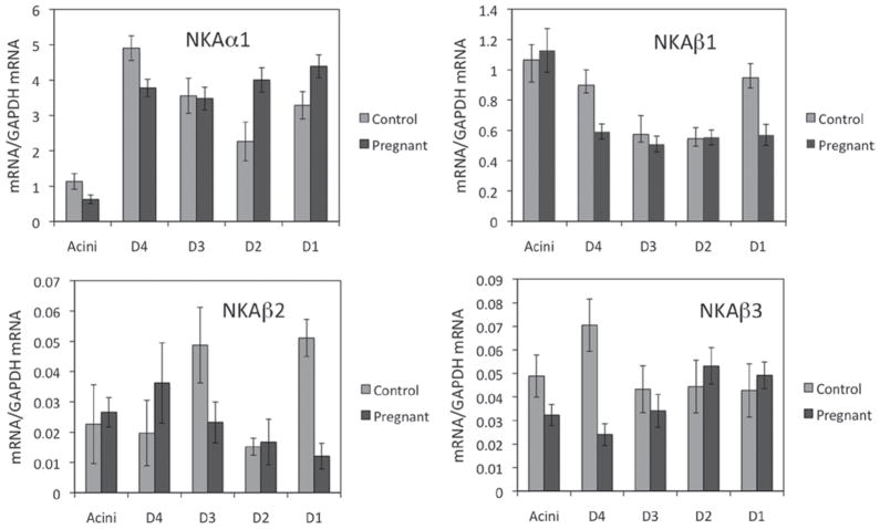
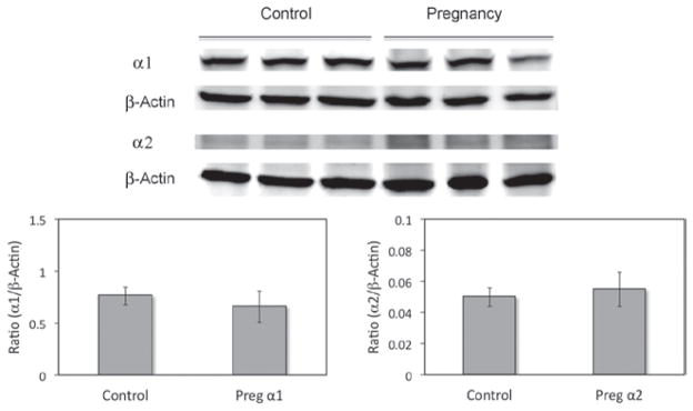
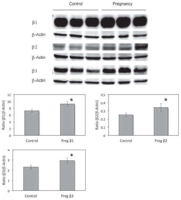
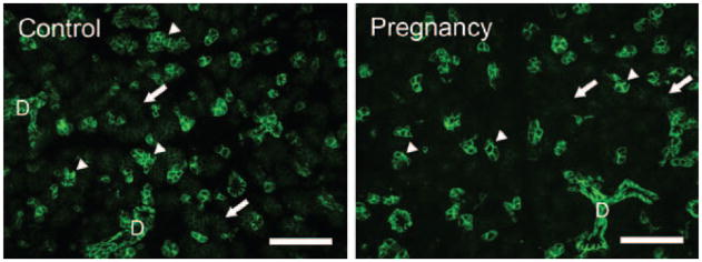
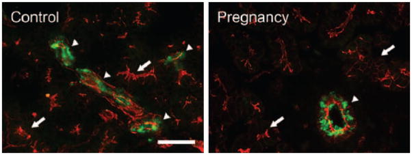
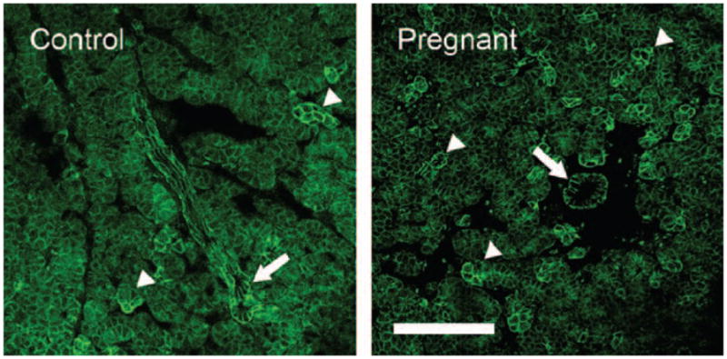
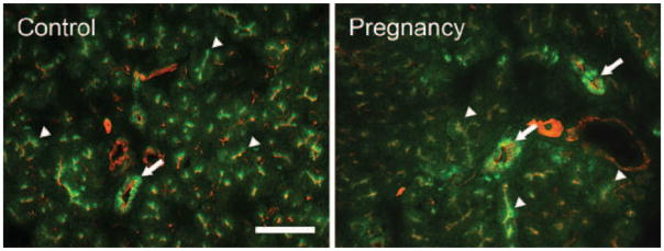
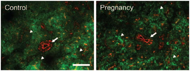
Results: Although only the mRNA for β3 from whole LG of pregnant rabbits was significantly different from that of normal controls, many mRNA levels for α1, α2, β1, β2, and β3 from acini and epithelial cells from various duct segments that were collected by LCM were significantly different from those of normal control rabbits. Western blots demonstrated that the expressions of all three β subunits were significantly higher in pregnant rabbits, while both α subunits remained unchanged during pregnancy. Interestingly, immunofluorescence results showed that the distribution patterns of all Na(+)/K(+)-ATPase subunits during pregnancy were similar to those of the control rabbits.

Conclusions: Changes were found in mRNA and protein expressions of Na(+)/K(+)-ATPase subunits in LG from term pregnant rabbits and these changes suggest a role in the pregnancy-related LG secretion changes and dry eye symptoms observed in these animals.


Conflict of interest statement

Declaration of interest: This work was supported by NIH grants EY017731 (CD) and EY03040.

Figures

FIGURE 1
FIGURE 1
Real-time RT-PCR of Na+/K+-ATPase subunits from LG epithelial cells of pregnant rabbits collected by LCM. Data from control rabbits is from our previous publication (used with permission). α1, mRNA level for α1 was predominantly localized in the ducts, and during pregnancy, its level was significantly decreased in acini and intralobular duct (p < 0.05), while its level was significantly increased in intralobar and interlobar ducts (p < 0.05). α2, mRNA level for α2 was extremely low and we were unable to detect any α2 in epithelial cells collected by LCM. β1, mRNA level for β1 from pregnant rabbits was most abundant in acini and its level was significantly decreased in intralobular and interlobar ducts (p < 0.05). β2, During pregnancy, mRNA level for β2 was most abundant in intralobular duct while least abundant in interlobar duct and its level was significantly decreased in interlobular and interlobar ducts as compared to normal controls (p < 0.05). β3, mRNA level for β3 was most abundant in intralobar and interlobar ducts, while its level was significantly reduced in acini and intralobular duct during pregnancy (p < 0.05). D4, intralobular duct; D3, interlobular duct; D2:, intralobar duct; D1, interlobar duct. Data were presented as mean ± SEM.
FIGURE 2
FIGURE 2
Western blots of α subunits from whole LG homogenates. No significant difference was detected in either α1 or α2 subunits between normal control and pregnant rabbits (p > 0.05). β-actin was used as loading control. Data are representative images of at least three replicates from at least 3 different animals each. Preg, pregnant.
FIGURE 3
FIGURE 3
Western blots of β subunits from whole LG homogenates. All β subunits were significantly increased in LG from pregnant rabbits (p < 0.05). β-actin was used as loading control. Data are representative images of at least three replicates from at least three different animals each. Preg, pregnant.
FIGURE 4
FIGURE 4
Immunofluorescence of α1-IR. Control: α1-IR (green) was present in every acinar cell, most prominently in the basolateral membranes but not on the apical membranes. The α1-IR was much stronger in some acinar cells (arrowheads) while much weaker in other acini (arrows), giving the gland a “mosaic” pattern. Ducts (D) were all stained as strong as those acinar cells showed the most intense α1-IR. Pregnancy: the distribution pattern of α1-IR in LG from pregnant rabbits was similar to that of control rabbits and no significant difference was detected. At least three control rabbits and three pregnant rabbits were stained and repeated at least three times. Scale bar = 50 μm.
FIGURE 5
FIGURE 5
Immunofluorescence of α2-IR. Control: Intense staining of α2-IR was detected in duct cells as clustered punctate staining (arrowheads) while minimal staining was found in acinar cells (arrows). Pregnancy: In pregnant rabbits, the staining pattern of α2-IR was similar to that of control animals, i.e., α2-IR was mostly found in duct cells as clusters of punctate staining (arrowheads), while minimal staining was found in acini (arrows). No significant difference of α2-IR was observed between control and pregnant rabbits. At least three control rabbits and three pregnant rabbits were stained and repeated at least three times. Scale bar = 25 μm.
FIGURE 6
FIGURE 6
Immunofluorescence of β1-IR. Control: β1-IR was present in basolateral membranes of all acinar and ductal cells as well as numerous punctate staining within the cytoplasm. However, levels of β1-IR differed in a “mosaic” pattern, higher in some acinar cells and/or acini (arrowheads) than others. β1-IR in ductal cells was uniformly higher (arrow). Pregnant: like in control animals, β1-IR was also present in all acinar cells and demonstrated a similar “mosaic” pattern (arrowheads). Ducts also showed a much higher β1-IR (arrow). However, no significant difference of β1-IR was observed between control and pregnant animals. At least three control rabbits and three pregnant rabbits were stained and repeated at least three times. Scale bar = 50 μm.
FIGURE 7
FIGURE 7
Immunofluorescence of β2-IR. Control: β2-IR (green) was present in every acinar cell as numerous punctate staining that aggregate towards the apical cytoplasm (arrowheads). The intensity of β2-IR was much higher in ducts (arrow). Pregnant: the distribution pattern of β2-IR in LG from pregnant rabbits was similar to that of control rabbits, i.e., numerous punctate β2-IR was detected in the cytoplasm of all acinar cells (arrowheads) and β2-IR was much stronger in duct cells (arrow). Red: F-actin that has been stained with rhodamine. At least three control rabbits and three pregnant rabbits were stained and repeated at least three times. Scale bar = 50 μm.
FIGURE 8
FIGURE 8
Immunofluorescence of β3-IR. Control: β 3-IR (green) was present in every acinar cell as numerous punctate staining within the cytoplasm (arrowheads) in a diffuse pattern. However, minimal β3-IR was detected in the duct cells (arrow). Pregnant: the distribution pattern of β3-IR in LG from pregnant rabbits was similar to that of control rabbits, i.e., numerous punctate β3-IR was detected in the cytoplasm of every acinar cell (arrowheads) while minimal β3-IR was present in the duct cells (arrow). Red: F-actin that has been stained with rhodamine. At least three control rabbits and three pregnant rabbits were stained and repeated at least three times. Scale bar = 50 μm.

Similar articles

Cited by

  • Inflammation mechanism and anti-inflammatory therapy of dry eye.
    Chu L, Wang C, Zhou H. Chu L, et al. Front Med (Lausanne). 2024 Feb 14;11:1307682. doi: 10.3389/fmed.2024.1307682. eCollection 2024. Front Med (Lausanne). 2024. PMID: 38420354 Free PMC article. Review.
  • Should I Get LASIK If I'm Breastfeeding?
    Moshirfar M, Rosen DB, Heiland MB, Ronquillo YC, Hoopes PC. Moshirfar M, et al. Ophthalmol Ther. 2019 Sep;8(3):349-352. doi: 10.1007/s40123-019-0195-5. Epub 2019 Jun 28. Ophthalmol Ther. 2019. PMID: 31254257 Free PMC article.
  • Ion channels in dry eye disease.
    Ashok N, Khamar P, D'Souza S, Gijs M, Ghosh A, Sethu S, Shetty R. Ashok N, et al. Indian J Ophthalmol. 2023 Apr;71(4):1215-1226. doi: 10.4103/IJO.IJO_3020_22. Indian J Ophthalmol. 2023. PMID: 37026252 Free PMC article. Review.

References

    1. Ding C, Parsa L, Nandoskar P, Zhao P, Wu K, Wang Y. Duct system of the rabbit lacrimal gland: structural characteristics and role in lacrimal secretion. Invest Ophthalmol Vis Sci. 2010;51:2960–2967. - PMC - PubMed
    1. Mircheff AK. Lacrimal fluid and electrolyte secretion: a review. Curr Eye Res. 1989;8:607–617. - PubMed
    1. Dartt DA, Møller M, Poulsen JH. Lacrimal gland electrolyte and water secretion in the rabbit: localization and role of (Na+ +K+)-activated ATPase. J Physiol (Lond) 1981;321:557–569. - PMC - PubMed
    1. Herok GH, Millar TJ, Anderton PJ, Martin DK. Characterization of an inwardly rectifying potassium channel in the rabbit superior lacrimal gland. Invest Ophthalmol Vis Sci. 1998;39:308–314. - PubMed
    1. Herok GH, Millar TJ, Anderton PJ, Martin DK. Role of chloride channels in regulating the volume of acinar cells of the rabbit superior lacrimal gland. Invest Ophthalmol Vis Sci. 2008;49:5517–5525. - PubMed

Publication types

MeSH terms

Substances

LinkOut - more resources