. 2012 Sep 25:12:16.
doi: 10.1186/1472-6890-12-16.

Illustration of extensive extracellular matrix at the epithelial-mesenchymal interface within the renal stem/progenitor cell niche

Affiliations

Illustration of extensive extracellular matrix at the epithelial-mesenchymal interface within the renal stem/progenitor cell niche

Will W Minuth et al. BMC Clin Pathol. .

Abstract

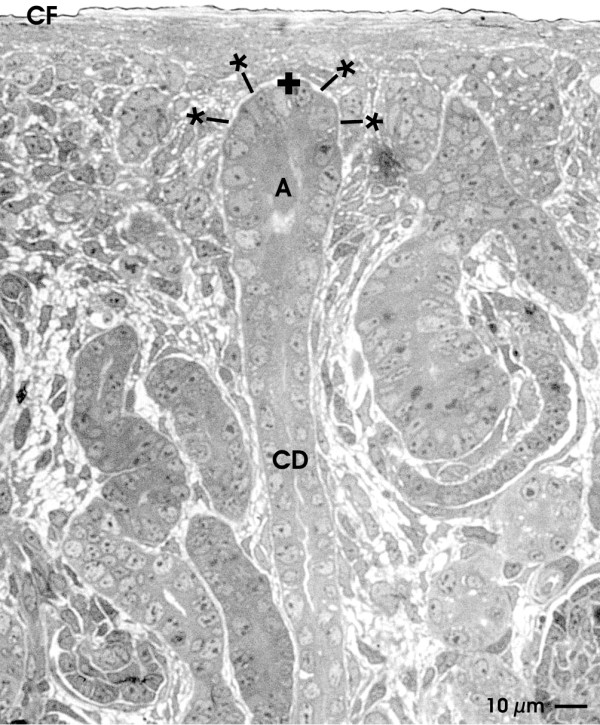
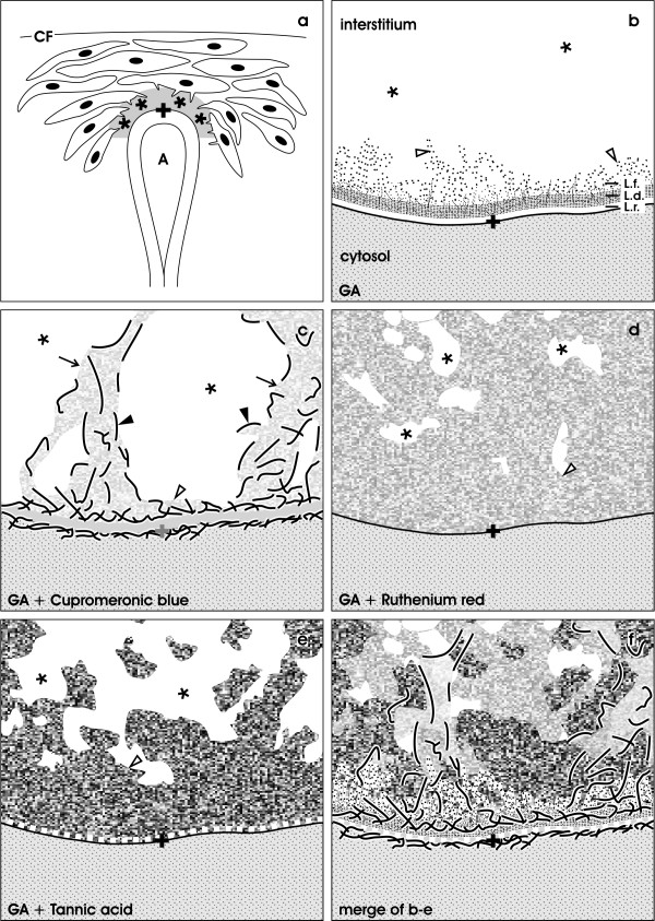
Background: Stem/progenitor cells are promising candidates to treat diseased renal parenchyma. However, implanted stem/progenitor cells are exposed to a harmful atmosphere of degenerating parenchyma. To minimize hampering effects after an implantation investigations are in progress to administer these cells within an artificial polyester interstitum supporting survival. Learning from nature the renal stem/progenitor cell niche appears as a valuable model. At this site epithelial stem/progenitor cells within the collecting duct ampulla face mesenchymal stem/progenitor cells. Both cell types do not have close contact but are separated by a wide interstitium.

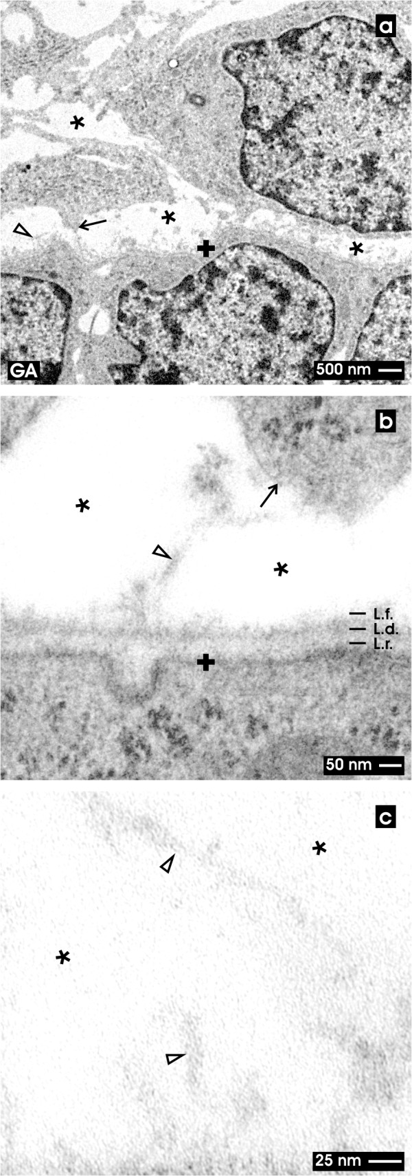
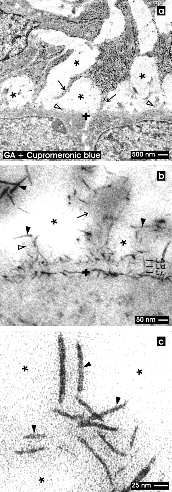
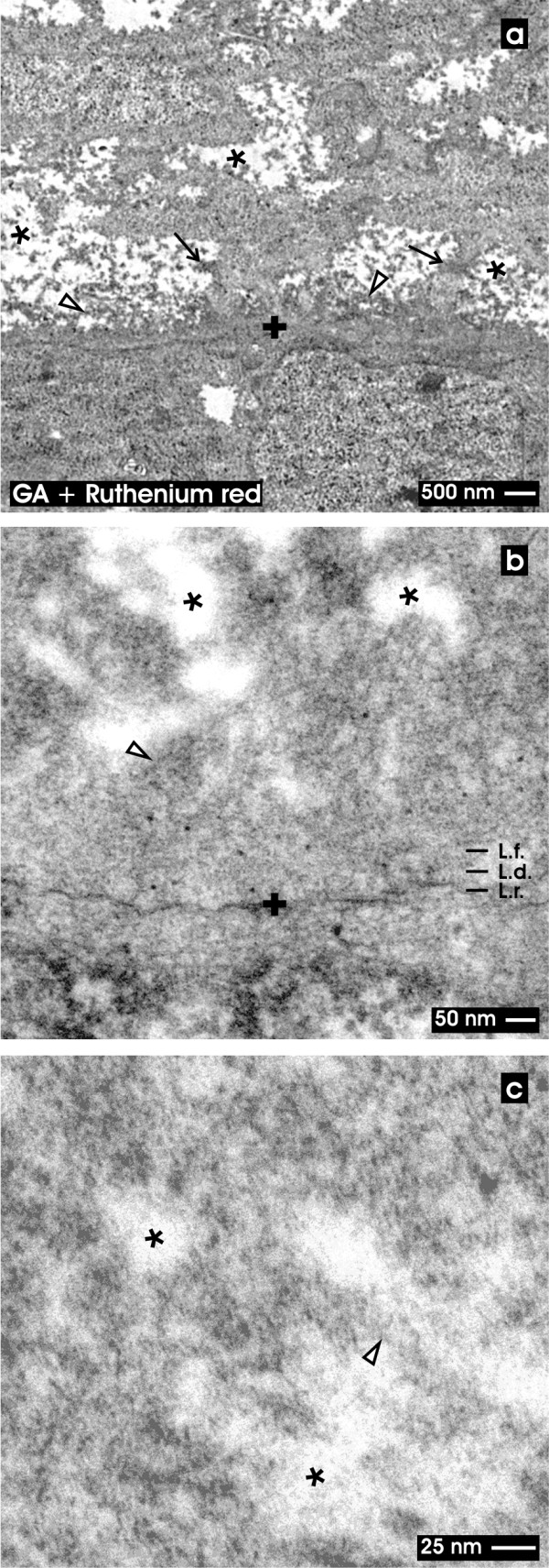
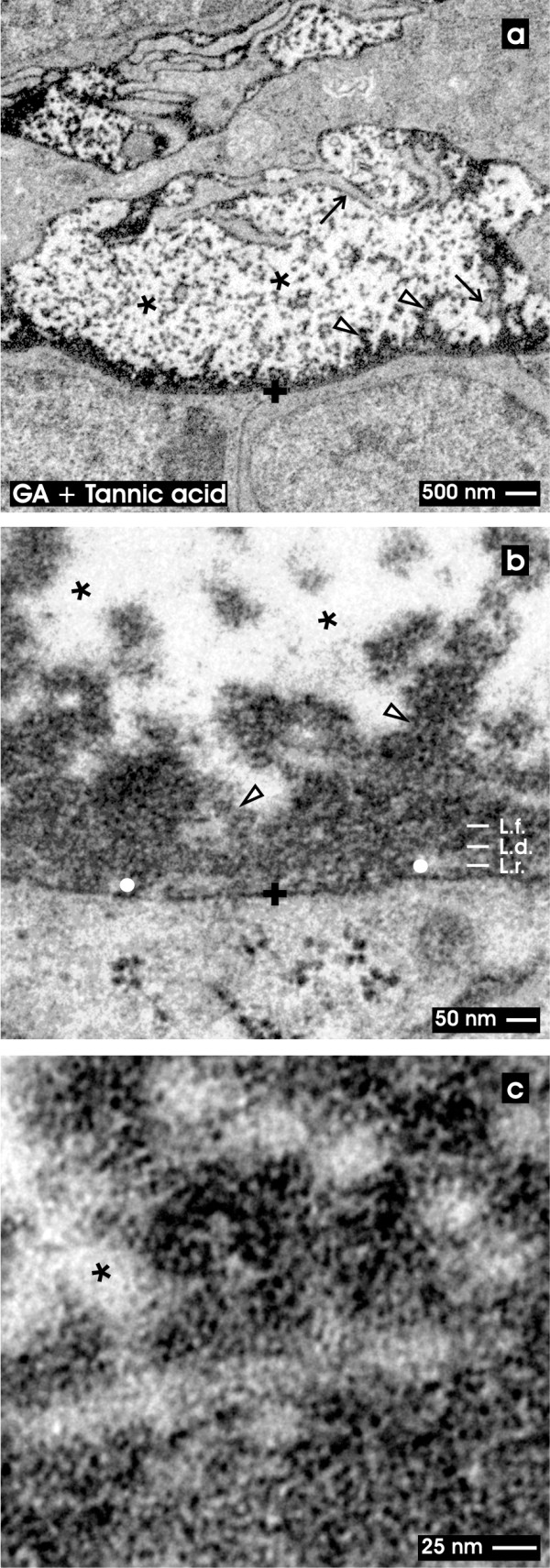
Methods: To analyze extracellular matrix in this particular interstitium, special contrasting for transmission electron microscopy was performed. Kidneys of neonatal rabbits were fixed in solutions containing glutaraldehyde (GA) or in combination with cupromeronic blue, ruthenium red and tannic acid.

Results: GA revealed a basal lamina at the ampulla and a bright but inconspicuously looking interstitial space. In contrast, GA containing cupromeronic blue exhibits numerous proteoglycan braces lining from the ampulla towards the interstitial space. GA containing ruthenium red or tannic acid demonstrates clouds of extracellular matrix protruding from the basal lamina of the ampulla to the surface of mesenchymal stem/progenitor cells.

Conclusions: The actual data show that the interstitium between epithelial and mesenchymal stem/progenitor cells contains much more and up to date unknown extracellular matrix than earlier observed by classical GA fixation.


Figures

Figure 1
Figure 1
Semithin section of the renal stem/progenitor cell niche underneath the organ capsule (CF). Accurate orientation of parenchyma is obtained by sectioning in parallel to the lumen of a collecting duct (CD). Epithelial stem/progenitor cells are found within the tip of a ureteric bud derived CD ampulla (A). A thin layer of nephrogenic mesenchymal stem/progenitor cells is found between the tip of a CD ampulla and the organ capsule. Epithelial and mesenchymal stem/progenitor cells are separated by a wide interstitial space (lined asterisk). Coordinate for electron microscopy is labeled by a cross (+).
Figure 2
Figure 2
TEM of the renal stem/progenitor cell niche after fixation in GA. Low (a), higher (b) and high (c) magnifications illustrate the interstitial interface (asterisk) at the tip of a CD ampulla labeled by a cross (+). (a) Low magnification illustrates that epithelial stem/progenitor cells within the CD ampulla are separated from mesenchymal stem/progenitor cells by a consistently developed basal lamina and a bright interstitial space (asterisk). Protrusions from mesenchymal stem/progenitor cells line towards the CD ampulla (arrow). (b) Higher magnification shows that a basal lamina at the tip of the CD ampulla borders the interstitial interface. The basal lamina consists of a lamina rara (L.r.), lamina densa (L.d.) and lamina fibroreticularis (L.f.) with single fibers (clear arrow head) protruding to the interstitial space. (c) High magnification depicts the interstitial interface (asterisk) with single tiny fibers of extracellular matrix (clear arrow head). Other compounds cannot be recognized after staining with GA.
Figure 3
Figure 3
TEM of the renal stem/progenitor cell niche after fixation in GA and cupromeronic blue. Low (a), higher (b) and high (c) magnifications illustrate the interstitial interface (asterisk) at the tip of a CD ampulla labeled by a cross (+). (a) Low magnification depicts that numerous protrusions (arrow) from mesenchymal stem/progenitor cells contact the basal lamina at the tip of a CD ampulla covering epithelial stem/progenitor cells. The interstitial space between both kind of tissues appears bright (asterisk). (b) Higher magnification demonstrates that braces of proteoglycans line along the basal lamina of epithelial stem/progenitor cells within the CD ampulla. At the the lamina rara (L.r.) and lamina densa (L.d.) label is lacking, while along the plasma membrane and the lamina fibroreticularis (L.f.) numerous cupromeronic blue labeled molecules (filled arrow head) can be seen. The label is lining to adjacent cell protrusions of mesenchymal stem/progenitor cells (arrow). The interstitial interface appears bright. (c) High magnification demonstrates cupromeronic blue labeled proteoglycans (filled arrow head) at the bright interstitial interface (asterisk). Clear arrow head marks fibers at the lamina fibroreticularis.
Figure 4
Figure 4
TEM of the renal stem/progenitor cell niche after fixation in GA and ruthenium red. Low (a), higher (b) and high (c) magnifications illustrate the interstitial interface (asterisk) at the tip of a CD ampulla labeled by a cross (+). (a) Low magnification shows a broad ruthenium red label lining along the basal lamina of the CD ampulla. Also neighboring mesenchymal stem/progenitor cells exhibit a punctuate label pattern at their surface. The cellular protrusions (arrow) show a coat labeled by ruthenium red and line through the interstitial space (asterisk) up to the lamina fibroreticularis (L.f.) of the CD ampulla. (b) Higher magnification illuminates that a three-laminar structure of the basal lamina of the CD ampulla cannot be seen. Instead numerous cloud-like bundles of ruthenium red labeled material are spanning from the outer surface of the CD ampulla through the interstitial space (asterisk) up to the surface of mesenchymal cells. (c) High magnification illustrates that the interstitial space (asterisk) is filled with clouds of extracellular material after ruthenium red staining. Clear arrow head marks fibers at the lamina fibroreticularis.
Figure 5
Figure 5
TEM of the renal stem/progenitor cell niche after fixation in GA and tannic acid. Low (a), higher (b) and high (c) magnifications illustrate the interstitial interface (asterisk) at the tip of a CD ampulla labeled by a cross (+). (a) Low magnification depicts that tannic acid covers as a dense coat the basal lamina of a CD ampulla. Further label lines along cellular protrusions of mesenchymal stem/progenitor cells (arrow). A punctuate pattern of tannic acid is found at the interstitial interface (asterisk). (b) Higher magnification illustrates a discontinuous label pattern at the lamina rara (L.r.; white dot), while the lamina densa (L.d.) and fibroreticularis (L.f.) depict an intense coat of tannic acid label. Further bundles of amorphous extracellular matrix are stained by tannic acid at the interstitial interface. (c) High magnification points out that label after tannic acid staining is not equally distributed but shows dense and less dense concentration. Clear arrow head marks fibers at the lamina fibroreticularis.
Figure 6
Figure 6
Illustration of detected structures at the interstitial interface within the renal stem/progenitor cell niche. (a) Mesenchymal stem/progenitor cells are separated from the epithelial stem/progenitor cells within the CD ampulla (A) by an extended interstitial interface (asterisk). Capsula fibrosa (CF). (b) After fixation with GA the interstitial interface (asterisk) is limited by the basal lamina of the CD ampulla. It consists of a lamina rara, lamina densa and lamina fibroreticularis. (c) After fixation in GA containing cupromeronic blue proteoglycans can be recognized lining along the basal lamina of the CD ampulla and protrusions of mesenchymal stem/progenitor cells. (d) Fixation with GA and ruthenium red shows a dense band along the basal lamina of the CD ampulla and numerous clouds of amorphous extracellular matrix within the interstitial space (asterisk). (e) Fixation with GA and tannic acid exhibits label at the basal lamina of the CD ampulla and bundles of extracellular matrix in the interstial interface (asterisk). (f) Merge of information found in illustrations b-e. It can be recognized that fixation with GA (b) does not depict extracellular matrix within the interstitium of the renal stem/progenitor cell niche. Label with cupromeronic blue (c), ruthenium red (d) and tannic acid (e) illustrates that the interstitial interface contains extracellular matrix. Cupromeronic blue (c), ruthenium red (d) and tannic acid (e) do not label the same but different kinds of molecules. Clear arrow head marks fibers at the lamina fibroreticularis. Arrow labels protrusions of mesenchymal stem/progenitor cells, filled arrow head illustrates labeled proteoglycans

Similar articles

Cited by

References

    1. Perin L, Giuliani S, Sedrakyan S, Sacco S, de Filippo RE. Stem cell and regenerative science applications in the development of bioengineering of renal tissue. Pediatr Res. 2008;63:467–471. doi: 10.1203/PDR.0b013e3181660653. - DOI - PubMed
    1. Ross EA, Williams MJ, Hamazaki T, Terada N, Clapp WL, Adin C, Ellison GW, Jorgensen M, Batich CD. Embryonic stem cells proliferate and differentiate when seeded into kidney scaffolds. J Am Soc Nephrol. 2009;20:2338–2347. doi: 10.1681/ASN.2008111196. - DOI - PMC - PubMed
    1. Benigni A, Morigi M, Remuzzi G. Kidney regeneration. Lancet. 2010;375:1310–1317. doi: 10.1016/S0140-6736(10)60237-1. - DOI - PubMed
    1. Burst VR, Gillis M, Pütsch F, Herzog R. Poor cell survival limits the beneficial impact of mesenchymal stem cell transplantation on acute kidney injury. Nephron Exp Nephrol. 2010;114:e107–e116. doi: 10.1159/000262318. - DOI - PubMed
    1. Yeagy BA, Cherqui S. Kidney repair and stem cells: a complex and controversial process. Pediatr Nephrol. 2011;26:1427–1434. doi: 10.1007/s00467-011-1789-x. - DOI - PubMed

LinkOut - more resources