. 2012 Sep 25:5:530.
doi: 10.1186/1756-0500-5-530.

Structural attributes for the recognition of weak and anomalous regions in coiled-coils of myosins and other motor proteins

Affiliations

Structural attributes for the recognition of weak and anomalous regions in coiled-coils of myosins and other motor proteins

Margaret S Sunitha et al. BMC Res Notes. .

Abstract

Background: Coiled-coils are found in different proteins like transcription factors, myosin tail domain, tropomyosin, leucine zippers and kinesins. Analysis of various structures containing coiled-coils has revealed the importance of electrostatic and hydrophobic interactions. In such domains, regions of different strength of interactions need to be identified since they could be biologically relevant.

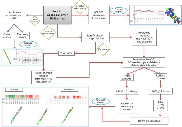
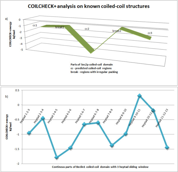
Findings: We have updated our coiled-coil validation webserver, now called COILCHECK+, where new features were added to efficiently identify the strength of interaction at the interface region and measure the density of charged residues and hydrophobic residues. We have examined charged residues and hydrophobic ladders, using a new algorithm called CHAHO, which is incorporated within COILCHECK + server. CHAHO permits the identification of spatial charged residue patches and the continuity of hydrophobic ladder which stabilizes and destabilizes the coiled-coil structure.

Conclusions: The availability of such computational tools should be useful to understand the importance of spatial clustering of charged residues and the continuity of hydrophobic residues at the interface region of coiled-coil dimers. COILCHECK + is a structure based tool to validate coiled-coil stability; it can be accessed at http://caps.ncbs.res.in/coilcheckplus.


Figures

Figure 1
Figure 1
Energy per residue for the coiled-coil dimers taken up for the current analysis. The structures used for the study are regular coiled-coils with identified knobs-into-holes packing. A set of 118 structural entries were used for the analysis (energies are sorted from smallest to largest). The energies of 95% of these structures fall below -kJ/mol. Any coiled-coil structure with an energy better than - kJ/mol would be a stable coiled-coil, thus COILCHECK + energy could be used as a validating tool to identify the stability of the dimer
Figure 2
Figure 2
Complete protocol of hydrophobic ladder and charged-patch algorithms
Figure 3
Figure 3
Hydophobic-ladder analysis on the 118 coiled-coil entries. All the structures were divided into different heptads based on SOCKET prediction. Each heptad was scored based on the residues present at a, d, a’ and d’ positions. a) Number of heptads assigned to respective group of hydrophobic ladder score based on the simple scoring scheme of the algorithm. b) Distribution of different combinations of hydrophobic residues at score 1 and score 0.5 using hydrophobic ladder scoring scheme
Figure 4
Figure 4
COILCHECK + analysis on known long-length coiled-coil structures and structures belonging to DNA binding zipper class proteins. a) COILCHECK + energies of different parts of Sec2p coiled-coil domain is shown. The structure (PDB id 2OCY) is divided into 5 parts based on SOCKET coiled-coil prediction and COILCHECK + energy for each part is identified. It can be clearly seen that the coiled-coil regions are assigned good energies and the break regions have poor energies. b) Beclin1 coiled-coil domain (PDB id 3Q8T), which has continuous heptad pattern, is divided into overlapped three heptad parts and each structure is checked for its COILCHECK + energies. It can be visualized that all the regions are less stable and it also couples with the irregular a-d pairing reported by Li and coworkers.
Figure 5
Figure 5
The results of CHAHO algorithm for tropomyosin structure (1C1G). a) Output of hydrophobic ladder shows the presence of alanine residues at the core positions are responsible for the flexibility of the protein (alanine staggers-yellow spheres, hydrophobic residues-magenta spheres). The hydrophobic ladder has eight specific breaks. b) The method was able to identify negatively charged residue patches on the structure which could help in interacting with actin (Magenta spheres-alanine clusters, Blue spheres-actin binding sites, Red box-predicted hot spot and the residue ranges for each of the boxed region are shown). c) 1C1G structure mapped with SB, SE, DB and DE residues (SB: stabilized buried charged patches, please see text for abbreviations). Boxed region shows the destabilizing negatively charged pairs which could be responsible for the bending of mid-region in the tropomyosin structure.
Figure 6
Figure 6
Hydro ladder and charge patch results for myosin VI medial tail. Hydrophobic ladder score plotted with medial tail region mapped with hydrophobic (magenta spheres) and non-hydrophobic (yellow spheres) residues and the destabilizing heptad identified using the buried charged-patch protocol for the medial tail is shown in parallel. A 5-heptad sliding window has been used for the calculations and for this graphical representation

Similar articles

Cited by

References

    1. Mckay DB, Steitz TA. Structure of cataboilite gene activator protein at 2.9Å resolution suggests binding to left-handed B-DNA. Nature. 1981;290:744–749. doi: 10.1038/290744a0. - DOI - PubMed
    1. Wilson IA, Skehel JJ, Wiley DC. Structure of the haemagglutinin membrane glycoprotein of influenza virus at 3 Å resolution. Nature. 1981;289:366–373. doi: 10.1038/289366a0. - DOI - PubMed
    1. Malashkevich VN, Kammerer RA, Efimov VP, Schulthess T, Engel J. The crystal structure of a five-stranded coiled coil in COMP: a prototype Ion channel. Science. 1996;274:761–765. doi: 10.1126/science.274.5288.761. - DOI - PubMed
    1. Liu J, Zheng Q, Deng Y, Cheng CS, Kallenbach NR, Lu M. A seven-helix coiled coil. Proc Natl Acad Sci USA. 2006;103:15457–15462. doi: 10.1073/pnas.0604871103. - DOI - PMC - PubMed
    1. Burkhard P, Stetefeld J, Strelkov SV. Coiled coils: a versatile protein folding motif. Trends Cell Biol. 2001;11:82–88. doi: 10.1016/S0962-8924(00)01898-5. - DOI - PubMed

Publication types

MeSH terms

LinkOut - more resources