

Wide-field spectral-domain optical coherence tomography in patients and carriers of X-linked retinoschisis
- PMID: 23009889
- PMCID: PMC3531562
- DOI: 10.1016/j.ophtha.2012.07.051
Wide-field spectral-domain optical coherence tomography in patients and carriers of X-linked retinoschisis
Abstract
Purpose: To evaluate macular and extramacular retinal anatomy in patients and carriers of X-linked retinoschisis (XLRS) using a wide-field spectral-domain optical coherence tomography (SD-OCT) imaging technique.
Design: Case series.
Participants: Six XLRS-affected male subjects and 3 XLRS female carriers.
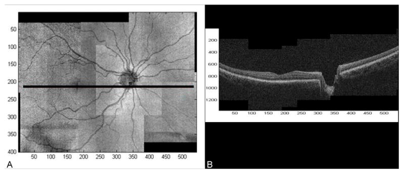
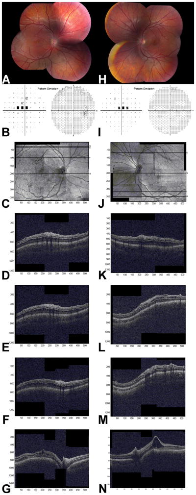
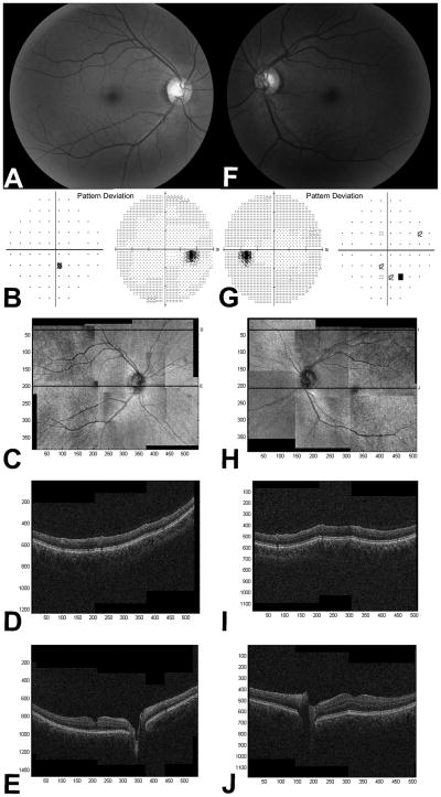
Methods: The subjects prospectively underwent XLRS DNA genotyping and comprehensive ophthalmic examination, including visual acuity, 30-2 Humphrey visual field, fundus photography, and wide-field SD-OCT, a montage technique to generate SD-OCT images spanning approximately 50 degrees horizontally and 35 degrees vertically of the posterior pole.
Main outcome measures: Distribution and location of schisis cavities.
Results: Among male subjects affected by XLRS, asymmetric bilateral schisis was seen in all eyes imaged with montage SD-OCT (11 eyes). Wide-field OCT images demonstrated schisis cavities only in the central macula in 6 eyes (55%), throughout the macula extending to the outside of the temporal arcades in 3 eyes (27%), and throughout the macula extending nasal to the optic nerve in 2 eyes (18%). Cystoid spaces accounting for macular splitting were present in the inner nuclear layer (INL) in all 11 eyes and in the outer nuclear layer (ONL) in 4 eyes. A few small cysts were seen parafoveally in the ganglion cell layer (GCL) or nerve fiber layer (NFL) in 4 eyes. Subclinical extramacular schisis spaces were seen (n=5 eyes) within the INL in 1 eye, the ONL in 1 eye, the INL/GCL/NFL in 1 eye, the ONL/GCL/NFL in 1 eye, and the INL/ONL/GCL/NFL in 1 eye. Schisis was rarely seen nasal to the optic nerve (2 eyes). Central/paracentral visual field defects were seen in 9 eyes. Female carriers did not show schisis on examination or OCT.
Conclusions: Wide-field SD-OCT is a useful tool for evaluating complex retinal anatomy. In patients with XLRS, the foveomacular schisis was seen most frequently in the INL. Subclinical extramacular schisis was seen in 45% of eyes and was equally prevalent in the INL, ONL, and GCL/FNL. The GCL/FNL cystoid spaces were small and seen near the fovea and the arcades only. Carriers were schisis-free.
Financial disclosure(s): The author(s) have no proprietary or commercial interest in any materials discussed in this article.
Copyright © 2013 American Academy of Ophthalmology. Published by Elsevier Inc. All rights reserved.
Conflict of interest statement
No conflicting relationships exist for other authors.
References
-
- Sieving PA, MacDonald IM, Meltzer MR, Smaoui N. Pagon RA, Bird TD, Dolan CR, editors. X-linked juvenile retinoschisis. [Accessed June 7, 2012];GeneReviews [database online] Updated May 12, 2009. Available at: http://www.ncbi.nlm.nih.gov/books/NBK1222/
-
- Yanoff M, Kertesz Rahn E, Zimmerman LE. Histopathology of juvenile retinoschisis. Arch Ophthalmol. 1968;79:49–53. - PubMed
-
- Manschot WA. Pathology of hereditary juvenile retinoschisis. Arch Ophthalmol. 1972;88:131–8. - PubMed
-
- Apushkin MA, Fishman GA, Janowicz MJ. Correlation of optical coherence tomography findings with visual acuity and macular lesions in patients with X-linked retinoschisis. Ophthalmology. 2005;112:495–501. - PubMed
-
- Brucker AJ, Spaide RF, Gross N, et al. Optical coherence tomography of X-linked retinoschisis. Retina. 2004;24:151–2. - PubMed
Publication types
MeSH terms
Grants and funding
LinkOut - more resources
Full Text Sources
Other Literature Sources
