Comparative Study
. 2012 Nov;49(11):943-54.
doi: 10.1016/j.fgb.2012.09.002. Epub 2012 Sep 23.

A Paracoccidioides brasiliensis glycan shares serologic and functional properties with cryptococcal glucuronoxylomannan

Affiliations
Comparative Study

A Paracoccidioides brasiliensis glycan shares serologic and functional properties with cryptococcal glucuronoxylomannan

Priscila C Albuquerque et al. Fungal Genet Biol. 2012 Nov.

Abstract

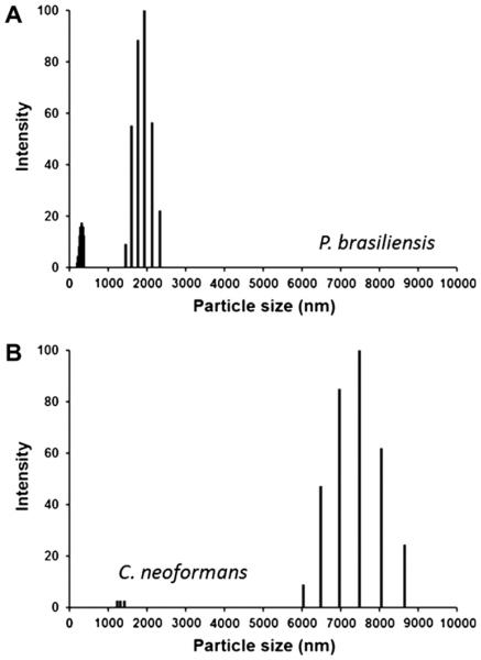
The cell wall of the yeast form of the dimorphic fungus Paracoccidioides brasiliensis is enriched with α1,3-glucans. In Cryptococcus neoformans, α1,3-glucans interact with glucuronoxylomannan (GXM), a heteropolysaccharide that is essential for fungal virulence. In this study, we investigated the occurrence of P. brasiliensis glycans sharing properties with cryptococcal GXM. Protein database searches in P. brasiliensis revealed the presence of sequences homologous to those coding for enzymes involved in the synthesis of GXM and capsular architecture in C. neoformans. In addition, monoclonal antibodies (mAbs) raised to cryptococcal GXM bound to P. brasiliensis cells. Using protocols that were previously established for extraction and analysis of C. neoformans GXM, we recovered a P. brasiliensis glycan fraction composed of mannose and galactose, in addition to small amounts of glucose, xylose and rhamnose. In comparison with the C. neoformans GXM, the P. brasiliensis glycan fraction components had smaller molecular dimensions. The P. brasiliensis components, nevertheless, reacted with different GXM-binding mAbs. Extracellular vesicle fractions of P. brasiliensis also reacted with a GXM-binding mAb, suggesting that the polysaccharide-like molecule is exported to the extracellular space in secretory vesicles. An acapsular mutant of C. neoformans incorporated molecules from the P. brasiliensis extract onto the cell wall, resulting in the formation of surface networks that resembled the cryptococcal capsule. Coating the C. neoformans acapsular mutant with the P. brasiliensis glycan fraction resulted in protection against phagocytosis by murine macrophages. These results suggest that P. brasiliensis and C. neoformans share metabolic pathways required for the synthesis of similar polysaccharides and that P. brasiliensis yeast cell walls have molecules that mimic certain aspects of C. neoformans GXM. These findings are important because they provide additional evidence for the sharing of antigenically similar components across phylogenetically distant fungal species. Since GXM has been shown to be important for the pathogenesis of C. neoformans and to elicit protective antibodies, the finding of similar molecules in P. brasiliensis raises the possibility that these glycans play similar functions in paracoccidiomycosis.


Figures

Fig. 1
Fig. 1
A surface glycan of P. brasiliensis (Pb18) yeast cells shares serologic properties with cryptococcal GXM. (A) Reactivity of P. brasiliensis surface components with different monoclonal antibodies (mAbs 18B7, 2D10 and 13F1) generated to C. neoformans GXM is shown. Fluorescent reactions were not observed when primary antibodies were replaced with isotype matched irrelevant antibodies, as illustrated for IgG1 in the ‘None’ panel. Similar results were obtained with irrelevant IgMs and TRITC-labeled anti-IgM antibodies (not shown). P. brasiliensis cells yeast cells are shown under differential interferential contrast (DIC) and fluorescence (anti-GXM) modes. Image merging (‘merge’ panel) demonstrates that the molecules recognized by the anti-GXM antibodies are externally associated with the cell wall. (B) Immunofluorescence analysis of P. brasiliensis yeast cells incubated with mAbs 18B7 and 2D10 in the presence of varying concentrations of the GXM-related glycan fraction. Increasing concentrations of the glycan efficiently inhibited recognition of yeast cells by the GXM antibodies. P. brasiliensis cells yeast cells are shown under DIC and fluorescence (mAbs 18B7 and 2D10) modes. Scale bar, 20 μm.
Fig. 2
Fig. 2
Serologic reactivity of P. brasiliensis glycans with antibodies originally raised to cryptococcal GXM. (A) Dose-dependent serologic reactivity of a DMSO extract from P. brasiliensis with different monoclonal antibodies raised to cryptococcal GXM (18B7, IgG; 2D10 and 13F1, IgMs) as measured by ELISA. Negative controls included similar serologic tests with DMSO extracts obtained from S. cerevisiae or C. albicans. (B) Reactivity of fungal glycans with mAb 18B7 after de-O-acetylation by alkali treatment. De-O-acetylation affected the recognition of cryptococcal GXM (Cn glycan) by mAb 18B7. The P. brasiliensis fraction (Pb glycan) was not affected by treatment with alkali. (C) DMSO extracts from yeast cells and mycelia show similar profiles of recognition by mAb 18B7. Results in β and C are representative of three independent dot blot analyses.
Fig. 3
Fig. 3
Analysis of P. brasiliensis extracellular vesicles. (A) Detection of sterols by HPTLC in extracellular vesicle fractions isolated from culture supernatants. (B) Dose-dependent recognition of P. brasiliensis vesicle (Pb vesicles) and cellular glycan (Pb glycan) fractions by mAb 18B7.
Fig. 4
Fig. 4
Monosaccharide composition of the polysaccharide fraction obtained from P. brasiliensis Pb18 by DMSO extraction. Methanolysis and derivatization of the components extracted with DMSO followed by separation by gas chromatography (A) revealed the presence of different peaks (1–9) corresponding to carbohydrate units. Mass spectrometry analysis of each of these peaks (B) resulted in the identification of abundant mannose and galactose, as well as of trace amounts of glucose, xylose and rhamnose. The relative molar distribution of each monosaccharide in the DMSO extract is shown in C.
Fig. 5
Fig. 5
Size distribution of glycan fractions obtained after DMSO extraction of P. brasiliensis Pb18 (A) and encapsulated C. neoformans H99 (B) cells.
Fig. 6
Fig. 6
Incorporation of P. brasiliensis Pb18 (Pb) molecules by an acapsular mutant (Cap67) of C. neoformans. Cap67 cells were treated with the Pb extract or with GXM purified from C. neoformans H99 cultures followed by incubation with mAbs raised to GXM. Positive reactions were observed for all antibodies, but the pattern of reactivity depended on which polysaccharide fraction was used to coat Cap67 cells. Scale bar, 20 μm.
Fig. 7
Fig. 7
Coating of Cap67 C. neoformans cells with the P. brasiliensis Pb18 extract results in the formation of capsule-like structures. (A and B) Analysis of the C. neoformans acapsular mutant after incubation in the absence of polysaccharide fractions (control) followed by exposure to mAb 18B7 by fluorescence microscopy. No serologic reactivity was observed. Incubation of Cap67 cells with the DMSO extract from P. brasiliensis results in the formation of extracellular aggregates (C, arrows) that resembled capsular structures when observed by fluorescence microscopy (D). Differential interferential contrast (A and C) and fluorescence (B and D) modes are shown. Results shown in C–D were confirmed by scanning electron microscopy of control (E) or glycan-coated (F) Cap 67 cells. Scale bars correspond to 20 μm in panels for A–D, 1 μm in E and 2 μm in F.
Fig. 8
Fig. 8
Coating of C. neoformans Cap67 (Cn) acapsular cells with P. brasiliensis Pb18 (Pb) molecules renders yeast cells less efficient in their ability to associate with macrophage-like cells. To measure the association of the Cap67 mutant with host cells, fungi were stained with FITC and incubated with the phagocytes, which were then analyzed by flow cytometry. (A) Comparison between the indices of association of macrophages with control Cap67 cells (no coating) or with the GXM-coated mutant. (B) Coating of Cap67 cells with the P. brasiliensis glycan fraction caused a decrease in the index of association of the acapsular mutant with macrophages. (C) Analysis of the efficacy of coating Cap67 cells with C. neoformans GXM or with the P. brasiliensis glycan fraction to inhibit the association of yeast cells with phagocytes. To differentiate adhered and internalized yeast cells, infected macrophages were treated with trypan blue, an extinguisher of extracellular FITC-derived fluorescence (D–F). The shift of histograms to regions of lower fluorescence after exposure to trypan blue denotes the presence of extracellularly-associated yeast cells. This figure is representative of three different experiments producing similar results.

References

    1. Araujo Gde S, et al. Capsules from pathogenic and non-pathogenic Cryptococcus spp. manifest significant differences in structure and ability to protect against phagocytic cells. PLoS One. 2012;7:e29561. - PMC - PubMed
    1. Barbosa FM, et al. Glucuronoxylomannan-mediated interaction of Cryptococcus neoformans with human alveolar cells results in fungal internalization and host cell damage. Microbes Infect. 2006;8:493–502. - PubMed
    1. Bertini S, et al. Expression of antibodies directed to Paracoccidioides brasiliensis glycosphingolipids during the course of paracoccidioidomycosis treatment. Clin. Vaccine Immunol. 2007;14:150–156. - PMC - PubMed
    1. Carbonell LM. Cell wall changes during the budding process of Paracoccidioides brasiliensis and Blastomyces dermatitidis. J. Bacteriol. 1967;94:213–223. - PMC - PubMed
    1. Carbonell LM, et al. Chemical morphology of glucan and chitin in the cell wall of the yeast phase of Paracoccidioides brasiliensis. J. Bacteriol. 1970;101:636–642. - PMC - PubMed

Publication types

MeSH terms

LinkOut - more resources