. 2012 Sep-Oct;31(7-8):398-411.
doi: 10.1016/j.matbio.2012.09.003. Epub 2012 Sep 23.

A disintegrin-like and metalloprotease domain containing thrombospondin type 1 motif-like 5 (ADAMTSL5) is a novel fibrillin-1-, fibrillin-2-, and heparin-binding member of the ADAMTS superfamily containing a netrin-like module

Affiliations

A disintegrin-like and metalloprotease domain containing thrombospondin type 1 motif-like 5 (ADAMTSL5) is a novel fibrillin-1-, fibrillin-2-, and heparin-binding member of the ADAMTS superfamily containing a netrin-like module

Hannah L Bader et al. Matrix Biol. 2012 Sep-Oct.

Abstract

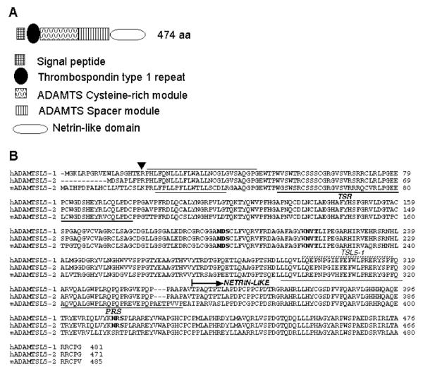
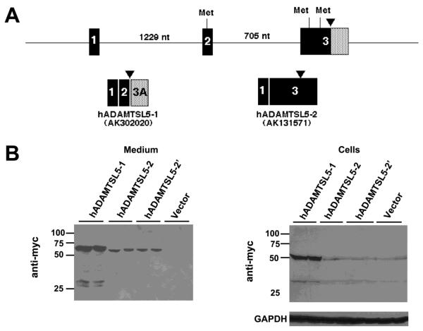
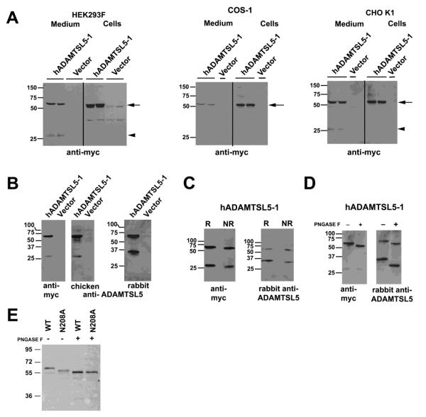
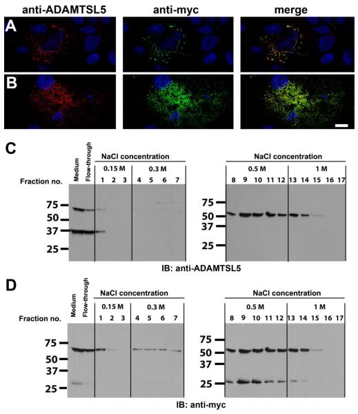
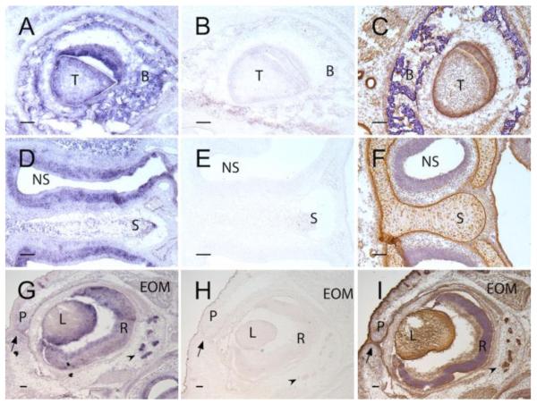
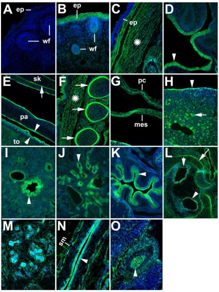
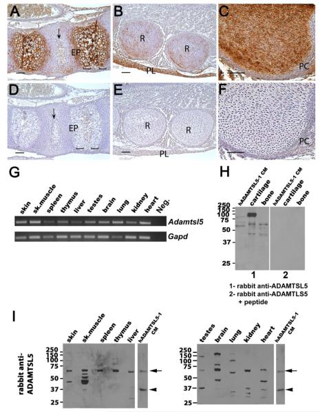
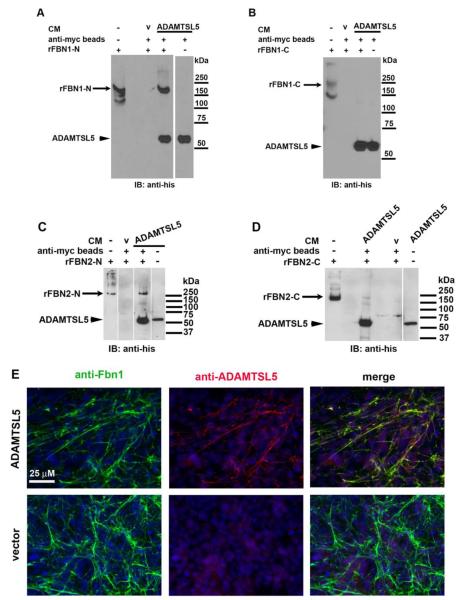
ADAMTS-like proteins are related to ADAMTS metalloproteases by their similarity to ADAMTS ancillary domains. Here, we have characterized ADAMTSL5, a novel member of the superfamily with a unique modular organization that includes a single C-terminal netrin-like (NTR) module. Alternative splicing of ADAMTSL5 at its 5' end generates two transcripts that encode different signal peptides, but the same mature protein. These transcripts differ in their translational efficiency. Recombinant ADAMTSL5 is a secreted, N-glycosylated 60kDa glycoprotein located in the subcellular matrix, on the cell-surface, and in the medium of transfected cells. RT-PCR and western blot analysis of adult mouse tissues showed broad expression. Western blot analysis suggested proteolytic release of the NTR module in transfected cells as well as in some mouse tissues. Immunostaining during mouse organogenesis identified ADAMTSL5 in musculoskeletal tissues such as skeletal muscle, cartilage and bone, as well as in many epithelia. Affinity-chromatography demonstrated heparin-binding of ADAMTSL5 through its NTR-module. Recombinant ADAMTSL5 bound to both fibrillin-1 and fibrillin-2, and co-localized with fibrillin microfibrils in the extracellular matrix of cultured fibroblasts, but without discernible effect on microfibril assembly. ADAMTSL5 is the first family member shown to bind both fibrillin-1 and fibrillin-2. Like other ADAMTS proteins implicated in microfibril biology through identification of human and animal mutations, ADAMTSL5 could have a role in modulating microfibril functions.


Figures

Figure 1
Figure 1. Domain organization and annotated primary structure of ADAMTSL5
A. Domain organization (not drawn to scale) The key to the modules is shown at the bottom. B. Primary structure of human and mouse ADAMTSL5 (GenBank accession numbers AK302020 (hADAMTSL5-1), AK131571 (hADAMTSL5-2) and NM_001113548.1 (mADAMTSL5-2): The sequences of alternatively spliced forms of human ADAMTSL5 and of mouse ADAMTSL5 splice variant 2 were aligned using the MegAlign program (Lasergene software). The arrowhead indicates the location upstream of which the primary structures diverge and the adjacent lines indicate the hydrophobic cores of human and mouse signal peptides. The single TSR is underlined with the bold line. N-glycosylation sequons are in bold type. The proline-rich segment (PRS) is indicated and underlined. The peptide immunogen of the rabbit polyclonal antibody (TSL5-1) is indicated by the stippled line. The netrin-like module is indicated.
Figure 2
Figure 2. Alternative splicing at the 5′ end of ADAMTSL5
A. Genomic region of the 5′ end of ADAMTSL5 (human gene) illustrating exons 1-3 and the intervening introns (size in nucleotides (nt)). The arrowhead indicates the location upstream of which the primary structures diverge (see Fig. 1B for sequences). Putative methionine codons (Met) within an acceptable Kozak consensus sequence are shown at the top of the figure. The pattern of exon splicing to generate the two alternatively spliced transcripts is shown below. Note that there are two potential exon 3 encoded start codons in the ADAMTSL5-2 transcript (Accession no. AK131571). B. Western blots of medium and lysate of HEK293F cells transfected with myc-His6 tagged ADAMTSL5-1 and ADAMTSL5-2 constructs. Lower levels of the expected 60 kDa protein species were obtained with ADAMTSL5-2 constructs in both cell lysate and medium. Abrogation of the upstream start codon in ADAMTSL5-2 (construct hADAMTSL5-2′) did not alter the protein level in cells or medium. The western blots show duplicate transfections for each construct.
Figure 3
Figure 3. ADAMTSL5 undergoes proteolysis and is N-glycosylated
A. Cells were transiently transfected with myc-His6 tagged hADAMTSL5-1. The panels show western blots from the lysate and medium of HEK293F cells, COS-1 cells and CHO-K1 cells as indicated above each blot, with anti-myc antibody. The arrow points to intact ADAMTSL-5, and the arrowhead shows a 27 kDa fragment in the medium. B. Characterization of rabbit and chicken polyclonal antibodies by western blot and comparison with anti-myc. Note that the chicken antibody, raised against a C-terminal peptide, gives essentially similar results as anti-myc. The rabbit polyclonal identified a 60 kDa band corresponding to intact ADAMTSL5, and a 33 kDa species corresponding to the N-terminal fragment resulting from proteolysis. C. Comparison of medium of transfected HEK293F cells immunoblotted after reducing (R) or non-reducing (NR) electrophoresis using anti-myc or rabbit polyclonal antibody to ADAMTSL5. Both antibodies react with same 60 kDa band representing intact ADAMTSL5. In contrast to anti-myc, which detects a 27 kDa ADAMTSL5 fragment, the rabbit polyclonal antibody reacts with a 33 kDa species. There is enhanced migration of each detected species under non-reducing conditions. D. Enzymatic deglycosylation of ADAMTSL5 in conditioned medium of stably transfected HEK293F cells. Increased electrophoretic mobility of anti-ADAMTSL5 reactive species is seen in the presence of PNGAse F (+) compared to the untreated control (−). Digestion with PNGAse F results in more rapid migration of the 60 kDa and 33 kDa (anti-ADAMTSL5) species, whereas migration of the 27 kDa myc-reactive band is unaffected. E. ADAMTSL5-N208A migrates more rapidly than wild-type (WT) protein. Deglycosylation of ADAMTSL5-N208A using PNGase F further enhances migration, suggestive of at least one other N-glycosylation site. Molecular weight markers (in kDa) are shown at left of each gel.
Figure 4
Figure 4. ADAMTSL5 is located at the cell-surface and in the ECM of transiently transfected COS-1 cells and binds heparin
A,B. Staining of non-permeabilized cells with anti-myc and anti-ADAMTSL5 antibodies gave identical staining patterns. Cos-1 cells were transiently transfected with myc-tagged hADAMTSL5-1 and were co-stained with rabbit anti-ADAMTSL5 (red) and anti-myc (green) antibody with the non-permeabilized staining technique. Nuclei were stained with DAPI and appear blue. Representative confocal images showing cell surface association (A, optical plane through mid-cell region) and ECM association (B, subcellular optical plane) are shown (see supplemental movie). Note that the staining pattern obtained with the two antibodies coincides, indicating that the signal is derived from full-length ADAMTSL5. Scale bar=10 μm. C,D. Heparin-agarose beads were incubated with conditioned medium from HEK293 cells stably transfected with hADAMTSL5-1. After incubation, the beads were washed and eluted with increasing salt concentrations as indicated. ADAMTSL5 was detected in the fractions by western blot using the rabbit anti-ADAMTSL5 antibody (C). After stripping, the blots were reprobed with a myc-antibody (D). Full-length ADAMTSL5 was eluted with 0.5–1 M NaCl, indicating strong binding to heparin. Furthermore, the C-terminal NTR-module containing 27 kDa fragment, which is myc-tagged, bound to the heparin-agarose with similar affinity as the full-length protein, whereas the N-terminal cleavage product of ADAMTSL5 (37 kDa fragment in G) was not retained by the heparin column (compare C and D). This suggests that the NTR-module mediated heparin binding.
Figure 5
Figure 5. In situ hybridization (ISH) and immunohistochemistry identify ADAMTSL5 in specific developing mouse craniofacial structures
Panels A,D and G show ISH using an antisense Adamtsl5 cRNA probe (purple stain), panels B,E,H, show ISH with the corresponding sense (control) cRNA probe. Panels C, F, I show immunostaining (brown signal) with rabbit anti-ADAMTSL5 polyclonal antibody. A-C. Developing molar tooth. Note specific signal in tooth (T) and surrounding alveolar bone (B). Panel C shows the corresponding immunostaining image, with signal additionally seen in epithelium of salivary gland ducts at the left-hand edge of the panel. D-F. Nasal septum and nasal sinus. In D, note ISH signal in nasal epithelium lining the sinus (NS) and in the cartilage of the nasal septum (S). In F, ADAMTSL5 immunostaining is seen in nasal septal cartilage (S), but there is relatively weak signal in epithelium lining the nasal septum (NS). In contrast, strong immunostaining was noted in the perichondrium of nasal septal cartilage (S). Panels G-I show a developing eye. ISH signal is seen in the lens (L), retina (R), epithelium of the fused palpebrae (P, arrow shows fusion seam) and in lacrimal gland epithelium (arrowhead). Note the similar distribution of ADAMTSL5 protein in panel I.
Figure 6
Figure 6. Detection of ADAMTSL5 by immunofluorescence in cryosections of 14.5 and 16.5 day-old mouse embryos
The expression pattern was essentially identical at both developmental stages. Shown are epifluorescence images of sagittal (A – K, M-O) or transverse (L) cryosections of E16.5 embryos (A – L) or E14.5 embryos (M – O). The sections were stained with rabbit anti-ADAMTSL5 (green) and nuclei were stained with DAPI (blue). Sections stained with secondary antibody only (A) did not give any signal. ADAMTSL5 was detected in the follicles of the developing whiskers (wf) (B), and in the epidermis (ep) throughout most of the body, e.g. head (B),but not in the epidermis of the back (C). ADAMTSL5 was also detected in the skeletal muscle throughout the body, including the skeletal muscle of the back (C, asterisk). ADAMTSL5 signal was observed in the nasal mucosa (D, arrowhead), the tongue musculature (E), and the skeletal muscle overlying the ribs (F). Oral epithelium of the palate (pa) (E) and tongue (E, to) were strongly stained; arrowheads indicate epithelial staining in D and E. ADAMTSL5 antibody stained the perichondrium of diverse skeletal elements such as skull bones (sk, basophenoid bone, E), ribs (F) and the bones of the inner ear (L); arrows indicate perichondrial staining. Note that these sections were not hyaluronidase treated, a procedure necessary to detect ADAMTSL5 in cartilage. In the thorax and abdomen, pericardium (pc) and mesothelium (mes) were also stained (G). In the liver, ADAMTSL5 antibody stained the liver capsule (arrowhead, H) and interspersed cells in the liver mesenchyme (arrow, H). In the lung, ADAMTSL5 was expressed in the bronchial eptihelium (arrowhead, I), and in the kidney, ADAMTSL5 was detected in the medullary tubular epithelium(J, arrowhead). The epithelium of the stomach was strongly positive (arrowhead, K). ADAMTSL5 was also detected in the sensory epithelium of the developing inner ear (L, arrowheads). ADAMTSL5 antibody stained both the epithelium and the stroma of the E14.5 salivary gland (M). Epithelial linings of the oesophagus (N), and bile duct (arrowhead in O) were stained. Note that anti-ADAMTSL5 stained both epithelium and smooth muscle layer (sm) of the esophagus (N), and bile duct (O). Smooth muscle staining in oesophagus and lung was confirmed by co-staining with a-smooth muscle actin (data not shown). Scale bars indicate 100 μm (C – H, J-M, O) or 50 μm (A, B, I, N).
Figure 7
Figure 7. ADAMTSL5 mRNA and protein are widely distributed in mouse tissues
A-F. Localization of ADAMTSL5 in E17.5 mouse cartilage, bone and intervertebral disc (IVD). Panels A-C show immunostaining in the spine (A), ribs (B) and hip (C). Panels D-F show corresponding sections stained with anti-ADAMTSL5 pre-incubated with the immunogenic peptide as a control. In the spine (A), antibody reactivity (brown color) is seen in extracellular matrix around hypertrophic chondrocytes of the endplate (EP) as well as in vertebral bone and IVD (arrow). In panel B, staining is seen in ECM of the ribs (R), as well as in the pleura (PL) lining the thoracic cavity. In panel C, note the strong staining in cartilage extracellular matrix and weaker staining in perichondrium (PC). No signal is seen in controls (D-F). Scale bars indicate 50 μm. G. Using RT-PCR, Adamtsl5 mRNA is detected in all tissues analyzed. As a negative control, PCR was performed without template (neg.), and as a positive control, PCR with Gapd primers was performed. The identity of the Adamtsl5 PCR product was confirmed by sequencing. H. Panel 1 shows western blot of adult mouse cartilage and bone extracts using recombinant ADAMTSL5 from conditioned medium (CM) of HEK293F cells as a control. Panel 2 show that pre-incubation of the rabbit polyclonal antibody with the peptide immunogen leads to loss of all immunoreactivity. I. Western blots of adult mouse tissue extracts using rabbit polyclonal anti-ADAMTSL5 antibody. Conditioned medium (CM) from stably transfected HEK293 cells was used as a positive control; vertical white lines separate lanes that were run on the same gel and regrouped for presentation. Full-length recombinant ADAMTSL5 (arrow) migrated at approximately 60 kDa with minor differences attributable to variable N-glycosylation (see also differences in the relative size of ADAMTSL5 expressed in HEK293F, COS-1 and CHO-K1 cells in Fig. 3A). The arrowhead indicates a 37 kDa fragment. Bands of similar molecular weight as the observed fragment with the rabbit antibody were present in testes, skeletal muscle, kidney and heart. Additional lower molecular weight bands, which likely correspond to different proteolytic products, are present in some tissues. In some extracts, bands with a higher molecular weight than expected are also observed, i.e., in the brain (70 kDa and 160 kDa) and in lung and testes, (90 kDa or 140 kDa). The origin of these bands is presently unknown.
Figure 8
Figure 8. ADAMTSL5 binds to fibrillin-1 and fibrillin-2, and to fibrillin microfibrils assembled by fibroblast cultures
A-D. Affinity pull-down with anti-myc agarose beads was done using myc-tagged ADAMTSL5 conditioned medium from HEK293F cells mixed with, or without polypeptides corresponding to fibrillin-1 N-terminal fragment rFBN1-N (panel A), fibrillin-1 C-terminal fragment rFBN1-C (panel B), fibrillin-2 N-terminal fragment (panel C; rFBN2-N) and fibrillin-2 C-terminal fragment (panel D; rFBN2-C) as indicated. Anti-His6 monoclonal antibody was used for detection by immunoblotting, since all the recombinant proteins used had C-terminal His6 tags. The N-terminal half of fibrillin-1, but not its C-terminal half, bound specifically to ADAMTSL5 (panels A and B). The N-terminal half of fibrillin-2 bound more robustly than the C-terminal half (panels C and D). Molecular weight markers are shown to the right of each panel. Vertical white lines on gels indicate removal of intervening lanes from the gels to bring together the lanes of interest. CM, conditioned medium; v, vector control (e.g. CM from vector-transfected cells E. Co-localization of ADAMTSL5 with fibrillin-1 containing microfibrils formed in fBNL cells co-cultured with ADAMTSL5 transfected or empty pcDNA vector-transfected HEK293F cells (V) for 6 days. Co-localization is evident from the yellow signal seen in merged image (top right).

References

    1. Ahram D, Sato TS, Kohilan A, Tayeh M, Chen S, Leal S, Al-Salem M, El-Shanti H. A homozygous mutation in ADAMTSL4 causes autosomal-recessive isolated ectopia lentis. Am J Hum Genet. 2009;84:274–278. - PMC - PubMed
    1. Apte SS. A disintegrin-like and metalloprotease (reprolysin-type) with thrombospondin type 1 motif (ADAMTS) superfamily: functions and mechanisms. J Biol Chem. 2009;284:31493–31497. - PMC - PubMed
    1. Bader HL, Ruhe AL, Wang LW, Wong AK, Walsh KF, Packer RA, Mitelman J, Robertson KR, O’Brien DP, Broman KW, Shelton GD, Apte SS, Neff MW. An ADAMTSL2 founder mutation causes Musladin-Lueke Syndrome, a heritable disorder of beagle dogs, featuring stiff skin and joint contractures. PLoS One. 2010;5 - PMC - PubMed
    1. Banyai L, Patthy L. The NTR module: domains of netrins, secreted frizzled related proteins, and type I procollagen C-proteinase enhancer protein are homologous with tissue inhibitors of metalloproteases. Protein Sci. 1999;8:1636–1642. - PMC - PubMed
    1. Bekhouche M, Kronenberg D, Vadon-Le Goff S, Bijakowski C, Lim NH, Font B, Kessler E, Colige A, Nagase H, Murphy G, Hulmes DJ, Moali C. Role of the netrin-like domain of procollagen C-proteinase enhancer-1 in the control of metalloproteinase activity. J Biol Chem. 2010;285:15950–15959. - PMC - PubMed

Publication types

MeSH terms