. 2013 Feb;49(2):93-101.
doi: 10.1016/j.oraloncology.2012.08.001. Epub 2012 Sep 23.

DSG3 as a biomarker for the ultrasensitive detection of occult lymph node metastasis in oral cancer using nanostructured immunoarrays

Affiliations

DSG3 as a biomarker for the ultrasensitive detection of occult lymph node metastasis in oral cancer using nanostructured immunoarrays

Vyomesh Patel et al. Oral Oncol. 2013 Feb.

Abstract

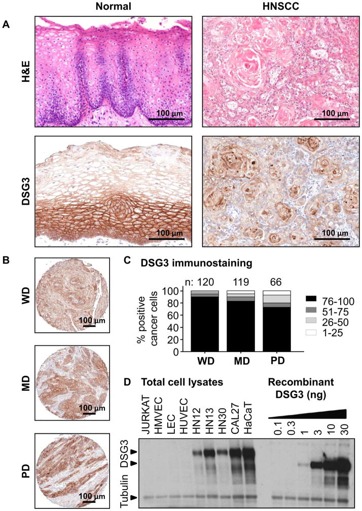
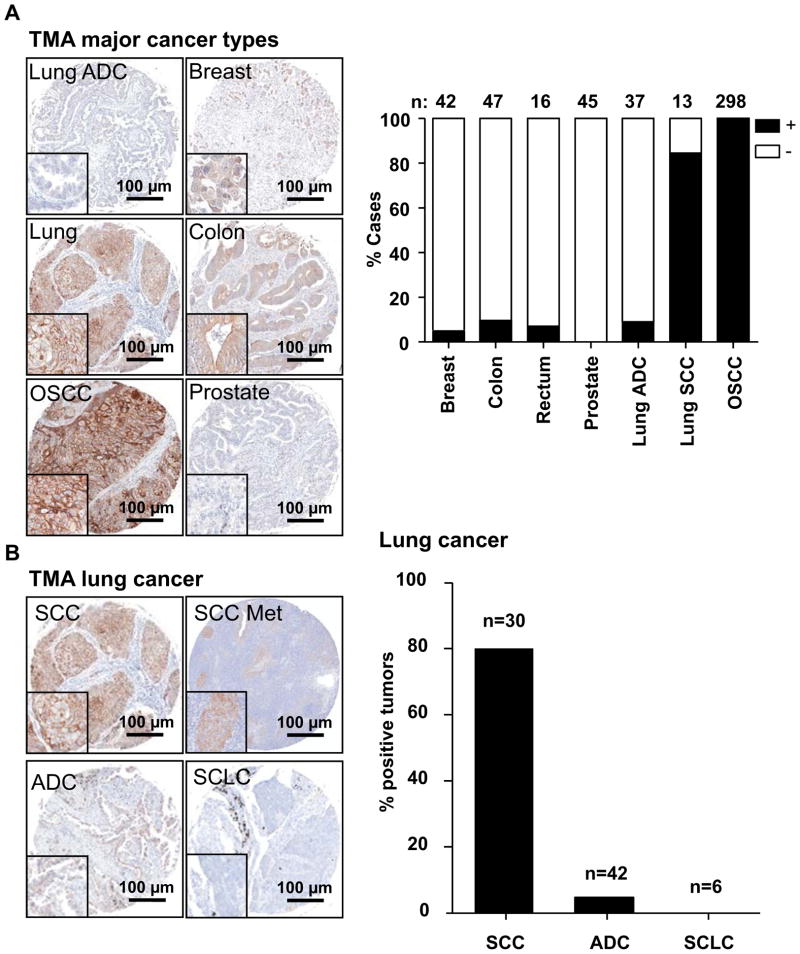
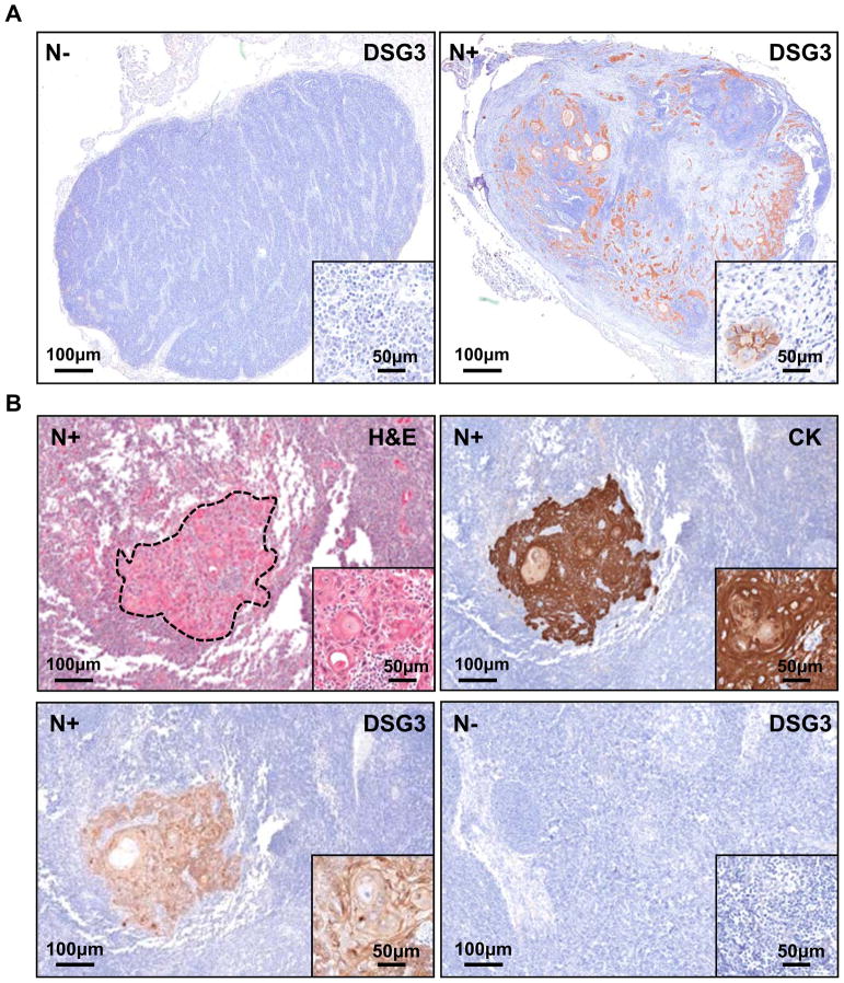
Objectives: The diagnosis of cervical lymph node metastasis in head and neck squamous cell carcinoma (HNSCC) patients constitutes an essential requirement for clinical staging and treatment selection. However, clinical assessment by physical examination and different imaging modalities, as well as by histological examination of routine lymph node cryosections can miss micrometastases, while false positives may lead to unnecessary elective lymph node neck resections. Here, we explored the feasibility of developing a sensitive assay system for desmoglein 3 (DSG3) as a predictive biomarker for lymph node metastasis in HNSCC.

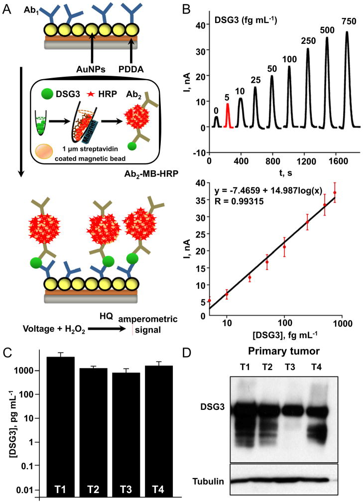
Materials and methods: DSG3 expression was determined in multiple general cancer- and HNSCC-tissue microarrays (TMAs), in negative and positive HNSCC metastatic cervical lymph nodes, and in a variety of HNSCC and control cell lines. A nanostructured immunoarray system was developed for the ultrasensitive detection of DSG3 in lymph node tissue lysates.

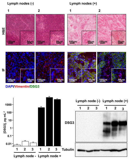
Results: We demonstrate that DSG3 is highly expressed in all HNSCC lesions and their metastatic cervical lymph nodes, but absent in non-invaded lymph nodes. We show that DSG3 can be rapidly detected with high sensitivity using a simple microfluidic immunoarray platform, even in human tissue sections including very few HNSCC invading cells, hence distinguishing between positive and negative lymph nodes.

Conclusion: We provide a proof of principle supporting that ultrasensitive nanostructured assay systems for DSG3 can be exploited to detect micrometastatic HNSCC lesions in lymph nodes, which can improve the diagnosis and guide in the selection of appropriate therapeutic intervention modalities for HNSCC patients.


Conflict of interest statement

Conflict of interest statement

None declared.

Figures

Figure 1
Figure 1. Validation of DSG3 expression in normal and malignant HNSCC
Normal oral mucosa biopsies and HNSCC were evaluated for DSG3 by IHC. A. DSG3 is expressed throughout the normal epithelium, but is stronger in the basal and parabasal layers. Diffuse expression was seen in the epithelial component of all HNSCC. B. Representative well (WD), moderate (MD) and poorly (PD) differentiated HSCC cases are shown. C. DSG3 was expressed in all tumors regardless of differentiation, with increased expression in WD cases. Numbers of cases analyzed is depicted. D. Total cell extracts from non-squamous (Jurkat, HMVEC, LEC, HUVEC) and oral-squamous (HN12, HN13, HN30, Cal27), and epidermal-squamous (HaCaT) were processed for Western blot analysis. Native DSG3 and its glycosylated forms were readily detected in squamous cells extracts, while absent from the non-squamous counterparts. These levels were compared with human recombinant DSG3 that was processed in a background of Jurkat cell lysate. Tubulin staining indicates equivalent loading and protein integrity.
Figure 2
Figure 2. Immunoreactivity of DSG3 in common tumor types
A. Multi-tumor TMAs (lung, breast, colon, prostate), and an oral specific TMA were assessed for DSG3 expression by IHC, and the staining scored for the presence of specific staining as (+) or (-). Most squamous cell lung cancers stained positive for DSG3, but very few of the adenocarcinomas gave positive reaction. All cores from the OSCC TMA scored positive. B. In lung cancer, DSG3 expression was positive in most squamous cell carcinomas (SCC) including lymph node metastasis (SCC Met), while few cases of adenocarcinomas (ADC) gave positive reaction, and all small cell lung carcinoma samples (SCLC), were negative.
Figure 3
Figure 3. Specific detection of DSG3 in human cervical lymph nodes
A. Formalin fixed and paraffin embedded tissue sections of non-metastatic (N−) and metastatic lymph nodes (N+) show DGS3 expression only in N+, with the staining localized to the malignant squamous cells (n=30). All N− cases were negative (n=5). B. The epithelial specificity of DSG3 immunoreactivity was further confirmed using simultaneous cytokeratin (CK) staining. A representative example is shown, whereby the H&E stained tumor island is matched with CK and DSG3 expression, with no non-specific staining. An example of a N− case stained for DSG3 is shown.
Figure 4
Figure 4. Rapid and ultrasensitive detection of DSG3 in human HNSCC samples using nanosensors
A. Scheme used for the ultrasensitive detection by the microfluidic immunoarray showing a single sensor in the array with capture DSG3 antibodies attached. Proteins are captured off-line on Ab2-magnetic bead (MB)-HRP bioconjugates, and after magnetic separation and washes, the MBs are injected into the immunoarray containing 8 sensors. A single immunoarray sensor is depicted. Following incubation, amperometric signals are generated by applying −0.3 V vs Ag/AgCl to the sensors by injecting a mixture of HRP-activator H2O2 and mediator hydroquinone (HQ). B. Varying recombinant DSG3 protein concentrations were used to generate a calibration plot. The sensitivity of DSG3 sensor using recombinant protein was 5646 nA mL [fg protein]−1 cm−2. C. Protein extracts of primary human oral squamous carcinomas (T 1–4) made with RIPA buffer were processed for detection of DSG3. High DSG3 levels were found to be present in all the samples, and this was confirmed by Western blot analysis of the same extracts for DSG3 (D). Tubulin was used as loading control.
Figure 5
Figure 5. Detection of DSG3 in metastatic human cervical lymph nodes
H&E stained cryosections of representative non-metastatic (−) and metastatic (+) human cervical lymph nodes were scanned and the total number of tumor cells per section was quantified (Table 1). Serial sections of these lymph nodes were evaluated by immunofluorescence for DSG3 and detected only in metastatic lymph nodes (green). Vimentin (red) was used to identify stromal tissue, and nuclei of all cells were stained blue with DAPI (Fig. 5A). Protein extracts made from single cryosections of lymph nodes were used for the detection of DSG3 by Western blot analysis and DSG3 quantification using nanosensors. DSG3 levels were similar to background for all non-metastatic samples, while DSG3 levels in all metastatic cases were proportional to the number of invading HNSCC cells (Fig. 5B).

References

    1. Ferlay J, Shin HR, Bray F, Forman D, Mathers C, Parkin DM. Estimates of worldwide burden of cancer in 2008: GLOBOCAN 2008. Int J Cancer. 2010;127(12):2893–917. - PubMed
    1. Siegel R, Naishadham D, Jemal A. Cancer statistics, 2012. CA Cancer J Clin. 2012;62(1):10–29. - PubMed
    1. Forastiere A, Koch W, Trotti A, Sidransky D. Head and neck cancer. N Engl J Med. 2001;345(26):1890–900. - PubMed
    1. Clark JR, Naranjo N, Franklin JH, de Almeida J, Gullane PJ. Established prognostic variables in N0 oral carcinoma. Otolaryngol Head Neck Surg. 2006;135(5):748–53. - PubMed
    1. Kuriakose MA, Trivedi NP. Sentinel node biopsy in head and neck squamous cell carcinoma. Current opinion in otolaryngology & head and neck surgery. 2009;17(2):100–10. - PubMed

Publication types