. 2012 Oct 9;109(41):16522-7.
doi: 10.1073/pnas.1210899109. Epub 2012 Sep 25.

Superstructure of the centromeric complex of TubZRC plasmid partitioning systems

Affiliations

Superstructure of the centromeric complex of TubZRC plasmid partitioning systems

Christopher H S Aylett et al. Proc Natl Acad Sci U S A. .

Abstract

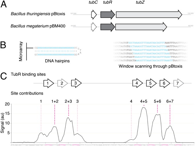
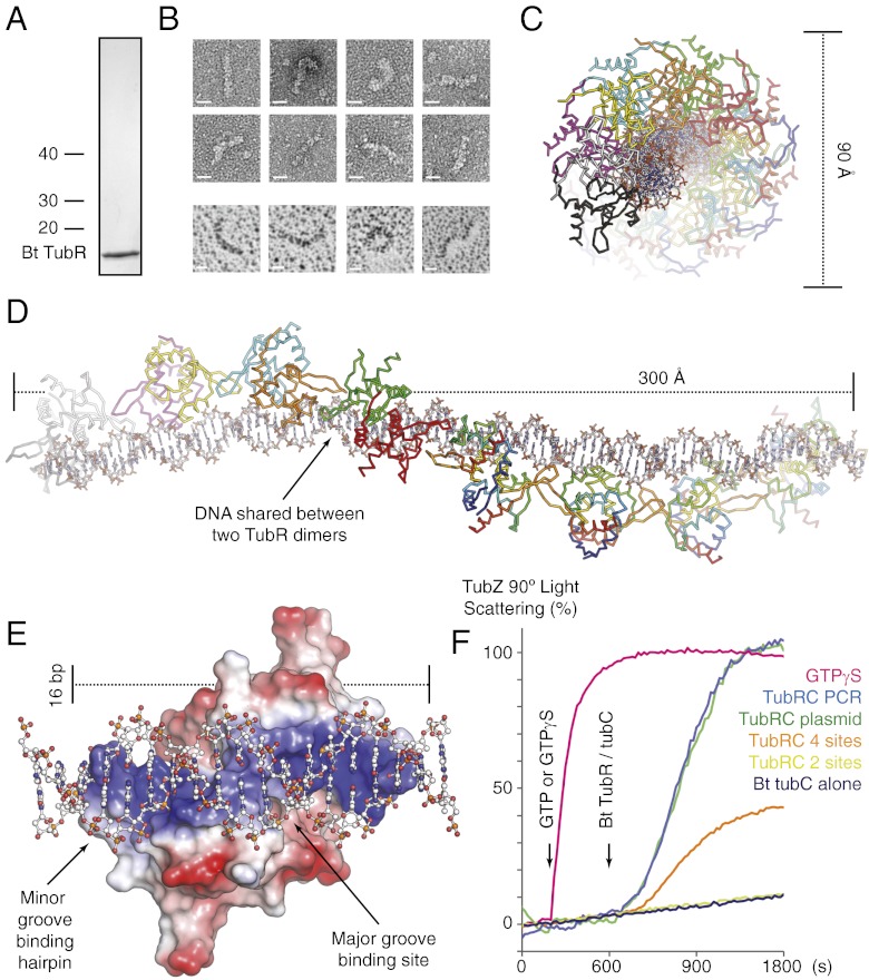
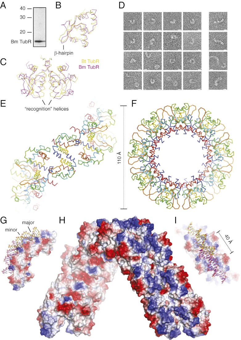
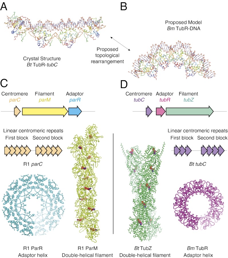
Bacterial plasmid partitioning systems segregate plasmids into each daughter cell. In the well-understood ParMRC plasmid partitioning system, adapter protein ParR binds to centromere parC, forming a helix around which the DNA is externally wrapped. This complex stabilizes the growth of a filament of actin-like ParM protein, which pushes the plasmids to the poles. The TubZRC plasmid partitioning system consists of two proteins, tubulin-like TubZ and TubR, and a DNA centromere, tubC, which perform analogous roles to those in ParMRC, despite being unrelated in sequence and structure. We have dissected in detail the binding sites that comprise Bacillus thuringiensis tubC, visualized the TubRC complex by electron microscopy, and determined a crystal structure of TubR bound to the tubC repeat. We show that the TubRC complex takes the form of a flexible DNA-protein filament, formed by lateral coating along the plasmid from tubC, the full length of which is required for the successful in vitro stabilization of TubZ filaments. We also show that TubR from Bacillus megaterium forms a helical superstructure resembling that of ParR. We suggest that the TubRC DNA-protein filament may bind to, and stabilize, the TubZ filament by forming such a ring-like structure around it. The helical superstructure of this TubRC may indicate convergent evolution between the actin-containing ParMRC and tubulin-containing TubZRC systems.


Conflict of interest statement

The authors declare no conflict of interest.

Figures

Fig. 1.
Fig. 1.
Bt tubC is composed of seven repeats, which bind TubR in a cooperative fashion. (A) Schematic comparing the tubZRC loci of Bt pBtoxis and Bm pBM400. (B) Illustration of the DNA hairpins produced on a microarray to sample the sequence of pBtoxis, and a schematic indicating how this window was scanned through a region of the plasmid sequence by successive single base pair movements. The variable window is shown in cyan, and all other base pairs in black. (C) Plot of the recorded signal for each microarray spot in a 1-bp scan over the region of Bt tubC (bp 126688 to 126496). Each point is plotted over the 12th bp of the 24-bp hairpin, with the sequence shown below. The assigned binding sites for Bt TubR are shown above, and the corresponding sequences are colored magenta below. The site(s) resulting in each peak have been annotated above the graph.
Fig. 2.
Fig. 2.
Bt TubR bound to tubC forms a DNA–protein filament that effects TubZ polymerization. (A) Coomassie-stained SDS/PAGE of purified Bt TubR. (B) Electron micrographs of negatively stained (Left) and rotary shadowed (Right) Bt TubR bound to full-length tubC showing the morphology of the DNA–protein filaments formed. (Scale bars, 10 nm.) (C and D) Crystal structure of Bt TubR (Cα ribbon representation, the first four dimers are colored by chain, the second four a continuum between blue at the N terminus and red at the C terminus) bound to two repeats of tubC (stick representation, C in white/CPK colors). Dimensions in angstroms are shown alongside the structure, whereas the filament has been rotated by 90° between the two plates. (E) Single Bt TubR dimer (surface charge representation, red negative, blue positive) from the structure, with B-DNA [ball and stick representation, C in white/Corey, Pauling, Koltun (CPK) coloring] extended from the 12-bp section used in refinement in order to show the interaction with the full 16-bp region covered by the dimer. (F) Effect of Bt TubRC on the polymerization of TubZ measured using 90° light scattering. GTP or GTPγS was added to all reactions at 200 s, and the complexes indicated to the Right of the graph were added at 600 s (color coded). The four- and two-site versions of Bt tubC encompassed binding sites 4–7 and 6–7.
Fig. 3.
Fig. 3.
Bm TubR forms helices of similar appearance in crystals and bound to DNA. (A) Coomassie-stained SDS/PAGE of purified Bm TubR. (B) Structural superimposition of monomers of Bt and Bm TubR (Cα ribbon representation, colored as indicated). (C) Structural superimposition of dimers of Bt and Bm TubR (Cα ribbon representation, colored as indicated). (D) Electron micrographs of negatively stained Bm TubR bound to Bt tubC DNA. (Scale bars, 10 nm.) (E and F) Crystal structure of Bm TubR helix (Cα ribbon representation, continuum between blue at the N terminus and red at the C terminus). Dimensions in angstroms are shown alongside the structure, whereas the helix has been rotated by 90° between the two plates. (GI) Four Bm TubR dimers (surface charge representation, red negative, blue positive) with the DNA from Protein Data Bank (PDB) ID code 1HW2 (stick representation, C in yellow or magenta/CPK colors) shown after the two structures have been superimposed for the two central dimers. The two plates are rotated by 90° relative to one another. (H) Surface charge representation of the Bm TubR helix (red negative, blue positive).
Fig. 4.
Fig. 4.
Bm TubR helix suggests further convergent evolution of type II and III partitioning systems. (A) Structure of Bt TubR (Cα ribbon representation, blue at N terminus, red at C terminus) bound to tubC (stick representation, C in white/CPK colors). (B) Structure of Bm TubR (Cα ribbon representation, blue at N terminus, red at C terminus) shown with the DNA (stick representation, C in white/CPK colors) from PDB ID code 1HW2 after superimposition of the protein chains. (C and D) Comparison of operon structure (3, 10, 12), centromeric structure (20, 21), filament superstructure (9, 14, 16), and adapter complex superstructure (, , this study) for the (actin-like) ParMRC and (tubulin-like) TubZRC plasmid partitioning systems.

References

    1. Gerdes K, Howard M, Szardenings F. Pushing and pulling in prokaryotic DNA segregation. Cell. 2010;141:927–942. - PubMed
    1. Salje J. Plasmid segregation: How to survive as an extra piece of DNA. Crit Rev Biochem Mol Biol. 2010;45:296–317. - PubMed
    1. Gerdes K, Møller-Jensen J, Bugge Jensen R. Plasmid and chromosome partitioning: Surprises from phylogeny. Mol Microbiol. 2000;37:455–466. - PubMed
    1. Austin S, Abeles A. Partition of unit-copy miniplasmids to daughter cells. II. The partition region of miniplasmid P1 encodes an essential protein and a centromere-like site at which it acts. J Mol Biol. 1983;169:373–387. - PubMed
    1. Ogura T, Hiraga S. Partition mechanism of F plasmid: Two plasmid gene-encoded products and a cis-acting region are involved in partition. Cell. 1983;32:351–360. - PubMed

Publication types

MeSH terms

LinkOut - more resources