. 2013 Jan;21(1):251-9.
doi: 10.1038/mt.2012.202. Epub 2012 Sep 25.

Type I IFN counteracts the induction of antigen-specific immune responses by lipid-based delivery of mRNA vaccines

Affiliations

Type I IFN counteracts the induction of antigen-specific immune responses by lipid-based delivery of mRNA vaccines

Charlotte Pollard et al. Mol Ther. 2013 Jan.

Abstract

The use of DNA and viral vector-based vaccines for the induction of cellular immune responses is increasingly gaining interest. However, concerns have been raised regarding the safety of these immunization strategies. Due to the lack of their genome integration, mRNA-based vaccines have emerged as a promising alternative. In this study, we evaluated the potency of antigen-encoding mRNA complexed with the cationic lipid 1,2-dioleoyl-3trimethylammonium-propane/1,2-dioleoyl-sn-glycero-3-phosphoethanolamine (DOTAP/DOPE ) as a novel vaccination approach. We demonstrate that subcutaneous immunization of mice with mRNA encoding the HIV-1 antigen Gag complexed with DOTAP/DOPE elicits antigen-specific, functional T cell responses resulting in specific killing of Gag peptide-pulsed cells and the induction of humoral responses. In addition, we show that DOTAP/DOPE complexed antigen-encoding mRNA displays immune-activating properties characterized by secretion of type I interferon (IFN) and the recruitment of proinflammatory monocytes to the draining lymph nodes. Finally, we demonstrate that type I IFN inhibit the expression of DOTAP/DOPE complexed antigen-encoding mRNA and the subsequent induction of antigen-specific immune responses. These results are of high relevance as they will stimulate the design and development of improved mRNA-based vaccination approaches.


Figures
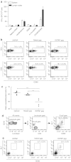
Figure 1
Figure 1
DOTAP/DOPE complexed antigen-encoding mRNA induces potent antigen-specific immune responses. (a) Mice were immunized s.c. with PBS, 20 µg of Gag-encoding mRNA, either as such (“naked”) or complexed with the indicated carriers. At week 4, mice were boosted with the same formulation. Spleens and lymph nodes were isolated at week 8, and the number of Gag-specific interferon-γ secreting cells was determined by enzyme-linked immunosorbent spot. Means ± SEM are shown of a total of 5 mice per group. PBAE, poly(β-amino ester). (b) Frequencies of CD69+ CD4+ and CD8+ T cells in draining lymph nodes 1 day after footpad injection of 16 µl DOTAP/DOPE (DOTAP), 4ug of naked Gag-encoding mRNA (naked gag) or 4 µg DOTAP/DOPE-complexed Gag-encoding mRNA (DOTAP gag). Means ± SD are shown of a total of 6 mice per group. (c) In vivo killing assay was performed by adoptive transfer of carboxyfluorescein diacetate succinimdyl ester-labeled peptide-loaded splenocytes to mice immunized with 20 µg of naked gag or DOTAP gag. The ratio of Gag-pulsed versus unpulsed cells in 4 mice is shown. ***P < 0.001. (d) Gating strategy for B cells and germinal center (GC) B cells. (e) The frequencies of B cells and GC B cells were determined by flow cytometry 7 days post footpad injection of 16 µl DOTAP, 4 µg naked gag or DOTAP gag. B cells were defined as CD45+ CD19+ CD3− cells, and GC B cells as GL-7+ CD95+ B cells. Means ± SD are shown of a total of 6 mice per group.
Figure 2
Figure 2
Prime-boost strategy enhances antigen-specific immune responses. Mice were immunized by 2 s.c. injections of 20 µg of naked or DOTAP gag with a 3-week interval. At week 8, some mice were boosted with 20 µg of the capsid protein p24 either subcutaneously (a and b) or intratracheally (c). T cell and B cell responses were analyzed 10 days after the p24 protein boost. (a) Number of Gag-specific interferon (IFN)-γ and interleukin-2 secreting T cells was determined by enzyme-linked immunosorbent spot (ELISPOT) on isolated spleen and lymph node cells. Means ± SEM are shown of a total of 5 mice per group. (b) Anti-p24 immunoglobulin (Ig)M, IgG1, and IgG2c titers were determined by serum enzyme-linked immunosorbent assay. Means ± SEM are shown for 5 mice, immunized with naked gag and 5 with DOTAP gag, each followed by p24 boost. (c) Number of Gag-specific IFN-γ secreting CD4 and CD8 T cells was determined by ELISPOT on isolated lung cells. Means ± SEM are shown of a total of 5 mice per group.
Figure 3
Figure 3
DOTAP/DOPE complexed mRNA displays intrinsic adjuvant activity. (a) Expression pattern of maturation markers of bone marrow-derived DCs (BMDCs) transfected with DOTAP gag. BMDCs were transfected with 2.5 µg DOTAP gag and the expression of major histocompatibility complex (MHC)-II, CD80, CD86, and CD40 on Gag+ and Gag-CD11c+ cells was analyzed 24 hours later by flow cytometry. (b) Transcript levels of interleukin (IL)-6, IL-1β, IFN-α and interferon (IFN)-β mRNA were determined by RT-qPCR 24 hours (IL-6 and IL-1β) or 6 hours (IFN-α and IFN- β) after transfection of BMDCs with 2.5 µg DOTAP gag. Results represent mean n-fold induction levels compared to unstimulated control cells ± SD from triplicate PCR reactions. (c and d) Nuclear translocation of IFN regulatory factors (IRF)-3 and IRF-7 was visualized by confocal microscopy at different time points after stimulation of BMDCs with 2.5 µg DOTAP gag. (c) Confocal images taken 5 minutes after incubation with DOTAP or DOTAP gag and (d) ratios of protein present in the nucleus to total protein. Results represent mean ± SD of 100 cells. (e) Transcript levels of IFN-α and IFN-β mRNA were determined by RT-qPCR 6 hours after transfection of WT, MyD88−/− or TRIF−/− DCs with 2.5 µg DOTAP gag. Results represent mean n-fold induction levels compared to unstimulated control cells ± SD from triplicate PCR reactions. TRIF, TIR-domain-containing adapter-inducing interferon-β. (f) Mice were injected i.v. with 20 µg naked gag or DOTAP gag. Serum levels of IFN-α were determined 1, 3, 6, and 24 hours later by enzyme-linked immunosorbent assay. Mean ± SEM of 4 mice per group is shown. (g) Flowcytometric analysis of DC subsets and inflammatory monocytes present in the draining lymph nodes 1 and 3 days after footpad injection of 4µg of DOTAP gag. Conventional DCs were defined as CD11chi MHCIIint, Tissue-derived DCs were CD11cint MHCIIhi and inflammatory monocytes were MHC-II+ Ly6chi CD11bhi. Mean ± SEM 6 mice per group is shown. ***P < 0.001 as compared with the corresponding inflammatory monocyte numbers in DOTAP injected control mice and mice injected with naked gag.
Figure 4
Figure 4
Type I IFN inhibitexpression of antigen-encoding mRNA and subsequent induction of immune responses. (a) Wild-type (WT) and interferon (IFN)αR−/− mice were immunized with 20µg DOTAP gag as previously described. Gag-specific IFN-γ and interleukin-2 secreting T cells were determined by enzyme-linked immunosorbent spot on isolated spleens. Mean of 5 mice per group is shown. ***P < 0.001. (b and c) WT, MyD88−/−, TRIF−/−, and IFNαR−/− bone marrow-derived DCs were transfected with 2.5 µg of DOTAP/DOPE-complexed Gag-encoding mRNA. Mean percentage ± SD of Gag+ CD11c+ cells (b) and expression pattern of the maturation markers major histocompatibility complex (MHC)-II, CD80, CD86, and CD40 on Gag+ and Gag- CD11c+ cells (c) was determined 24 hours later by flowcytometry. TRIF, TIR-domain-containing adapter-inducing interferon-β. (d) Flowcytometric analysis of DC subsets and inflammatory monocytes present in the draining lymph nodes of WT and IFNαR−/− mice 1 day post footpad injection of 4µg DOTAP gag. Conventional DCs were defined as CD11chi MHCIIint, tissue-derived DCs were CD11cint MHCIIhi and inflammatory monocytes were MHC-II+ Ly6chi CD11bhi. Mean ± SEM of 6 mice per group is shown.

References

    1. Pascolo S. Vaccination with messenger RNA (mRNA) Handb Exp Pharmacol. 2008;183:221–235. - PubMed
    1. Bettinger T, Carlisle RC, Read ML, Ogris M., and, Seymour LW. Peptide-mediated RNA delivery: a novel approach for enhanced transfection of primary and post-mitotic cells. Nucleic Acids Res. 2001;29:3882–3891. - PMC - PubMed
    1. Kreiter S, Selmi A, Diken M, Koslowski M, Britten CM, Huber C.et al. (2010Intranodal vaccination with naked antigen-encoding RNA elicits potent prophylactic and therapeutic antitumoral immunity Cancer Res 709031–9040. - PubMed
    1. Kreiter S, Diken M, Selmi A, Diekmann J, Attig S, Hüsemann Y.et al. (2011FLT3 ligand enhances the cancer therapeutic potency of naked RNA vaccines Cancer Res 716132–6142. - PubMed
    1. Van Lint S, Goyvaerts C, Maenhout S, Goethals L, Disy A, Benteyn D.et al. (2012Preclinical evaluation of TriMix and antigen mRNA-based antitumor therapy Cancer Res 721661–1671. - PubMed

Publication types