. 2012 Oct 9;109(41):E2794-802.
doi: 10.1073/pnas.1205742109. Epub 2012 Sep 24.

TCR signaling via Tec kinase ITK and interferon regulatory factor 4 (IRF4) regulates CD8+ T-cell differentiation

Affiliations

TCR signaling via Tec kinase ITK and interferon regulatory factor 4 (IRF4) regulates CD8+ T-cell differentiation

Ribhu Nayar et al. Proc Natl Acad Sci U S A. .

Abstract

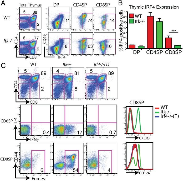
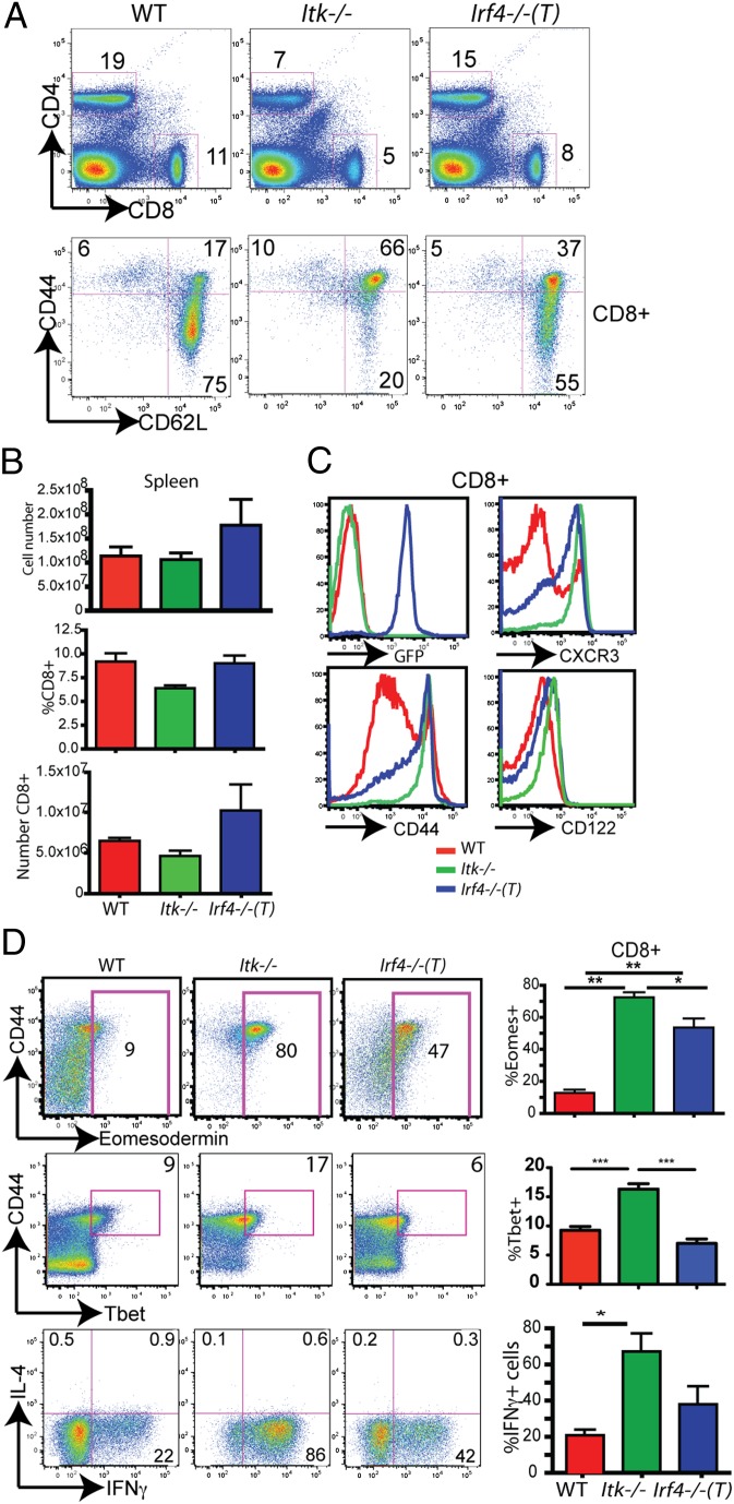
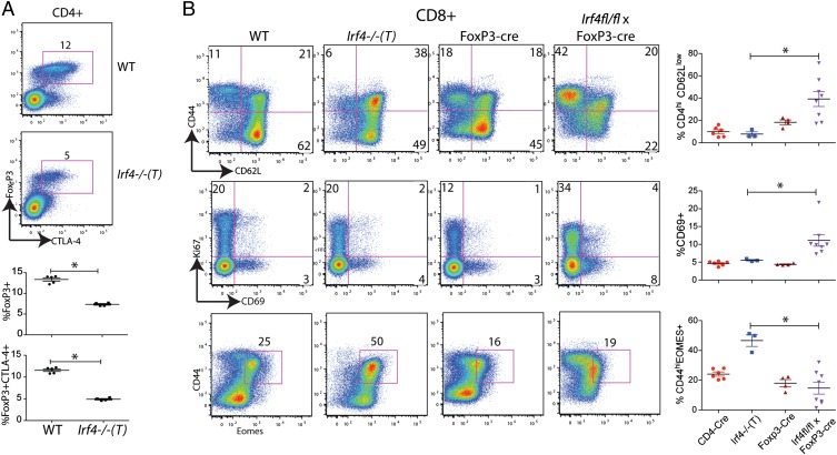
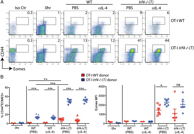
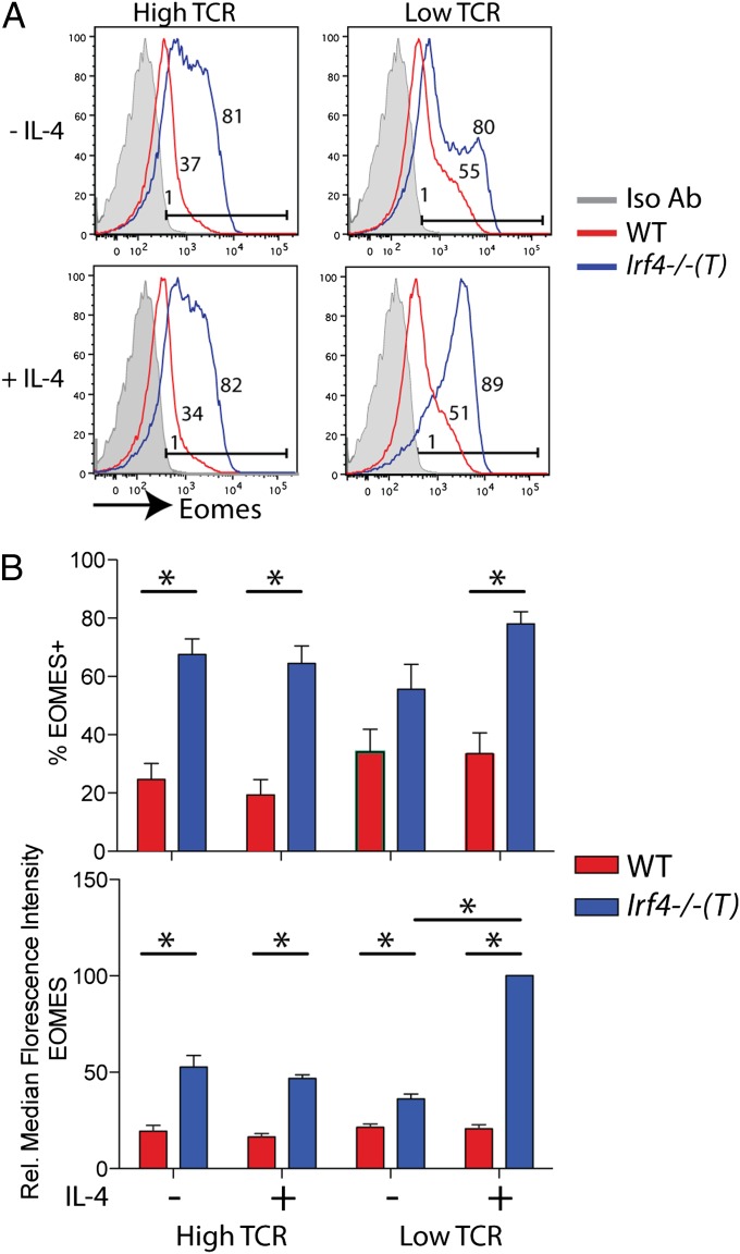
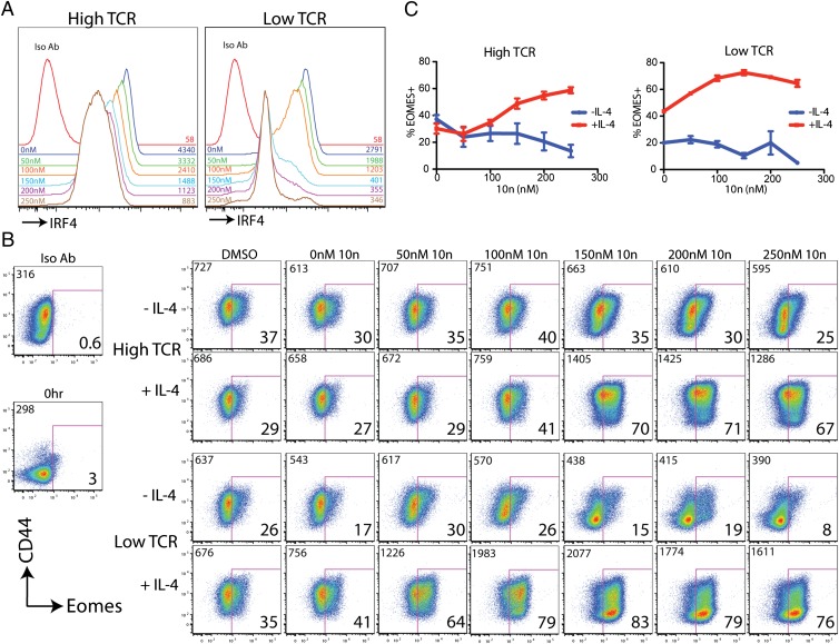
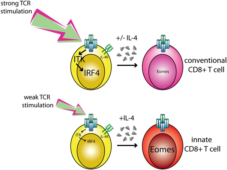
CD8(+) T-cell development in the thymus generates a predominant population of conventional naive cells, along with minor populations of "innate" T cells that resemble memory cells. Recent studies analyzing a variety of KO or knock-in mice have indicated that impairments in the T-cell receptor (TCR) signaling pathway produce increased numbers of innate CD8(+) T cells, characterized by their high expression of CD44, CD122, CXCR3, and the transcription factor, Eomesodermin (Eomes). One component of this altered development is a non-CD8(+) T cell-intrinsic role for IL-4. To determine whether reduced TCR signaling within the CD8(+) T cells might also contribute to this pathway, we investigated the role of the transcription factor, IFN regulatory factor 4 (IRF4). IRF4 is up-regulated following TCR stimulation in WT T cells; further, this up-regulation is impaired in T cells treated with a small-molecule inhibitor of the Tec family tyrosine kinase, IL-2 inducible T-cell kinase (ITK). In contrast to WT cells, activation of IRF4-deficient CD8(+) T cells leads to rapid and robust expression of Eomes, which is further enhanced by IL-4 stimulation. In addition, inhibition of ITK together with IL-4 increases Eomeso up-regulation. These data indicate that ITK signaling promotes IRF4 up-regulation following CD8(+) T-cell activation and that this signaling pathway normally suppresses Eomes expression, thereby regulating the differentiation pathway of CD8(+) T cells.


Conflict of interest statement

The authors declare no conflict of interest.

Figures

Fig. 1.
Fig. 1.
IRF4 is up-regulated during thymic selection but is not required for normal T-cell development. (A) Thymocytes from WT or Itk−/− mice were isolated and surface-stained for CD4, CD8, and CD69, and then for intracellular IRF4 protein. Thymocyte subsets (DP, CD4SP, and CD8SP) were gated on, and IRF4 vs. CD69 is displayed. Numbers on dot-plots represent the percentage of cells expressing IRF4, based on comparison with an isotype control. Data are representative of six or more independent experiments. (B) Compilation of the percentage of IRF4-expressing cells for each thymocyte subset (n = 15; ***P < 0.001 based on the Mann–Whitney test). (C) Thymocytes from WT, Itk−/−, and Irf4−/−(T) mice were stained with antibodies to CD4, CD8, CD44, intracellular Eomes, CXCR3, and CD124; stimulated for 4 h with phorbol 12-myristate 13-acetone (PMA) and ionomycin; and stained for intracellular IL-4 and IFN-γ. CD4 vs. CD8 profiles are shown (Top) along with gated CD8SP cells (Middle and Bottom).
Fig. 2.
Fig. 2.
Peripheral IRF4-deficient T cells share the innate phenotype of Itk−/− CD8+ T cells. (A) Splenocytes were isolated from WT, Itk−/−, and Irf4−/−(T) mice, and they were then stained with antibodies to CD4, CD8, CD44, and CD62L. CD4 vs. CD8 staining of total splenocytes (Upper) and CD44 vs. CD62L staining on gated CD8+ T cells (Lower) are shown. (B) Compilations of the total splenocyte numbers, percentages of CD8+ T cells, and absolute numbers of CD8+ T cells in the spleens are shown (n = 8). (C) CD8+ splenic T cells from WT, Itk−/−, and Irf4−/−(T) mice were analyzed for GFP, CXCR3, CD44, and CD122 expression. (D) CD8+ splenic T cells from WT, Itk−/−, and Irf4−/−(T) mice stained with antibodies to CD44, Eomes, and Tbet or were stimulated for 4 h with phorbol 12-myristate 13-acetone and ionomycin, and they were then analyzed for IL-4 vs. IFN-γ expression. Compilations of the data show the percentages of Eomes+ cells (n = 12), Tbet+ cells (n = 6–10), and IFN-γ+ cells (n = 10). Statistically significant differences are indicated by *(P < 0.05), **(P < 0.001), or ***(P < 0.0001) based on the Mann–Whitney test.
Fig. 3.
Fig. 3.
Aberrant FoxP3+ regulatory T cells in Irf4−/−(T) mice. (A) CD4+ T cells from the spleens of WT and Irf4−/−(T) mice were analyzed for intracellular FoxP3 and CTLA-4. Numbers on the dot-plots represent the percentages of CD4+ T cells that are FoxP3+CTLA-4+. Below are compilations of the percentages of CD4+ T cells that are FoxP3+ or FoxP3+CTLA-4+. Data are representative of two independent experiments. Statistical significance was determined by the Mann–Whitney test. (B) Splenic CD8+ T cells from WT, Irf4−/−(T), FoxP3-cre control, and Irf4fl/fl × FoxP3-cre mice were analyzed for expression of CD44, CD62L, CD69, Ki67, and intracellular Eomes. Dot-plots display gated CD8+TCR-β+ cells; numbers indicate the percentages of cells in the indicated quadrants or gates. (Right) Compilations of data for the four groups of mice are shown. Statistical significance was determined by the Mann–Whitney test (*P = 0.02).
Fig. 4.
Fig. 4.
High expression of Eomes by IRF4-deficient CD8+ T cells is dependent on both cell-intrinsic and cell-extrinsic factors in Irf4−/−(T) mice. Splenocytes from OT-I WT and OT-I Irf4−/−(T) mice were mixed 1:1 and adoptively transferred into WT or Irf4−/−(T) hosts and then injected i.p. with 1 mg of αIL-4 neutralizing antibody or PBS 2–4 h before adoptive transfer. CD8+ T cells were analyzed 44 h later with a viability stain, along with antibodies to CD8, Vα2, Vβ5, CD45.1, CD45.2, CD44, CD62L, and intracellular Eomes or an isotype control. (A) Dot-plots show Eomes vs. CD44 staining on gated live CD8+, Vα2+, and Vβ5+ cells distinguished by CD45.1 (WT) vs. CD45.2 [Irf4−/− (T)+] staining. Numbers indicate the percentages of CD44hiEomes+ cells. Iso Ctr, antibody control for Eomes staining; 0 h, cells analyzed directly ex vivo before adoptive transfer. (B) Compilations of data show percentages of CD44hiEomes+ cells (Left) and raw MFI of Eomes staining (Right) on live CD8+, Vα2+, and Vβ5+ cells distinguished by CD45.1 (WT) vs. CD45.2 [Irf4−/−(T)+] staining from three independent experiments with two to three host mice per group per experiment. Statistical significance was determined by a t test (*P = 0.01; **P = 0.001; ***P < 0.0001). ns, not significant.
Fig. 5.
Fig. 5.
Naive Irf4−/−(T) CD8SP thymocytes up-regulate high levels of Eomes following TCR stimulation. (A) WT and Irf4−/−(T) CD8SP thymocytes were isolated and stimulated with 1 μg/mL CD3 plus 4 μg/mL CD28 (“high TCR”) or 0.1 μg/mL CD3 plus 0.4 μg/mL CD28 (“low TCR”) antibodies in the presence or absence of IL-4 (10 ng/mL) for 38 h. Cells were stained with antibodies to CD4, CD8, CD24, CD44, and intracellular Eomes or an isotype control. Histograms show Eomes expression relative to the isotype control (Iso Ab; gray-filled histograms) on gated CD8+CD24loCD44hi cells; numbers indicate the percentages of Eomes+ cells. (B) Compilations of data indicate percentages of Eomes+ cells (Upper) and relative (Rel.) MFIs of Eomes staining (Lower) for n = 4 experiments. Statistical significance was determined by the Mann–Whitney test (*P < 0.03).
Fig. 6.
Fig. 6.
Inhibition of ITK synergizes with IL-4 to up-regulate high levels of Eomes following TCR stimulation. Naive peripheral CD8+ T cells were isolated from OT-I WT mice and were stimulated with 1 μg/mL CD3 plus 4 μg/mL CD28 (“high TCR”) or 0.1 μg/mL CD3 plus 0.4 μg/mL CD28 (“low TCR”) antibodies in the presence or absence of IL-4 (10 ng/mL) for 31 h. Cultures were supplemented with the small-molecule ITK inhibitor, 10n, at the indicated concentrations or with DMSO alone at the highest concentration used. Cells were stained with a viability dye, and antibodies to CD8, CD69, CD44, intracellular IRF4, Eomes, or isotype controls. (A) Histograms show IRF4 expression relative to the isotype control (Iso Ab). The concentrations of 10n (Left) and the MFI of IRF4 staining (Right) are indicated in each histogram. All data are from cells stimulated in high TCR (Left) or Low TCR (Right) in the absence of exogenous IL-4. (B) Dot-plots show Eomes vs. CD44 staining in cells stimulated with high TCR (Upper two rows) or low TCR (Lower two rows) and supplemented with IL-4 or 10n as indicated. Numbers at the upper left of each dot-plot indicate the MFI of Eomes staining. Numbers at the lower right of each dot-plot indicate the percentage of Eomes+ cells. Data are representative of four experiments. (C) Compilations of data indicate percentages of Eomes+ cells for each condition.
Fig. P1.
Fig. P1.
Model depicts the development of conventional vs. innate CD8+ T cells in the thymus. The strength of TCR signaling regulates Eomesodermin expression via the Tec family tyrosine kinase ITK and the transcription factor IRF4. Strong TCR signaling promotes robust activation of ITK, leading to high levels of expression of the transcription factor IRF4. Either in the presence or the absence of IL-4, CD8+ T cells stimulated under these conditions express low levels of Eomesodermin (Eomes). During T-cell development in the thymus, this pathway produces conventional T cells. In contrast, following weak TCR stimulation, ITK activation is modest and cells produce low amounts of IRF4.These conditions, together with IL-4 receptor signaling, promote maximal up-regulation of Eomesodermin, leading to the development of innate CD8+ T cells.

References

    1. Berg LJ. Signalling through TEC kinases regulates conventional versus innate CD8(+) T-cell development. Nat Rev Immunol. 2007;7:479–485. - PubMed
    1. Bendelac A, Savage PB, Teyton L. The biology of NKT cells. Annu Rev Immunol. 2007;25:297–336. - PubMed
    1. Atherly LO, et al. The Tec family tyrosine kinases Itk and Rlk regulate the development of conventional CD8+ T cells. Immunity. 2006;25:79–91. - PubMed
    1. Broussard C, et al. Altered development of CD8+ T cell lineages in mice deficient for the Tec kinases Itk and Rlk. Immunity. 2006;25:93–104. and correction (2006) 25:849. - PubMed
    1. Dubois S, Waldmann TA, Müller JR. ITK and IL-15 support two distinct subsets of CD8+ T cells. Proc Natl Acad Sci USA. 2006;103:12075–12080. - PMC - PubMed

Publication types

MeSH terms