Review
. 2012 Dec;21(12):1792-807.
doi: 10.1002/pro.2165. Epub 2012 Nov 13.

LacZ β-galactosidase: structure and function of an enzyme of historical and molecular biological importance

Affiliations
Review

LacZ β-galactosidase: structure and function of an enzyme of historical and molecular biological importance

Douglas H Juers et al. Protein Sci. 2012 Dec.

Abstract

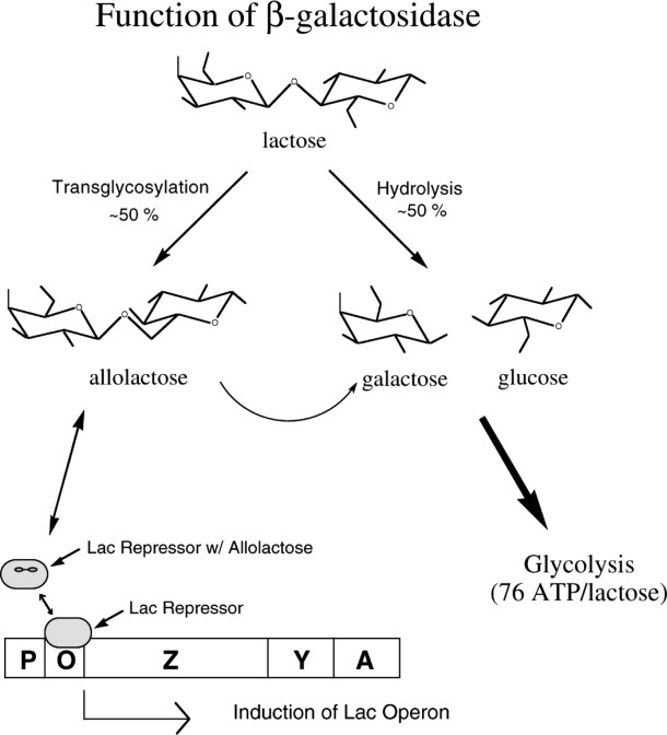
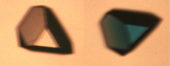
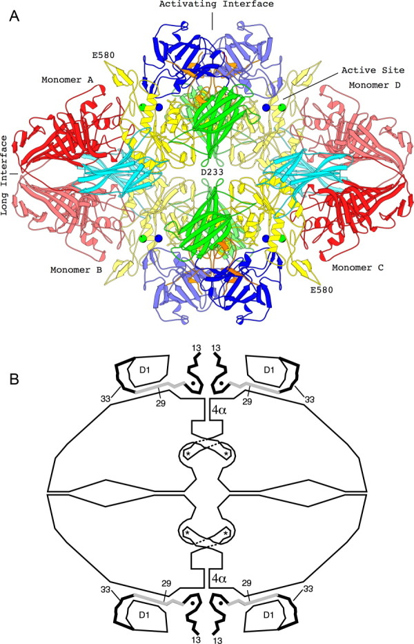
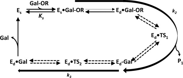
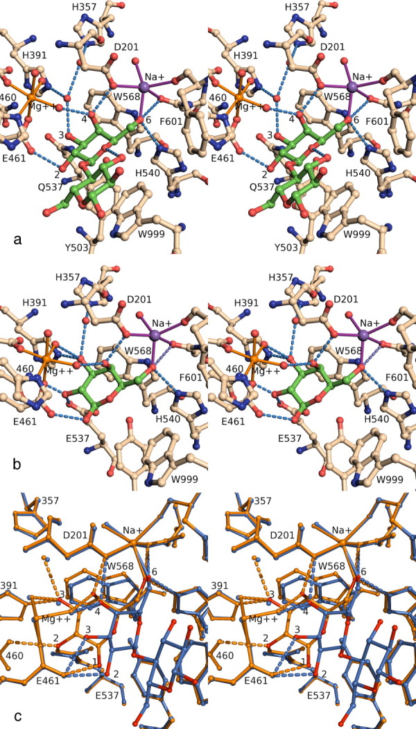
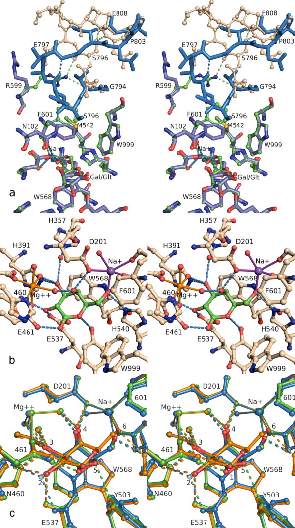
This review provides an overview of the structure, function, and catalytic mechanism of lacZ β-galactosidase. The protein played a central role in Jacob and Monod's development of the operon model for the regulation of gene expression. Determination of the crystal structure made it possible to understand why deletion of certain residues toward the amino-terminus not only caused the full enzyme tetramer to dissociate into dimers but also abolished activity. It was also possible to rationalize α-complementation, in which addition to the inactive dimers of peptides containing the "missing" N-terminal residues restored catalytic activity. The enzyme is well known to signal its presence by hydrolyzing X-gal to produce a blue product. That this reaction takes place in crystals of the protein confirms that the X-ray structure represents an active conformation. Individual tetramers of β-galactosidase have been measured to catalyze 38,500 ± 900 reactions per minute. Extensive kinetic, biochemical, mutagenic, and crystallographic analyses have made it possible to develop a presumed mechanism of action. Substrate initially binds near the top of the active site but then moves deeper for reaction. The first catalytic step (called galactosylation) is a nucleophilic displacement by Glu537 to form a covalent bond with galactose. This is initiated by proton donation by Glu461. The second displacement (degalactosylation) by water or an acceptor is initiated by proton abstraction by Glu461. Both of these displacements occur via planar oxocarbenium ion-like transition states. The acceptor reaction with glucose is important for the formation of allolactose, the natural inducer of the lac operon.


Figures

Figure 1
Figure 1
Schematic summarizing the roles of β-galactosidase in the cell. The enzyme can hydrolyze lactose to galactose plus glucose, it can transgalactosylate to form allolactose, and it can hydrolyze allolactose. The presence of lactose results in the synthesis of allolactose which binds to the lac repressor and reduces its affinity for the lac operon. This in turn allows the synthesis of β-galactosidase, the product of the lacZ gene.
Figure 2
Figure 2
Demonstration that β-galactosidase in crystals is catalytically active. Crystal of β-galactosidase (orthorhombic; ca. 0.2 mm) in the absence (left) and in the presence, after about 2 h, of the substrate X-gal (right).
Figure 3
Figure 3
The tetrameric structure of β-galactosidase. (a) The backbone structure of the enzyme. Domain 1, blue; Domain 2, green; Domain 3, yellow; Domain 4, cyan; Domain 5, red. Lighter and darker shading is used to differentiate equivalent domains in different subunits. Metal ions are shown as spheres, Na+, green; Mg++, blue. Interactive views are available in the electronic version of the article (see below). (b) Sketch of the tetramer, aligned as in (a) showing features particularly relevant to α-complementation. The four active sites (each highlighted with an asterisk) are located toward the center of the figure. In each case a loop including residues 272–288 extends from one subunit to complete the active site of a neighboring subunit. The “activation interface” extends vertically through the center of the tetramer. Part of the interface comprises a bundle of four α-helices labeled 4α. Residues 13–50, shown as thick lines, pass through a tunnel between the first domain (labeled D1) and the rest of the protein. The region shaded gray (residues 23–31) is deleted in one of the α-donors. Magnesium ions (small solid circles) bridge between the complementation peptide and the rest of the protein (from Ref. 8). An interactive view is available in the electronic version of the article.
Figure 4
Figure 4
Overall reaction scheme for β-galactosidase. A galactoside (Gal-OR) binds to free enzyme (Es) initially in a shallow mode (Es•Gal-OR) which progresses to a deep mode (Ed•Gal-OR). The first chemical step (galactosylation, k2) is shown here to begin with Es•Gal-OR and follows through the transition state Ed•TS1 to a covalent intermediate, Ed-Gal, normally releasing the first product HOR (not shown). The intermediate is released to water (degalactosylation, k3) through the second transition state Ed•TS2 to a product complex Ed•Gal, finally releasing the second product, galactose. In the case of allolactose production, the first product (glucose) is not released and acts as the acceptor for the degalactosylation reaction, releasing the disaccharide, allolactose.
Figure 5
Figure 5
(a) Shallow mode binding illustrated by a stereoview of active site complex between E537Q β-galactosidase and lactose (PDB code 1jyn). H-bonds less than 3.0 Å involving the lactose molecule or key water molecules are shown as dashed lines. Protein residues are shown with wheat colored carbons and the ligand with green colored carbons. Figures 5 and 6 were prepared with PYMOL. (b) Deep mode binding of substrate illustrated by a stereoview of the active site complex between native enzyme and galactose (1jz7). (c) Comparison of shallow mode and deep mode binding. The complexes shown are E537Q/lactose (1jyn, shallow, in blue) and native/galactose (1jz7 deep, in orange). Progression from shallow to deep involves rotation of the galactosyl moiety. The 6- and 4-hydroxyls maintain their interactions, but the other hydroxyls shift. The 3-hydroxyl moves to occupy the position formerly occupied by a water molecule. An interactive view is available in the electronic version of the article.
Figure 6
Figure 6
(a) Loop switching. Stereoview comparing the galactose complex (ball-and-stick) to the lactone complex (stick only), emphasizing the conformation changes in Arg599, Phe601, and the Gly794-Pro803 loop. In the galactose complex (1jz7), the loop is in the open conformation (wheat ball-and-stick), with H-bonds to Arg599 and intraloop H-bonds to Ser796. In the lactone complex (1jz5), the ligand is deeper in the active site, Phe601 closes toward the Na+, and the loop follows (blue sticks), which breaks the Arg599 and Ser796 H-bonds and creates a new H-bond to Asn102, and a new intraloop H-bond. The side chain for Arg800 has been omitted for clarity. (b) Transition state analog. Stereoview of active site complex between native enzyme and galactonolactone (1jz5). (c) Comparison of the binding modes for galactose (orange), galactonolactone (green) and a 2-F-galactosyl-enzyme intermediate (blue). All complexes are with native enzyme (1jz7, 1jz5, and 1jz4). Each hydroxyl makes similar interactions in the three different complexes, with the greatest differences at the 6-OH, 2-OH, ring oxygen, and C5 positions. Progressing through the three complexes, the galactosyl ring is deeper in the active site, and Phe601 is in the closed conformation with the lactone and the intermediate. An interactive view is available in the electronic version of the article.

References

    1. Jacob F, Monod J. Genetic regulatory mechanisms in the synthesis of proteins. J Mol Biol. 1961;3:318–356. - PubMed
    1. Huber RE, Kurz G, Wallenfels K. A quantitation of the factors which affect the hyrolase and trangalactosylase activities of β-galactosidase (E. coli) on lactose. Biochemistry. 1976;15:1994–2001. - PubMed
    1. Wyckoff HW, Doscher M, Tsernoglou D, Inagami T, Johnson LN, Hardman KD, Allewell NM, Kelly DM, Richards FM. Design of a diffractometer and flow cell system for X-ray analysis of crystalline proteins with applications to the crystal chemistry of ribonuclease-S. J Mol Biol. 1967;27:563–578. - PubMed
    1. Matthews BW. Determination of molecular weight from protein crystals. J Mol Biol. 1974;82:513–526. - PubMed
    1. Fowler A, Zabin I. Amino acid sequence of β-galactosidase. XI. Peptide ordering procedures and the complete sequence. J Biol Chem. 1978;253:5521–5525. - PubMed

LinkOut - more resources