. 2012 Nov 15;120(20):4205-14.
doi: 10.1182/blood-2012-01-402545. Epub 2012 Sep 25.

Terminal myeloid differentiation in vivo is induced by FLT3 inhibition in FLT3/ITD AML

Affiliations

Terminal myeloid differentiation in vivo is induced by FLT3 inhibition in FLT3/ITD AML

Amy Sexauer et al. Blood. .

Abstract

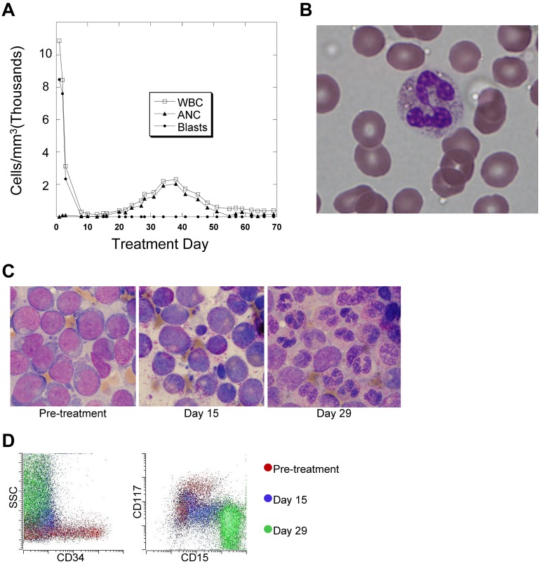
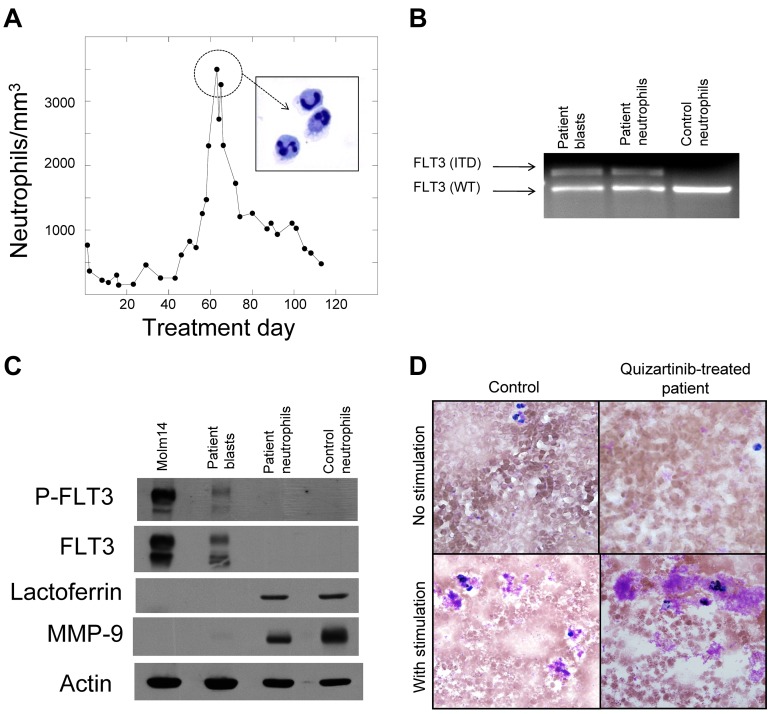
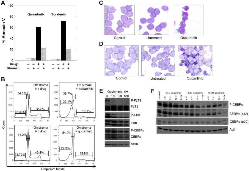
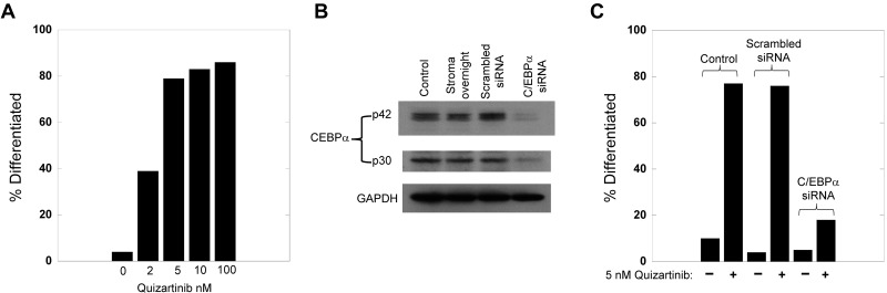
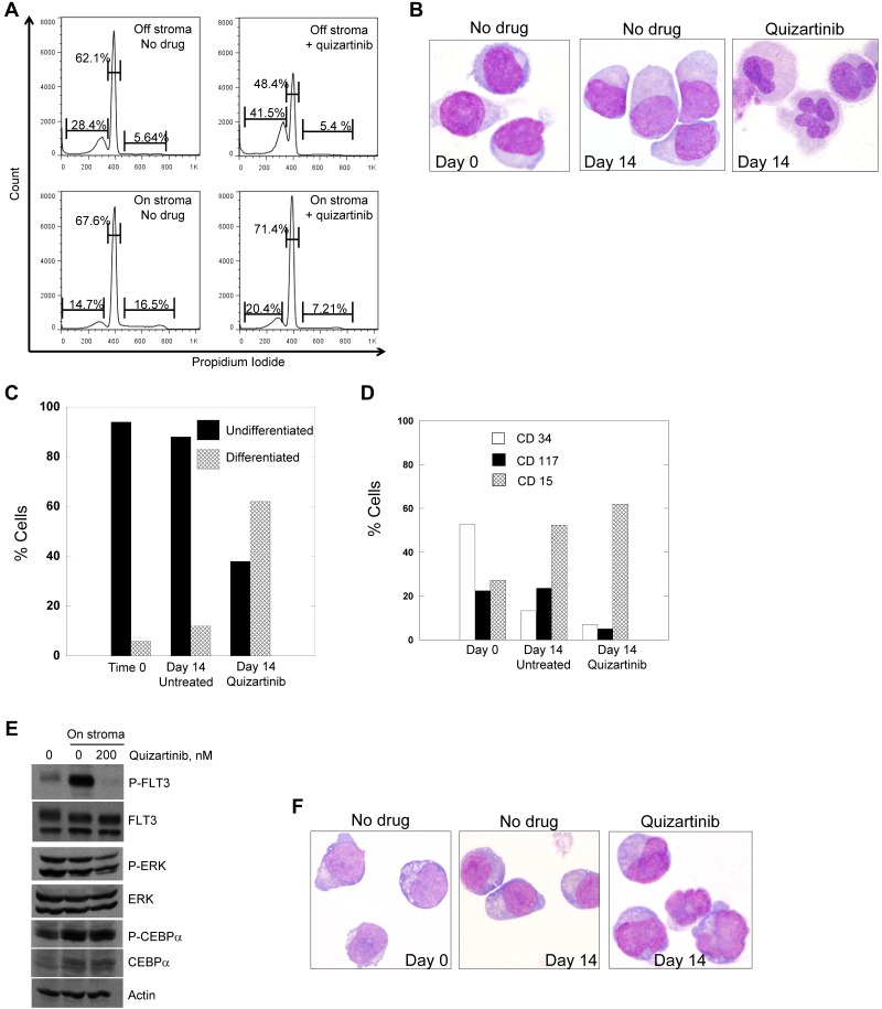
A hallmark of cancer is the disruption of differentiation within tumor cells. Internal tandem duplication mutations of the FLT3 kinase (FLT3/ITD) occur commonly in acute myeloid leukemia (AML) and are associated with poor survival, leading to efforts to develop FLT3 kinase inhibitors. However, FLT3 inhibitors have thus far met with limited success, inducing only a clearance of peripheral blasts with minimal BM responses. Quizartinib is a novel potent and selective FLT3 inhibitor currently being studied in clinical trials. In 13 of 14 FLT3/ITD AML patients with normal karyotype treated with quizartinib, we observed terminal myeloid differentiation of BM blasts in association with a clinical differentiation syndrome. The single patient whose blasts failed to differentiate had a preexisting C/EBPα mutation and another developed a C/EBPα mutation at disease progression, suggesting a mechanism of resistance to FLT3 inhibition. In vitro, in primary blasts cocultured with human BM stroma, FLT3 inhibition with quizartinib induced cell-cycle arrest and differentiation rather than apoptosis. The present study is the first description of terminal differentiation of cancer cells in patients treated with a tyrosine kinase inhibitor. These data highlight the importance of the differentiation block in the patho-genesis of AML.


Figures

Figure 1
Figure 1
In vivo terminal myeloid differentiation in patients treated with quizartinib. (A) Graph of peripheral total WBC count, absolute neutrophil count (ANC), and absolute blast count in a representative patient (Table 1 patient 2) during the first 2 months of treatment with quizartinib. (B) Photomicrograph of a peripheral blood neutrophil from a whole blood sample collected from the patient shown in panel A on day 35 of treatment. (C) BM aspirates collected from a representative patient (Table 1 patient 7) 1 week before and on days 15 and 29 of treatment with quizartinib. (D) Flow cytometry analysis of BM aspirate specimens collected pretreatment and on days 15 and 29 from patient 7. The analysis was performed on the day of collection, and the dot plots are overlaid, organized by color.
Figure 2
Figure 2
Neutrophils are derived from the leukemic blasts. (A) Graph of peripheral blood absolute neutrophil count from patient 4 during treatment with quizartinib. Neutrophils from day 60 were isolated to > 95% homogeneity as described in “Methods,” which was confirmed by cytospin (inset). (B) Genomic DNA samples isolated from pretreatment blasts, day 60 neutrophils, and neutrophils from a healthy donor were analyzed by PCR for the FLT3/ITD mutation, as described in “Methods.” Shown is an ethidium bromide–stained agarose gel. WT indicates wild-type. (C) Whole-cell lysates were prepared from Molm14 cells, pretreatment blasts from patient 4, day 60 neutrophils from patient 4, and neutrophils from a healthy donor. Lysates were analyzed for total FLT3, phosphorylated FLT3, lactoferrin, and MMP9, as described in “Methods.” (D) NBT reduction assay of peripheral blood collected from a healthy donor (“Control”) compared with a quizartinib-treated patient during the neutrophil surge. Cells “with stimulation” were exposed to bacterial extract to induce respiratory burst activity.
Figure 3
Figure 3
Treatment of Molm14 cells with FLT3 inhibitors. (A-B) Molm14 cells were exposed to quizartinib (10nM) or sorafenib (100nM) in suspension culture or coculture with BM stroma and analyzed for annexin V binding (A) and propidium iodide staining (B) by flow cytometry. (C-D) Molm14 cells were cocultured with stroma in the presence and absence of 10nM quizartinib for 24 hours, and then cells were collected and analyzed for morphology (C) and NBT reduction activity (D). (E) Molm14 cells were cocultured on stroma in the presence and absence of 10nM quizartinib. After 1 hour, cells were collected and lysates were analyzed by immunoblotting as described in “Immunoblotting.” (F) Molm14 cells were cocultured with stroma and quizartinib at the indicated concentrations. Cells were collected after 0, 4, 8, 12, and 24 hours of drug exposure and analyzed by immunoblotting for phosphorylated and total C/EBPα.
Figure 4
Figure 4
Knockdown of C/EBPa blocks the differentiation induced by FLT3 inhibition. (A) Molm14 cells were cocultured with stroma in the presence of increasing concentrations of quizartinib for 24 hours, and then cells were collected and analyzed for morphology. For each condition, 100 viable cells were counted and categorized as undifferentiated or differentiated. (B) Molm14 cells (2 million per sample) were incubated with siRNA for C/EBP-α (or scrambled control), electroporated, and then cocultured with stroma. After 24 hours, the cells were harvested, lysed, and analyzed for C/EBPα protein levels by Western blotting. Incubation for 48 hours led to identical results (not shown). (C) Molm14 cells were subjected to siRNA C/EBPa knockdown as in panel B, cocultured with stroma for 24 hours, and then treated with 5nM quizartinib. After 24 hours, the cells were harvested and examined and scored for morphologic changes as in panel A.
Figure 5
Figure 5
Treatment of primary patient blasts with FLT3 inhibitors. (A) Blasts from patient 7 were cocultured with stroma or grown in suspension culture overnight and then 200nM quizartinib was added to both cultures. Cells were incubated with drug for 48 hours and then stained with propidium iodide and analyzed by flow cytometry. Blasts from patient 7 were cocultured with stroma in the presence and absence of 200nM quizartinib for 14 days, and cells were collected and analyzed by light microscopy (B) and scored for differentiation (C). (D) Cells from panel B were also analyzed for the presence of differentiation markers by flow cytometry as described in “Flow cytometry.” (E) Blasts from patient 7 were cocultured with stroma overnight and then 200nM quizartinib was added. After 1 hour, cells were collected and lysates were analyzed by immunoblotting as described in “Flow cytometry.” (F) Blasts from a 52-year-old man with relapsed, refractory FLT3/ITD AML were cocultured on stroma as in panel B. Shown are cells harvested after 9 days of culture with or without quizartinib.

References

    1. Hanahan D, Weinberg RA. The hallmarks of cancer. Cell. 2000;100(1):57–70. - PubMed
    1. Tenen DG. Disruption of differentiation in human cancer: AML shows the way. Nat Rev Cancer. 2003;3(2):89–101. - PubMed
    1. de Thé H, Lavau C, Marchio A, Chomienne C, Degos L, Dejean A. The PML-RAR alpha fusion mRNA generated by the t(15;17) translocation in acute promyelocytic leukemia encodes a functionally altered RAR. Cell. 1991;66(4):675–684. - PubMed
    1. Erickson P, Gao J, Chang KS, et al. Identification of breakpoints in t(8;21) acute myelogenous leukemia and isolation of a fusion transcript, AML1/ETO, with similarity to Drosophila segmentation gene, runt. Blood. 1992;80(7):1825–1831. - PubMed
    1. Liu P, Tarle SA, Hajra A, et al. Fusion between transcription factor CBF beta/PEBP2 beta and a myosin heavy chain in acute myeloid leukemia. Science. 1993;261(5124):1041–1044. - PubMed

Publication types

MeSH terms