. 2012 Oct 9;109(41):16672-7.
doi: 10.1073/pnas.1213802109. Epub 2012 Sep 10.

Structure of the cleavage-activated prefusion form of the parainfluenza virus 5 fusion protein

Affiliations

Structure of the cleavage-activated prefusion form of the parainfluenza virus 5 fusion protein

Brett D Welch et al. Proc Natl Acad Sci U S A. .

Abstract

The paramyxovirus parainfluenza virus 5 (PIV5) enters cells by fusion of the viral envelope with the plasma membrane through the concerted action of the fusion (F) protein and the receptor binding protein hemagglutinin-neuraminidase. The F protein folds initially to form a trimeric metastable prefusion form that is triggered to undergo large-scale irreversible conformational changes to form the trimeric postfusion conformation. It is thought that F refolding couples the energy released with membrane fusion. The F protein is synthesized as a precursor (F0) that must be cleaved by a host protease to form a biologically active molecule, F1,F2. Cleavage of F protein is a prerequisite for fusion and virus infectivity. Cleavage creates a new N terminus on F1 that contains a hydrophobic region, known as the FP, which intercalates target membranes during F protein refolding. The crystal structure of the soluble ectodomain of the uncleaved form of PIV5 F is known; here we report the crystal structure of the cleavage-activated prefusion form of PIV5 F. The structure shows minimal movement of the residues adjacent to the protease cleavage site. Most of the hydrophobic FP residues are buried in the uncleaved F protein, and only F103 at the newly created N terminus becomes more solvent-accessible after cleavage. The conformational freedom of the charged arginine residues that compose the protease recognition site increases on cleavage of F protein.


Conflict of interest statement

The authors declare no conflict of interest.

Figures

Fig. 1.
Fig. 1.
Biochemical and structural characterization of PIV5 F-GCNt. (A) The major F-GCNt domains are highlighted by color. Residue ranges are also indicated. (B) SDS/PAGE analysis under reducing conditions of uncleaved (F0) and trypsin-cleaved (F1 + F2) PIV5 F-GCNt. The gel indicates the completeness of protease cleavage and purity of protein used for crystallization trials. The F1-GCNt and F2 fragments are the expected sizes, indicating that the trypsin cleavage is specific for the multibasic protease cleavage/activation site. (C and D) Electron microscopy of cleaved prefusion (C) and postfusion (D) FGCNt reveals characteristic “tree-like” and “golf tee” structures, respectively. Heat was used as a surrogate for HN activation to convert F-GCNt to the postfusion conformation, which assembles into rosettes. Arrowheads indicate molecules viewed from the side and in C point to the boundary between the treetop and trunk. (E) Cartoon representation of the crystal structure of the cleaved prefusion F-GCNt trimer viewed from the side and colored by domains as in A. Arrowheads indicate the locations of the termini resulting from protease cleavage. (F) Surface representation of the trimer oriented as in E with color-coded chains (A, yellow; B, red; C, orange) and the hydrophobic FP highlighted in blue. On cleavage, the FP remained sandwiched between DII and DIII of adjacent chains as in the uncleaved structure. The GCNt domain was omitted from the model because of insufficient electron density.
Fig. 2.
Fig. 2.
Cleavage site in the cleaved prefusion PIV5 F-GCNt structure. (A) Overlay of chains A (magenta), B (yellow), and C (blue) of the cleaved F-GCNt structure zoomed-in on the protease cleavage site. The F103 side chain, at the N terminus of F1 after cleavage, is shown as sticks, and the C-terminal alpha carbon of F2 is shown as a sphere for each chain. (B) Ribbon diagram overlay of chain A from the uncleaved (green) and cleaved (magenta) structures zoomed-in as in A. The cleaved structure is shown as a trimer making DII from the adjacent chain C (light red) visible. F103 side chains are shown as sticks. (C) Detailed view of interactions involving F103 of chain A (magenta). An intermolecular hydrogen bond is apparent between the carbonyl carbon of F103 and the NH1 nitrogen of R419 in DII (light red) of chain C. F103 of chain A is further stabilized by several intermolecular hydrophobic interactions with DII of chain C, including interactions with Q389, M388, and S419, as well as an intramolecular hydrophobic interaction with V106. (D) Detailed view of interactions involving F103 of chain B (yellow). Here intermolecular interactions with DII of the adjacent chain are minimal. Instead, F103 packs against nearby residues in chain B, including T97, R98, A104, and V106. The carbonyl carbon of F103 forms an intramolecular hydrogen bond with the nitrogen of G105, possibly shared with the nitrogen of V106. (E) Detailed view of interactions involving F103 of chain C (blue). F103 packs against the globular structure, making hydrophobic contact with P96, V107, V125, V128, and K129. A water-mediated hydrogen bond is observed between the NH2-terminal nitrogen and the NZ nitrogen of K129. (F) A portion of the model from the cleaved prefusion PIV5 F-GCNt crystal structure near the protease cleavage site showing the final fit to the 2mFo-DFc electron density map.
Fig. 3.
Fig. 3.
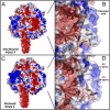
Electrostatic surface renderings of uncleaved and cleaved PIV5 F-GCNt. (A) Side view of uncleaved PIV5 F-GCNt trimer with electrostatic surface shown. (B) Close-up view of the furin cleavage loop (chain A) with a cartoon representation (gray) visible through the partially transparent surface. The F103 side chains are shown as sticks. (C and D) Similar views of the cleaved structure as shown in A and B. Electrostatic solvent-accessible surfaces were calculated at a salt concentration of 0.15 M using the Adaptive Poisson–Bolzmann Solver and PDB2PQR software (v1.3) via the CHARM algorithm and mapped onto the vdw surface in Pymol (v1.3). The electrostatic potential ranges from −2 (red) to +2 (blue) kT/e.
Fig. 4.
Fig. 4.
Comparison of FPs in prefusion PIV5 F-GCNt, influenza HA, and Ebola virus GP crystal structures. (A and B) Top-down view of ribbon diagrams of the uncleaved and cleaved influenza HA crystal structures, respectively (PDB ID codes: 1HA0 and 2HMG) (28, 44) [Drawn from PDB ID: 1HA0 (45).] (C and D) A similar view of uncleaved and cleaved PIV5 F crystal structures, respectively (PDB ID code: 2B9B) (20). In A–D, the residues that move after protease cleavage are highlighted. Residues N-terminal to the cleavage site are highlighted in blue, and residues C-terminal to the cleavage site are shown in red. Additional residues composing the FP are shown in orange, and all FP residues have side chains, shown as sticks. (E) A similar view of cleaved prefusion Ebola GP (PDB ID code: 3CSY) (27). The entire fusion loop of Ebola virus is highlighted in orange with FP residue sidechains shown as sticks. (F) Close-up view of Ebola virus GP FP shown packed against the surface of an adjacent chain in the trimer. Color-coding is as in E. (G) Close-up view of PIV5 F FP buried at the interface between adjacent chains of the trimer. Color-coding is as in B, except the entire FP is colored orange. The C-alpha carbons at each terminus are shown as spheres, colored yellow for the new N- and C-termini created by protease cleavage (B, D, E, and G only). Ovals illustrate the area of FP.
Fig. 5.
Fig. 5.
Structure of distinct conformational states of the F protein ectodomain. Side-view ribbon diagrams of prefusion PIV5 F-GCNt protein in its uncleaved (PDB ID code: 2B9B) and protease-cleaved states, as well as the uncleaved postfusion structure of the hPIV3 F ectodomain (PDB ID code: 1ZTM). Arrowheads indicate the position of protease cleavage sites in the prefusion structures for two of the three chains (the third is hidden from view). Arrows indicate the progression of F through its various conformational states. An exception for the uncleaved postfusion hPIV3 F structure is noted by square brackets as cleavage activation is a biological requirement for fusion activity, although not for refolding of the majority of the F ectodomain into the postfusion form. The hPIV3 F protein lacked a GCNt trimerization domain, and it folded directly into a postfusion conformation even in the absence of protease cleavage. Each structure is color-coded by domain (DI, yellow; DII, red; DIII, magenta; HRB, blue).

Comment in

  • Structure of the primed paramyxovirus fusion protein.
    Steinhauer DA, Plemper RK. Steinhauer DA, et al. Proc Natl Acad Sci U S A. 2012 Oct 9;109(41):16404-5. doi: 10.1073/pnas.1214903109. Epub 2012 Oct 1. Proc Natl Acad Sci U S A. 2012. PMID: 23027936 Free PMC article. No abstract available.

References

    1. Lamb RA, Parks GD. Paramyxoviridae: The viruses and their replication. In: Knipe DM, Howley PM, editors. Fields Virology. 5th Ed. Philadelphia: Lippincott Williams & Wilkins; 2007. pp. 1449–1496.
    1. Lamb RA, Jardetzky TS. Structural basis of viral invasion: Lessons from paramyxovirus F. Curr Opin Struct Biol. 2007;17:427–436. - PMC - PubMed
    1. Russell CJ, Luque LE. The structural basis of paramyxovirus invasion. Trends Microbiol. 2006;14:243–246. - PMC - PubMed
    1. Harrison SC. Viral membrane fusion. Nat Struct Mol Biol. 2008;15:690–698. - PMC - PubMed
    1. Jardetzky TS, Lamb RA. Virology: A class act. Nature. 2004;427:307–308. - PubMed

Publication types

MeSH terms

Associated data

LinkOut - more resources