. 2012 Nov 13;109(46):18903-8.
doi: 10.1073/pnas.1212579109. Epub 2012 Sep 24.

Crystal structures of two subtype N10 neuraminidase-like proteins from bat influenza A viruses reveal a diverged putative active site

Affiliations

Crystal structures of two subtype N10 neuraminidase-like proteins from bat influenza A viruses reveal a diverged putative active site

Xueyong Zhu et al. Proc Natl Acad Sci U S A. .

Abstract

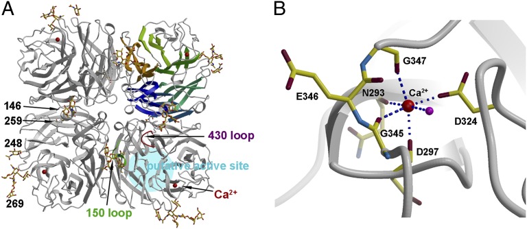
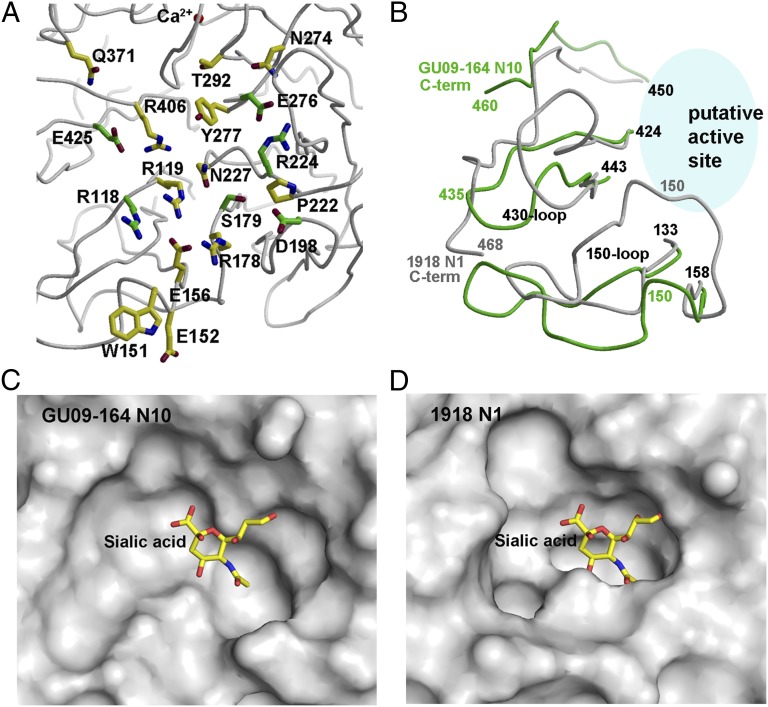
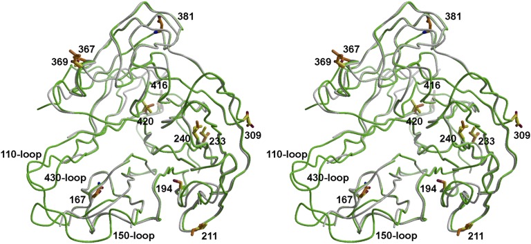
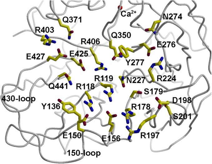
Recently, we reported a unique influenza A virus subtype H17N10 from little yellow-shouldered bats. Its neuraminidase (NA) gene encodes a protein that appears to be highly divergent from all known influenza NAs and was assigned as a new subtype N10. To provide structural and functional insights on the bat H17N10 virus, X-ray structures were determined for N10 NA proteins from influenza A viruses A/little yellow-shouldered bat/Guatemala/164/2009 (GU09-164) in two crystal forms at 1.95 Å and 2.5 Å resolution and A/little yellow-shouldered bat/Guatemala/060/2010 (GU10-060) at 2.0 Å. The overall N10 structures are similar to each other and to other known influenza NA structures, with a single highly conserved calcium binding site in each monomer. However, the region corresponding to the highly conserved active site of influenza A N1-N9 NA subtypes and influenza B NA differs substantially. In particular, most of the amino acid residues required for NA activity are substituted, and the putative active site is much wider because of displacement of the 150-loop and 430-loop. These structural features and the fact that the recombinant N10 protein exhibits no, or extremely low, NA activity suggest that it may have a different function than the NA proteins of other influenza viruses. Accordingly, we propose that the N10 protein be termed an NA-like protein until its function is elucidated.


Conflict of interest statement

The authors declare no conflict of interest.

Figures

Fig. 1.
Fig. 1.
Overall structure of GU09-164 N10 NAL with a conserved calcium binding site. (A) The NAL tetramer is viewed from above the viral surface and consists of four identical monomers in C4 symmetry. One monomer is represented in six different colors to illustrate the canonical β-propeller-fold of six four-stranded, antiparallel β-sheets. The active site region in influenza A and B NAs is highlighted in blue and designated here as the NAL putative active site as no activity has yet been found. This region is located on the membrane-distal surface (on top of the molecule). The putative active site 150-loop and 430-loop are highlighted, and four N-linked glycosylation sites are shown with attached carbohydrates. A single bound calcium ion is shown in red spheres. (B) A close-up view of the conserved calcium binding site in each NAL monomer. The calcium ion (large red sphere) is coordinated by Asn293, Asp297, Gly345, Asp324, and Gly347, and a water molecule (smaller magenta sphere).
Fig. 2.
Fig. 2.
Putative active site of GU09-164 N10 NAL and comparison with the active site of 1918 N1 NA. (A) Conserved catalytic and active site residues in other known NAs are shown on GU09-164 NAL putative active site. The six residues that are conserved in NA and NAL are colored with green carbon atoms, whereas the 11 active site residues that are not conserved are colored with yellow carbon atoms. (B) Comparison of GU09-164 NAL 150-loop, 430-loop, and C terminus (in green) with that of the corresponding loops in 1918 NA (in gray). GU09-164 NAL C terminus is 10-residues shorter than that 1918 NA [two residues are absent in the 1918 NA model (PDB ID code 3BEQ)]. The 150-loop and 430-loop of GU09-164 NAL adopt a more open conformation than 1918 NA. (C) Molecular surface of the putative active site of GU09-164 NAL. A “canonical” sialic acid is modeled into the NAL structure as observed in NA structures. Its glycerol moiety in this mode of sialic acid binding would collide with the NAL active site. However, the putative active site pocket of GU09-164 NAL is much wider than 1918 NA, as shown in D. Thus, the putative active site does not seem to be configured for conventional sialic acid binding and a ligand, if any, is currently unknown. (D) Molecular surface of the active site of 1918 NA in its apo form with the 150-loop in the open conformation. A canonical sialic acid model in other NA structures is superimposed to show its location. For comparison, all panels are generated in the same orientation.
Fig. 3.
Fig. 3.
Stereoview of the superimposed monomeric GU10-060 NAL (in gray) with a monomer from the GU09-164 NAL tetramer (in green) in ribbon presentation. Three loops of GU10-060 NAL including the 110-loop (from 103 to 110), 150-loop (from 141 to 151), and 430-loop (from 428 to 439), as well as an N-terminal fragment (82–90) and a C-terminal fragment (455–460) are not modeled because of poor electron density. The side chains of NAL residues that differ in sequence are shown with yellow carbons for GU09-164 and brown carbons for GU10-060. None of the changes are in the putative active site region.
Fig. 4.
Fig. 4.
Putative active or binding site of GU09-164 NAL showing the high number of polar and charged residues and conserved calcium binding site.

Comment in

References

    1. Tong S, et al. A distinct lineage of influenza A virus from bats. Proc Natl Acad Sci USA. 2012;109:4269–4274. - PMC - PubMed
    1. Air GM, Laver WG. The neuraminidase of influenza virus. Proteins. 1989;6:341–356. - PubMed
    1. Varghese JN, Laver WG, Colman PM. Structure of the influenza virus glycoprotein antigen neuraminidase at 2.9 Å resolution. Nature. 1983;303:35–40. - PubMed
    1. Baker AT, Varghese JN, Laver WG, Air GM, Colman PM. Three-dimensional structure of neuraminidase of subtype N9 from an avian influenza virus. Proteins. 1987;2:111–117. - PubMed
    1. Russell RJ, et al. The structure of H5N1 avian influenza neuraminidase suggests new opportunities for drug design. Nature. 2006;443:45–49. - PubMed

Publication types

LinkOut - more resources