. 2012 Oct 9;109(41):16684-9.
doi: 10.1073/pnas.1212809109. Epub 2012 Sep 24.

Impact of lactobacilli on orally acquired listeriosis

Affiliations

Impact of lactobacilli on orally acquired listeriosis

Cristel Archambaud et al. Proc Natl Acad Sci U S A. .

Abstract

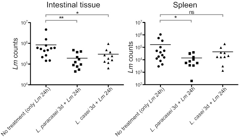
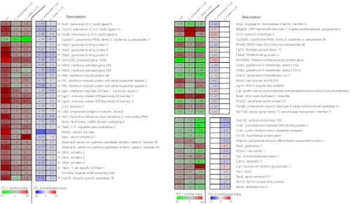
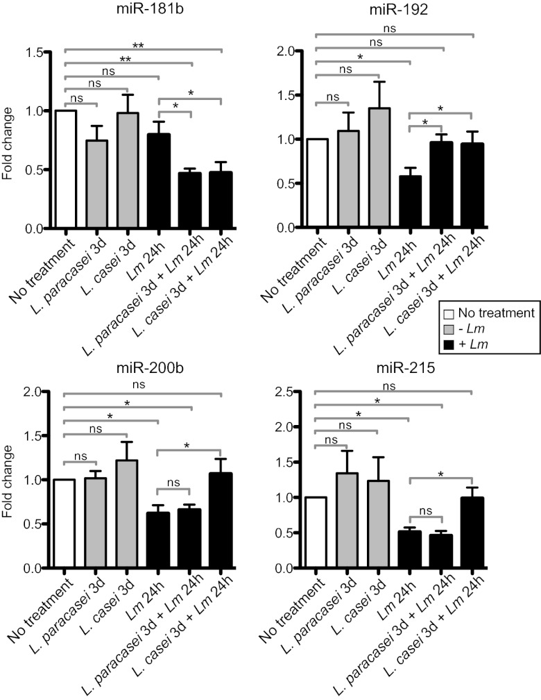
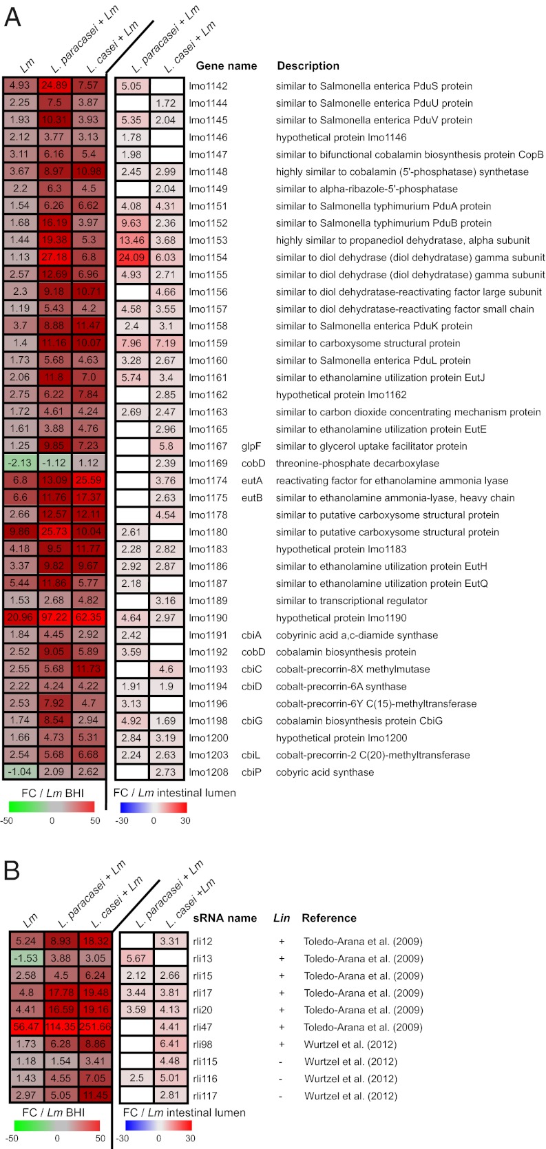
Listeria monocytogenes is a foodborne pathogen that crosses the intestinal barrier and disseminates within the host. Here, we report a unique comprehensive analysis of the impact of two Lactobacillus species, Lactobacillus paracasei CNCM I-3689 and Lactobacillus casei BL23, on L. monocytogenes and orally acquired listeriosis in a gnotobiotic humanized mouse model. We first assessed the effect of treatment with each Lactobacillus on L. monocytogenes counts in host tissues and showed that each decreases L. monocytogenes systemic dissemination in orally inoculated mice. A whole genome intestinal transcriptomic analysis revealed that each Lactobacillus changes expression of a specific subset of genes during infection, with IFN-stimulated genes (ISGs) being the most affected by both lactobacilli. We also examined microRNA (miR) expression and showed that three miRs (miR-192, miR-200b, and miR-215) are repressed during L. monocytogenes infection. Treatment with each Lactobacillus increased miR-192 expression, whereas only L. casei association increased miR-200b and miR-215 expression. Finally, we showed that treatment with each Lactobacillus significantly reshaped the L. monocytogenes transcriptome and up-regulated transcription of L. monocytogenes genes encoding enzymes allowing utilization of intestinal carbon and nitrogen sources in particular genes involved in propanediol and ethanolamine catabolism and cobalamin biosynthesis. Altogether, these data reveal that the modulation of L. monocytogenes infection by treatment with lactobacilli correlates with a decrease in host gene expression, in particular ISGs, miR regulation, and a dramatic reshaping of L. monocytogenes transcriptome.


Conflict of interest statement

The authors declare no conflict of interest.

Figures

Fig. 1.
Fig. 1.
Infection with L. monocytogenes (Lm) after Lactobacillus treatment. Lm counts in the intestinal tissue and in the spleen of gnotobiotic E16P mice that were monoassociated or not with the lactobacilli for 3 d and infected 3 d later with Lm for 24 h. Each dot represents one organ. Horizontal bars represent the mean for each condition. Statistical tests were performed using a Mann–Whitney test. Asterisks indicate a value considered statistically significant (*P < 0.05, **P < 0.01); NS, nonsignificant difference.
Fig. 2.
Fig. 2.
Transcriptomic analysis of the host response after Lactobacillus treatment and L. monocytogenes (Lm) infection. Effect of treatment with Lactobacillus on murine gene expression upon Lm infection. Mice were monoassociated or not monoassociated for 3 consecutive days (3 d) and infected 3 d later with Lm for 24 h. The heatmap presents a subset of host genes whose expression was significantly affected by the lactobacilli during Lm infection [false discovery rate, Benjamini and Hochberg approach (FDR-BH), P < 0.05]. Left, the three columns show values corresponding to the fold change (FC) of gene relative expression in Lm-infected mice (Lm), in mice treated with L. paracasei and infected by Lm (L. paracasei + Lm) and in mice treated with L. casei and infected by Lm (L. casei + Lm) compared with control mice (FC/control mice). Right, the two last columns show FC of gene relative expression in (L. paracasei + Lm) and (L. casei + Lm) mice compared with Lm-infected mice (FC/Lm-infected mice). White square in the heatmap indicates a gene whose expression was not significantly affected by the treatment with the lactobacilli. Asterisks indicate ISGs.
Fig. 3.
Fig. 3.
Detection of miRNA during infection. Relative expression of miR181b, miR-192, miR-200b, and miR-215 in mice monoassociated for 3 consecutive days (3 d) with L. paracasei or L. casei, or in mice that were monoassociated or not with the lactobacilli for 3 d and infected 3 d later with L. monocytogenes (Lm) for 24 h. Fold change is presented after standardization to the small nuclear RNA U6 and using control mice that have been normalized to 1 as reference. Data are represented as mean with SEM. Statistical tests were performed using a two-tailed Student t test. Asterisks indicate a value considered statistically significant (*P < 0.05, **P < 0.01); NS, nonsignificant difference.
Fig. 4.
Fig. 4.
Listeria tiling analysis. (A) Effect of treatment with Lactobacillus on L. monocytogenes (Lm) gene expression during infection. Mice were monoassociated or not monoassociated for 3 consecutive days (3 d) and infected 3 d later with Lm for 24 h. Heatmaps present a subset of Lm genes (A) and all sRNAs (B) whose expression was significantly affected by the lactobacilli during Lm infection (t test P < 0.05). Left, the three columns show values corresponding to the fold change (FC) of gene (A) and sRNA (B) relative expression in ileal-cecal content from Lm-infected mice (Lm), in mice treated with L. paracasei and infected by Lm (L. paracasei + Lm) and in mice treated with L. casei and infected by Lm (L. casei + Lm) compared with Lm growing in broth medium (FC/Lm BHI). Right, the two last columns show FC of gene relative expression in (L. paracasei + Lm) and (L. casei + Lm) mice compared with Lm-infected mice (FC/Lm-intestinal lumen). White square in the heatmap indicates a gene whose expression was not significantly affected by treatment with the lactobacilli. Absence (−) or presence (+) of sRNAs in L. innocua (Lin) is indicated.

Comment in

References

    1. Sekirov I, Russell SL, Antunes LC, Finlay BB. Gut microbiota in health and disease. Physiol Rev. 2010;90:859–904. - PubMed
    1. Gordon JI, Klaenhammer TR. A rendezvous with our microbes. Proc Natl Acad Sci USA. 2011;108(Suppl 1):4513–4515. - PMC - PubMed
    1. Khor B, Gardet A, Xavier RJ. Genetics and pathogenesis of inflammatory bowel disease. Nature. 2011;474:307–317. - PMC - PubMed
    1. Dominguez-Bello MG, Blaser MJ. Do you have a probiotic in your future? Microbes Infect. 2008;10:1072–1076. - PMC - PubMed
    1. Dobrogosz WJ, Peacock TJ, Hassan HM. Evolution of the probiotic concept from conception to validation and acceptance in medical science. Adv Appl Microbiol. 2010;72:1–41. - PubMed

Publication types

MeSH terms