. 2012;12(7):9530-50.
doi: 10.3390/s120709530. Epub 2012 Jul 11.

Synthetic biomimetic membranes and their sensor applications

Affiliations

Synthetic biomimetic membranes and their sensor applications

Young-Rok Kim et al. Sensors (Basel). 2012.

Abstract

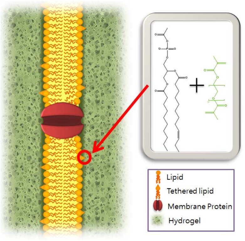
Synthetic biomimetic membranes provide biological environments to membrane proteins. By exploiting the central roles of biological membranes, it is possible to devise biosensors, drug delivery systems, and nanocontainers using a biomimetic membrane system integrated with functional proteins. Biomimetic membranes can be created with synthetic lipids or block copolymers. These amphiphilic lipids and polymers self-assemble in an aqueous solution either into planar membranes or into vesicles. Using various techniques developed to date, both planar membranes and vesicles can provide versatile and robust platforms for a number of applications. In particular, biomimetic membranes with modified lipids or functional proteins are promising platforms for biosensors. We review recent technologies used to create synthetic biomimetic membranes and their engineered sensors applications.

Keywords: biomimetic membranes; ion channel sensors; lipid bilayer.


Figures

Figure 1.
Figure 1.
When a hydrogel crosslinks with the head group of a modified lipid, the bilayer is covalently linked to the surrounding hydrogel matrix (right) [29].
Figure 2.
Figure 2.
Electron micrograph of didodecyldimethylammonium bromide vesicles. (240,000 X). The sample solution was sonicated in the presence of uranyl acetate. Reprinted with permission from Kunitak et al. © 2000 American Chemical Society [31].
Figure 3.
Figure 3.
Schematic representation of a DNA-loaded ABA copolymer vesicle. λ phage binds to a LamB protein, and the DNA is transferred across the block copolymer membrane. Reprinted with permission from Graff et al. Copyright (2002) PNAS [32].
Figure 4.
Figure 4.
The triblock copolymer PMOXA-PDMS-PMOXA. The central siloxane region mimics the hydrophobic center in lipid membranes while the hydrophilic PMOXA end blocks mimic polar lipid head groups.
Figure 5.
Figure 5.
Schematic representation of a planar polymer membrane made from tri-block copolymers: PMOXA (poly methyloxazoline)-PDMS (poly dimethylsiloxane)-PMOXA (poly methyloxazoline). Reprinted with permission from Nardin et al. © 2000 American Chemical Society [44].
Figure 6.
Figure 6.
pH responsive polypeptide vesicles. Schematic illustration of a polypeptide, KP160(L0.3/K0.7)40, and its assembly to a vesicle. Due to the conformational change with pH, molecules entrapped in the vesicle are released. Reprinted by permission from Macmillan Publishers Ltd: Nature Materials [49], copyright 2004.
Figure 7.
Figure 7.
(a) Two-site sandwich assay. Immobilized ion channels (GT), synthetic archaebacterial membrane-spanning lipids (MSL) and half-membrane-spanning tethered lipids (DLP) are attached to a gold surface. In addition, the membrane is composed of mobile half-membrane-spanning lipids (DPEPC/GDPE) and mobile ion channels (G). The mobile ion channels are coupled to biotinylated antibody fragments (Fab') using streptavidin (SA). When the target analyte is approached to the bilayer, the gramicidin A dimer is displaced upon the binding of the analyte to the binding sites of the lipid surface and of gramicidin, decreasing impedance across the bilayer. (b) Competitive assay. The membrane contains hapten-linked gramicidin, (Gh). When the addition of analyte competes with the happen for Fab', hapten-linked gramicidin will be liberated and associated with another gramicidin, increasing impedance across the bilayer. Reprinted by permission from Macmillan Publishers Ltd: Nature [54], copyright 1997.
Figure 8.
Figure 8.
(a) Schematic illustration of an electrofluidic lipid membrane biosensor. Red insets are microscope images of the SLB. (b) Left: microscope image of multiple corrals patterned on a glass substrate. Right: electrophoresis of multiple SLB patterns. Reprinted with permission from Lee et al. Copyright (2012) John Wiley & Sons, Inc. [58].
Figure 9.
Figure 9.
Schematic illustration of SPR and a typical sensorgram. A sensor chip measures the intensity of the reflection of the incident light due to the interaction between the target molecules (green spheres) in the flow solution and the probe molecules (pink diamonds).
Figure 10.
Figure 10.
(a) Schematic illustrations of an OmpF (3) containing lipid bilayer tethered to a solid surface (2) and ligand interaction with the TR domain of colicin N. Reprinted with permission from Stora et al. [64] Copyright (1999) John Wiley & Sons, Inc. (b) nAchR reconstituted in a tethered bilayer: determining receptor orientation and functionality. Reprinted from Sévin-Landais et al. [66] Copyright (2000) with permission from Elsevier.
Figure 11.
Figure 11.
Chemical structure of polydiacetylene and its chromatic properties. (a) Polymerized backbone of vesicles. (b) blue-red color transitions (drawn with ChemDraw, PerkinElmer, Inc.).
Figure 12.
Figure 12.
Blue to red color transition upon ligand binding to the receptor conjugated to the head group of PDA (drawn with ChemDraw, PerkinElmer, Inc.).
Figure 13.
Figure 13.
(A) aptamer-in-liposome composite (a) and liposomes without aptamer (b). (B) Chemical capturing efficiency of the aptamer-in-liposome composite and a liposome without an aptamer encapsulated for three small organic compounds. Reprinted with permission from Kim et al. Copyright (2011) John Wiley & Sons, Inc. [88].
Figure 14.
Figure 14.
PMOXA-PDMS-PMOXA polymersome with encapsulated β-lactamase. Left side: ampicillin cannot permeate polymersome membrane. Right side: ampicillin and ampicillinoic acid can transfer across the polymersome membrane through OmpF channel. Reprinted with permission from Nardin et al. Copyright (2001) John Wiley & Sons, Inc. [96].

References

    1. Eggeling C., Ringemann C., Medda R., Schwarzmann G., Sandhoff K., Polyakova S., Belov V.N., Hein B., von Middendorff C., Schönle A., et al. Direct observation of the nanoscale dynamics of membrane lipids in a living cell. Nature. 2008;457:1159–1162. - PubMed
    1. Verma A., Uzun O., Hu Y.H., Hu Y., Han H.-S., Watson N., Chen S., Irvine D.J., Stellacci F. Surface-structure-regulated cell-membrane penetration by monolayer-protected nanoparticles. Nat. Mater. 2008;7:588–595. - PMC - PubMed
    1. Kim S., Jeon T.J., Oberai A., Yang D., Schmidt J.J., Bowie J.U. Transmembrane glycine zippers: Physiological and pathological roles in membrane proteins. Proc. Natl. Acad. Sci. USA. 2005;102:14278–14283. - PMC - PubMed
    1. Jung S.H., Choi S., Kim Y.R., Jeon T.J. Storable droplet interface lipid bilayers for cell-free ion channel studies. Bioprocess Biosyst. Eng. 2012;35:241–246. - PubMed
    1. Poulos J.L., Jeon T.J., Schmidt J.J. Automatable production of shippable bilayer chips by pin tool deposition for an ion channel measurement platform. Biotechnol. J. 2010;5:511–514. - PubMed

Publication types

LinkOut - more resources