Case Reports
. 2012 Sep 27;367(13):1220-7.
doi: 10.1056/NEJMoa1203055.

Use of reprogrammed cells to identify therapy for respiratory papillomatosis

Affiliations
Case Reports

Use of reprogrammed cells to identify therapy for respiratory papillomatosis

Hang Yuan et al. N Engl J Med. .

Abstract

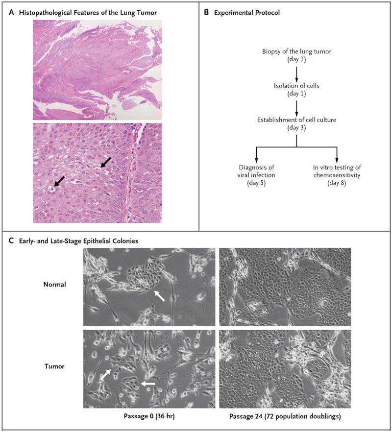
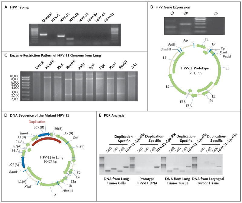
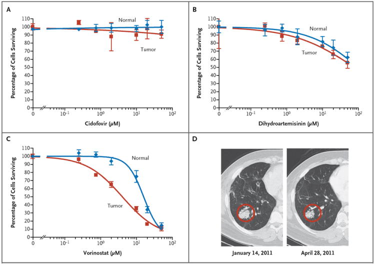
A patient with a 20-year history of recurrent respiratory papillomatosis had progressive, bilateral tumor invasion of the lung parenchyma. We used conditional reprogramming to generate cell cultures from the patient's normal and tumorous lung tissue. Analysis revealed that the laryngeal tumor cells contained a wild-type 7.9-kb human papillomavirus virus type 11 (HPV-11) genome, whereas the pulmonary tumor cells contained a 10.4-kb genome. The increased size of the latter viral genome was due to duplication of the promoter and oncogene regions. Chemosensitivity testing identified vorinostat as a potential therapeutic agent. At 3 months after treatment initiation, tumor sizes had stabilized, with durable effects at 15 months.


Figures

Figure 1
Figure 1. Generation of Conditionally Reprogrammed Cells from Normal and Neoplastic Lung
Panel A shows the histopathological features of the lung tumor (hematoxylin and eosin): typical papillomatosis without evidence of dysplasia (top) and cells with koilocytotic atypia (bottom, arrows). Panel B summarizes the experimental protocol. Panel C shows the morphologic features of conditionally reprogrammed lung tumor cells cocultured with feeder cells for 36 hours and after continued proliferation for 72 population doublings. The arrows at the left indicate the colonies of cells isolated from the patient.
Figure 2
Figure 2. Detection of Mutant HPV-11 in Lung Tumor and Derivative Cell Cultures
Panel A shows typing for human papillomavirus (HPV) in the lung tumor cells by means of a polymerase-chain-reaction (PCR) assay. DNA was isolated from cultured tumor cells, and a general HPV primer set (MY09/MY11), as well as type-specific primer sets, was used for HPV typing. Only the primers for HPV type 11 (HPV-11) yielded the expected PCR products, which were confirmed by DNA sequencing. Panel B shows HPV gene expression in the tumor cells. The messenger RNA (mRNA) transcripts of HPV-11 E6 and E7, but not L1, were detected in the tumor cells by reverse-transcriptase PCR. Panel C shows an altered enzyme-restriction pattern in the HPV-11 genome in the tumor cells. All indicated enzymes cut the prototype HPV-11 genome only once (GenBank accession number, M14119.1). After rolling circle amplification, HPV-11 from lung cells exhibited two digestion sites for enzymes BamHI, AatII, AgeI, FspI, XcmI, and PpuMI. Panel D shows the DNA sequence of the mutant HPV-11 in the tumor cells. Viral DNA was cloned and sequenced. Open reading frames were predicted with the use of ORF Finder (National Center for Biotechnology Information), and the genome map was constructed with the use of Vector NTI software (Invitrogen). The duplicated sequence included L1-LCR-E6-E7-E1 sequences, although the L1 and E1 sequences were only partially duplicated. We annotated the duplicated sequence as L1(B), LCR(B), E6(B), E7(B), and E1(B). Duplication-specific or general HPV-11 PCR primers, shown in Panel E, were used for the detection of corresponding HPV-11 in the DNA isolated from lung tumor cells or from paraffin-embedded lung and laryngeal tissues.
Figure 3
Figure 3. Sensitivity of the Lung Tumor Cells to Three Selected Drugs
Normal lung cells and lung tumor cells were treated with cidofovir (Panel A), dihydroartemisinin (Panel B), or vorinostat (Panel C) at the indicated concentrations for 48 hours. Cell viability was measured with the use of the CellTiter-Glo Luminescent Cell Viability Assay. Vorinostat had a median curative dose of 4.2 μM in tumor cells (95% confidence interval [CI], 3.2 to 5.4) versus 15.9 μM in normal cells (95% CI, 13.2 to 19.2), with selectivity for tumor versus normal cells (P<0.001). Panel D shows images from a computed tomographic scan of a representative lung tumor mass in the right lower lobe before and after a 3-month course of treatment with vorinostat. This mass remained reduced in size 9 months later, when the yearlong course of therapy was terminated.

Comment in

References

    1. Derkay CS, Wiatrak B. Recurrent respiratory papillomatosis: a review. Laryngoscope. 2008;118:1236–47. - PubMed
    1. Donne AJ, Hampson L, Homer JJ, Hampson IN. The role of HPV type in recurrent respiratory papillomatosis. Int J Pediatr Otorhinolaryngol. 2010;74:7–14. - PubMed
    1. Goon P, Sonnex C, Jani P, Stanley M, Sudhoff H. Recurrent respiratory papillomatosis: an overview of current thinking and treatment. Eur Arch Otorhinolaryngol. 2008;265:147–51. - PMC - PubMed
    1. Cook JR, Hill DA, Humphrey PA, Pfeifer JD, El-Mofty SK. Squamous cell carcinoma arising in recurrent respiratory papillomatosis with pulmonary involvement: emerging common pattern of clinical features and human papillomavirus serotype association. Mod Pathol. 2000;13:914–8. - PubMed
    1. Liu X, Ory V, Chapman S, et al. ROCK inhibitor and feeder cells induce the conditional reprogramming of epithelial cells. Am J Pathol. 2012;180:599–607. - PMC - PubMed

Publication types

MeSH terms

Supplementary concepts