Review
. 2012 Nov 8;55(21):8997-9008.
doi: 10.1021/jm3008618. Epub 2012 Oct 5.

Small-molecule structure correctors target abnormal protein structure and function: structure corrector rescue of apolipoprotein E4-associated neuropathology

Affiliations
Review

Small-molecule structure correctors target abnormal protein structure and function: structure corrector rescue of apolipoprotein E4-associated neuropathology

Robert W Mahley et al. J Med Chem. .

Abstract

An attractive strategy to treat proteinopathies (diseases caused by malformed or misfolded proteins) is to restore protein function by inducing proper three-dimensional structure. We hypothesized that this approach would be effective in reversing the detrimental effects of apolipoprotein (apo) E4, the major allele that significantly increases the risk of developing Alzheimer's disease and other neurodegenerative disorders. ApoE4's detrimental effects result from its altered protein conformation ("domain interaction"), making it highly susceptible to proteolytic cleavage and the generation of neurotoxic fragments. Here, we review apoE structure and function and how apoE4 causes neurotoxicity, and describe the identification of potent small-molecule-based "structure correctors" that induce proper apoE4 folding. SAR studies identified a series of small molecules that significantly reduced apoE4's neurotoxic effects in cultured neurons and a series that reduced apoE4 fragment levels in vivo, providing proof-of-concept for our approach. Structure-corrector-based therapies could prove to be highly effective for the treatment of many protein-misfolding diseases.


Figures
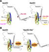
Figure 1
Figure 1
Models of apoE3 and apoE4. (A) ApoE4 displays a unique property called domain interaction caused by the ionic interaction between Arg-61 in the amino-terminal domain with Glu-255 in the carboxyl-terminal domain. ApoE3 is also a dynamic structure and undergoes domain interaction to a significantly less degree than apoE4. (B) ApoE4 domain interaction can be blocked by a small-molecule apoE4SC that disrupts the ionic interaction between Arg-61 and Glu-255. This converts apoE4 to an apoE3-like molecule both structurally and functionally.
Figure 2
Figure 2
ApoE synthesis is induced by injurious agents that stress or damage CNS neurons to participate in the transport of cholesterol and other lipids for membrane repair and synapse formation. ApoE3 progresses through the secretory pathway; however, apoE4, because of domain interaction, displays impaired trafficking through the ER and Golgi apparatus and is targeted to a neuron-specific protease that initially clips off the carboxyl-terminal 27–30 amino acids, generating neurotoxic fragments. These fragments escape the secretory pathway and enter the cytosol, where they target the mitochondria, resulting in mitochondrial dysfunction, and alter the cytoskeleton, enhancing tau phosphorylation and forming neurofibrillary-like tangles.
Figure 3
Figure 3
(A) Model describing the FRET construct. The FRET signal is generated between the donor and acceptor fluorophores. ApoE4 gives a higher FRET signal because of the closeness of the amino- and carboxyl-terminal domains. The apoE4 signal is reduced by the small-molecule apoE structure corrector (SC) through the disruption of domain interaction. (B) A small-molecule apoE4SC (1) inhibits the FRET signal in GFP-apoE4-eDHFR-expressing Neuro-2a cells, but has little effect on GFP-apoE3-eDHFR-expressing cells. (C) Dose-response analysis reveals the relative potencies of three active phthalazinones (2, 3, and 1) and an inactive one [5 (PH-008)] (see ref. for details). Modified from figure originally published in Chen H-K et al. Small molecule structure correctors abolish detrimental effects of apolipoprotein E4 in cultured neurons. J. Biol. Chem. 2012, 287:5253–5266. © the American Society for Biochemistry and Molecular Biology.
Figure 4
Figure 4
Structure of phthalazinones, displaying high potency (nanomolar) to low potency and inactivity with respect to the ability to block apoE4 domain interaction and to rescue apoE4 detrimental effects on neurons. Modified from figure originally published in Chen H-K et al. Small molecule structure correctors abolish detrimental effects of apolipoprotein E4 in cultured neurons. J. Biol. Chem. 2012, 287:5253–5266. © the American Society for Biochemistry and Molecular Biology.
Figure 5
Figure 5
MtCOX1 levels determined by the in-cell western assay demonstrated apoE4 is associated with decreased COX1 levels, which can be restored in apoE4-expressing Neuro-2a cells by apoE structure correctors. (A) Neuro-2a cells expressing apoE3 or apoE4-R61T, which lacks domain interaction, display mtCOX1 levels similar to control cells, whereas apoE4- expressing cells have significantly lower levels (mean ± SD in three separate experiments; p < 0.001). (B) The small-molecule structure corrector 1 increased mtCOX1 levels in apoE4-expressing cells but not in the apoE3- or apoE4-R61T-expressing cells. (C) Dose-response curves for phthalazinones 3 and 1 in Neuro-2a cells (expressed as a percent increase in mtCOX1 levels at nanomolar concentrations). 3 displayed an IC50 of 41 nM (see ref. for details). Modified from figure originally published in Chen H-K et al. Small molecule structure correctors abolish detrimental effects of apolipoprotein E4 in cultured neurons. J. Biol. Chem. 2012, 287:5253–5266. © the American Society for Biochemistry and Molecular Biology.
Figure 6
Figure 6
ApoE4SCs rescue apoE4-associated impairment of mitochondrial motility, dendritic spine formation, and neurite outgrowth in cultured neurons. (A) Mitochondrial motility was retarded in PC12 cells incubated with apoE4, whereas cells incubated with apoE3 or apoE4- R61T behaved like control cells (mean ± SE expressed as a percentage of motile mitochondria; p < 0.001 for apoE4 versus control cells). (B) Dose-response analysis reveals that mitochondrial motility was restored in apoE4-expressing PC12 cells treated with apoE4SC 2 (see ref. for details). (C) Dendritic spine density (spines per µm dendrite) was measured in hippocampal primary neurons from mice expressing apoE3 or apoE4 and transiently transfected with EGFP-β-actin to highlight the spines. ApoE4 expression resulted in a significant reduction in spines (p < 0.01). Spine density was restored in apoE4-expressing neurons following treatment with 3 (100 nM) without affecting apoE3-expressing cells (see ref. for details). Modified from figure originally published in Brodbeck J et al. Structure-dependent impairment of intracellular apolipoprotein E4 trafficking and its detrimental effects are rescued by small-molecule structure correctors. J. Biol. Chem. 2011, 286:17217–17226. © the American Society for Biochemistry and Molecular Biology. (D) ApoE4-expressing Neuro-2a cells demonstrated a 50–60% decrease in cells displaying neurites longer than the cell body diameter compared with apoE3-expressing cells. 3 restored neurite outgrowth in apoE4-expressing cells (100 nM; p < 0.01) (see ref. for details). (E) Active phthalazinones (2, 3, and 1) reverse the impaired neurite outgrowth seen in apoE4-expressing Neuro-2a cells in vitro (2 and 3 at 3 nM; p < 0.0005) compared with no addition control cells, and 1 at 30 nM (p < 0.02) compared with no addition control cells (see ref. for details). Modified from figure originally published in Chen H-K et al. Small molecule structure correctors abolish detrimental effects of apolipoprotein E4 in cultured neurons. J. Biol. Chem. 2012, 287:5253–5266. © the American Society for Biochemistry and Molecular Biology.
Figure 7
Figure 7
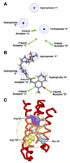
(A) The six-featured pharmacophore model possesses 3 hydrophobic spheres (blue: “A,” “E,” and “F”) and 3 hydrogen bond acceptors (green: “B,” “C,” and “D”). (B) The 3 phthalazinone is fitted to the six-featured pharmacophore model. (C) Predicted model of how the active 3 docks within the amino-terminal domain of apoE4. In this model, hydrophobe “F” interacts with Arg-61 (purple), hydrogen bond acceptor “B” interacts with Arg-119 (yellow), and hydrogen bond acceptor “C” interacts with Glu-50 (light blue). Modified from figure originally published in Chen H-K et al. Small molecule structure correctors abolish detrimental effects of apolipoprotein E4 in cultured neurons. J. Biol. Chem. 2012, 287:5253–5266. © the American Society for Biochemistry and Molecular Biology.
Figure 8
Figure 8
ApoE4 impairs fluorescence recovery in the ER and Golgi apparatus in Neuro-2a cells as determined by fluorescence recovery after photobleaching. Cells expressing EGFP-apoE3 or EGFP-apoE4 were exposed to a laser to photobleach the fusion proteins in the ER or Golgi apparatus and then followed for recovery over a period of 60 s. (A) Fluorescence recovery is decreased in the EGFP-apoE4-expressing cells compared with EGFP-apoE3-expressing cells. The bar graph reports data as a percent of the immobile fraction, which reflects the impaired trafficking of apoE4 (mean ± SD; p < 0.001). (B) The impaired trafficking of apoE4 is reversed in the apoE4-R61T mutant, which lacks domain interaction. ApoE4-R61T behaves like apoE3 (mean ± SD; p < 0.001). (C) Treatment of apoE4-expressing cells with 3 (100 nM) restored the trafficking of apoE4 through the ER and Golgi apparatus. 3 does not affect the trafficking of apoE3 or apoE4-R61T (see ref. for details). Modified from figure originally published in Brodbeck J et al. Structure-dependent impairment of intracellular apolipoprotein E4 trafficking and its detrimental effects are rescued by small-molecule structure correctors. J. Biol. Chem. 2011, 286:17217–17226. © the American Society for Biochemistry and Molecular Biology.

References

    1. Riordan JR. CFTR function and prospects for therapy. Annu. Rev. Biochem. 2008;77:701–726. - PubMed
    1. Van Goor F, Straley KS, Cao D, González J, Hadida S, Hazlewood A, Joubran J, Knapp T, Makings LR, Miller M, Neuberger T, Olson E, Panchenko V, Rader J, Singh A, Stack JH, Tung R, Grootenhuis PDJ, Negulescu P. Rescue of ΔF508-CFTR trafficking and gating in human cystic fibrosis airway primary cultures by small molecules. Am. J. Physiol. Lung Cell. Mol. Physiol. 2006;290:L1117–L1130. - PubMed
    1. Kalid O, Mense M, Fischman S, Shitrit A, Bihler H, Ben-Zeev E, Schutz N, Pedemonte N, Thomas PJ, Bridges RJ, Wetmore DR, Marantz Y, Senderowitz H. Small molecule correctors of F508del-CFTR discovered by structure-based virtual screening. J. Comput. Aided Mol. Des. 2010;24:971–991. - PMC - PubMed
    1. Pedemonte N, Lukacs GL, Du K, Caci E, Zegarra-Moran O, Galietta LJV, Verkman AS. Small-molecule correctors of defective ΔF508-CFTR cellular processing identified by high-throughput screening. J. Clin. Invest. 2005;115:2564–2571. - PMC - PubMed
    1. Pedemonte N, Tomati V, Sondo E, Galietta LJV. Influence of cell background on pharmacological rescue of mutant CFTR. Am. J. Physiol. Cell Physiol. 2010;298:C866–C874. - PubMed

Publication types

MeSH terms

Substances