. 2013 Feb;133(2):334-43.
doi: 10.1038/jid.2012.344. Epub 2012 Sep 27.

Endogenous retinoids in the pathogenesis of alopecia areata

Affiliations

Endogenous retinoids in the pathogenesis of alopecia areata

F Jason Duncan et al. J Invest Dermatol. 2013 Feb.

Abstract

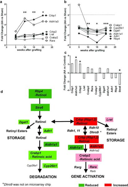
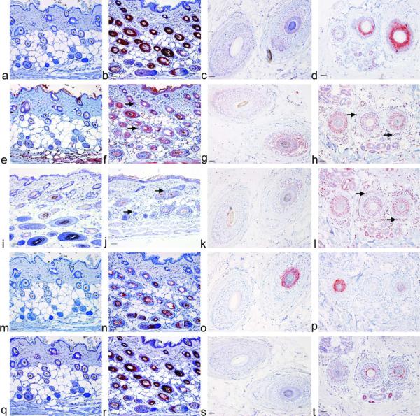
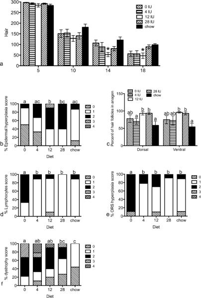
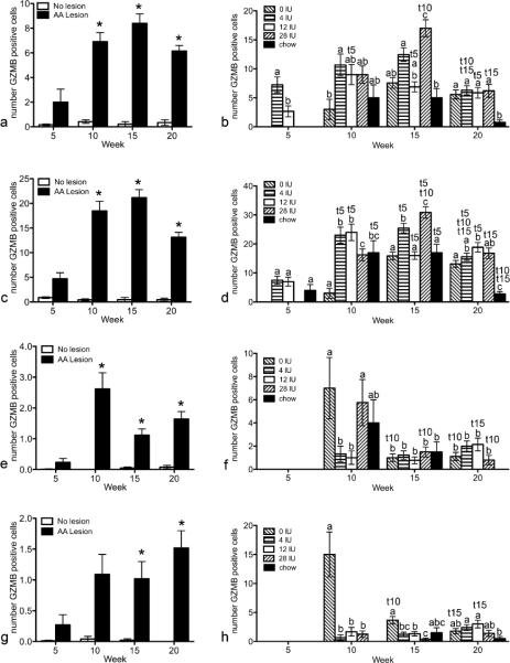
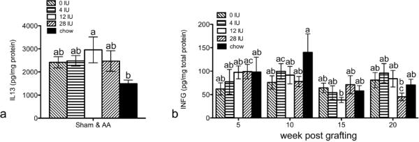
Alopecia areata (AA) is an autoimmune disease that attacks anagen hair follicles. Gene array in graft-induced C3H/HeJ mice revealed that genes involved in retinoic acid (RA) synthesis were increased, whereas RA degradation genes were decreased in AA compared with sham controls. This was confirmed by immunohistochemistry in biopsies from patients with AA and both mouse and rat AA models. RA levels were also increased in C3H/HeJ mice with AA. C3H/HeJ mice were fed a purified diet containing one of the four levels of dietary vitamin A or an unpurified diet 2 weeks before grafting and disease progression followed. High vitamin A accelerated AA, whereas mice that were not fed vitamin A had more severe disease by the end of the study. More hair follicles were in anagen in mice fed high vitamin A. Both the number and localization of granzyme B-positive cells were altered by vitamin A. IFNγ was also the lowest and IL13 highest in mice fed high vitamin A. Other cytokines were reduced and chemokines increased as the disease progressed, but no additional effects of vitamin A were seen. Combined, these results suggest that vitamin A regulates both the hair cycle and immune response to alter the progression of AA.


Figures

Figure 1
Figure 1. Retinoid metabolism is altered in alopecia areata
Fold changes (FC) in RA synthesis (a) and retinol degradation (b) proteins determined by microarray analysis between C3H/HeJ mice grafted with alopecia areata skin compared to sham controls 5, 10, 15, and 20 weeks after surgery, or between mice with spontaneous disease compared to unaffected mice (c). *p < 0.05, q < 0.05 all genes, ** p < 0.05, q < 0.05 all genes except Lrat, Rara, and Cyp26b1, *** p < 0.05, q < 0.05 Only Rbp4, Adh1, and Dgat1. d) Diagram of retinoid metabolism with genes significantly altered highlighted. Red, FC > + 3; Pink, FC 0 to +3; Dark green, FC < − 2; Light green, FC 0 to −2.
Figure 2
Figure 2. Retinoic acid synthesis enzymes and binding proteins are increased in alopecia areata
Immunohistochemistry (IHC) was performed on C3H/HeJ mouse skin collected 10 weeks after grafting with sham controls (a, e, i, m, q), Alopecia Areata (b, f, j, n, r), biopsies from human patients with tinea capitis controls (c, g, k, o, s) or alopecia areata (d, h, l, p, t) with antibodies against CRBP/RBP1 (a–d), DHRS9 (e–h), ALDH1A1 (i–l), ALDH1A3 (m–p), and CRABP2 (q–t). Bar = 50 μm. Arrow, positive immune cells.
Figure 3
Figure 3. Progression of AA was altered by dietary vitamin A
Mice were fed purified diets containing 0, 4, 12, or 28 IU dietary vitamin A/g diet, or a control chow diet starting two weeks prior to grafting and sacrificed 5, 10, 15 (b, c), or 20 (d, e, f) weeks post grafting. Ventral hair loss (a) was measured weekly with 300 representing a full coat of hair. Data are shown as mean ±SEM. H&E slides were scored by JP Sundberg on a scale of 0–4. Data are shown as percent of each score for epidermal hyperplasia (b), anagen (c), lymphocytes (d), outer root sheath hyperplasia (e), and follicular dystrophy (f). n=8–11. *=significantly different from chow fed mice, p<0.05, different letters are significantly different, p<0.05.
Figure 4
Figure 4. Granzyme B (GZMB) is significantly increased in mice with AA and altered by vitamin A
IHC was performed and GZMB positive cells enumerated in the infundibulum (a, b), isthmus (c, d), suprabulbar (e, f), and bulb (g, h). Mann Whitney tests were performed to compare mice with AA lesions to mice without lesions (a, c, e, g; n=40–50). Generalized linear model with the Poisson response function was then performed on mice that received the AA graft with hair follicles in mid-anagen thru midcatagen (b, d, f, h; n=1–10). Data are shown as mean ± SE. * p<0.05 vs no lesion; different letters within a time point are significantly different p<0.05; t5, t10, t15 significantly different from 5, 10, and 15 weeks respectively p<0.05.
Figure 5
Figure 5. Alterations in protein levels of cytokines by high vitamin A during AA progression in C3H/HeJ mice
Protein levels of cytokines IL13 (a) and IFNG (b, c) were determined by ELISA from dorsal lumbar skin. Data were natural log (ln) transformed and analysis of variance performed using SPSS, v19. Data are shown as mean ±SE. n=63–65 for a, and n=16–19 for b. different letters are significantly different p<0.05.

Comment in

References

    1. Afonina IS, Cullen SP, Martin SJ. Cytotoxic and non-cytotoxic roles of the CTL/NK protease granzyme B. Immunological Reviews. 2010;235:105–116. - PubMed
    1. Anzano MA, Lamb AJ, Olson JA. Growth, appetite, sequence of pathological signs and survival following the induction of rapid, synchronous vitamin A deficiency in the rat. J Nutr. 1979;109:1419–1431. - PubMed
    1. Arechalde A, Saurat JH. Retinoids: Unapproved uses or indications. Clinics in Dermatology. 2000;18:63–76. - PubMed
    1. Asseman C, Mauze S, Leach MW, Coffman RL, Powrie F. An essential role for interleukin 10 in the function of regulatory T cells that inhibit intestinal inflammation. J Exp Med. 1999;190:995–1004. - PMC - PubMed
    1. Asseman C, Powrie F. Interleukin 10 is a growth factor for a population of regulatory T cells. Gut. 1998;42:157–158. - PMC - PubMed

Publication types

MeSH terms

LinkOut - more resources