. 2012 Nov 15;18(22):6147-54.
doi: 10.1158/1078-0432.CCR-12-1008. Epub 2012 Sep 26.

Decreased NK cells in patients with head and neck cancer determined in archival DNA

Affiliations

Decreased NK cells in patients with head and neck cancer determined in archival DNA

William P Accomando et al. Clin Cancer Res. .

Abstract

Purpose: Natural killer (NK) cells are a key element of the innate immune system implicated in human cancer. To examine NK cell levels in archived bloods from a study of human head and neck squamous cell carcinoma (HNSCC), a new DNA-based quantification method was developed.

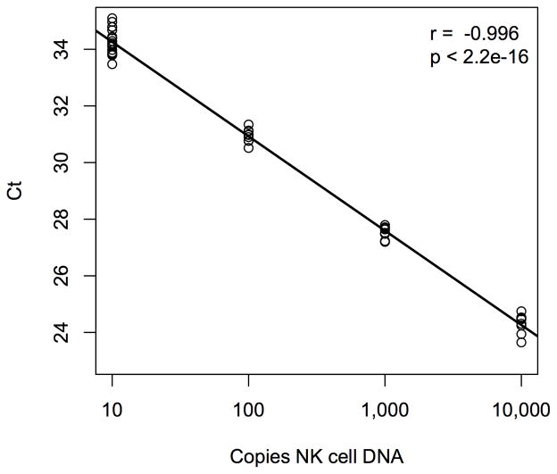
Experimental design: NK cell-specific DNA methylation was identified by analyzing DNA methylation and mRNA array data from purified blood leukocyte subtypes (NK, T, B, monocytes, granulocytes), and confirmed via pyrosequencing and quantitative methylation specific PCR (qMSP). NK cell levels in archived whole blood DNA from 122 HNSCC patients and 122 controls were assessed by qMSP.

Results: Pyrosequencing and qMSP confirmed that a demethylated DNA region in NKp46 distinguishes NK cells from other leukocytes, and serves as a quantitative NK cell marker. Demethylation of NKp46 was significantly lower in HNSCC patient bloods compared with controls (P < 0.001). Individuals in the lowest NK tertile had over 5-fold risk of being a HNSCC case, controlling for age, gender, HPV16 status, cigarette smoking, alcohol consumption, and BMI (OR = 5.6, 95% CI, 2.0 to 17.4). Cases did not show differences in NKp46 demethylation based on tumor site or stage.

Conclusions: The results of this study indicate a significant depression in NK cells in HNSCC patients that is unrelated to exposures associated with the disease. DNA methylation biomarkers of NK cells represent an alternative to conventional flow cytometry that can be applied in a wide variety of clinical and epidemiologic settings including archival blood specimens.


Conflict of interest statement

Conflicts of Interest: WPA, JKW, EAH, and KTK share a relevant patent pending.

Figures

Figure 1
Figure 1
Loci in NKp46 chosen from candidate NK cell-specific differential DNA methylation markers, selected by DNA methylation and mRNA expression criteria. Linear mixed effects modeling of DNA methylation microarray data from MACS isolated human leukocytes generated a coefficient estimating differential methylation in NK cells relative to other cell subtypes, shown on the x-axis. Linear modeling of mRNA microarray data from the same isolated cells determined log-fold change in expression between NK cells and each of the following subtypes: T-cells, B-cells, granulocytes and monocytes. The average of these four log-fold change values is shown on the y-axis. Significance for a particular gene region was achieved when q < 0.1 for all four mRNA expression linear models as well as the DNA methylation mixed effects model. Candidates for NK cell-specific DNA methylation biomarkers were limited to significant gene loci exhibiting decreased methylation in NK cells (methylation estimate < 0) and within genes that exhibited increased RNA expression (log fold change > 1). These candidate loci are marked with asterisks in the top left quadrant, and NKp46 loci are marked with red asterisks.
Figure 2
Figure 2
Demethylation status of NKp46 determined by methylation specific quantitative PCR (qMSP) of isolated human leukocyte populations. Individual samples of MACS purified white blood cell subtypes were subjected to a qMSP assay that detects demethylated copies of NKp46 DNA. Extent of NKp46 methylation is illustrated in this heatmap where yellow indicates that all copies of DNA in particular sample were demethylated in the targeted region of NKp46, while blue indicates that all copies were methylated.
Figure 3
Figure 3
Linearity of NKp46 qMSP Calibration. Bisulfite converted universal methylated DNA was used to keep total amount of DNA in all samples constant. At least three replicates of each standard are plotted. Real time PCR Ct values decrease linearly with ten-fold increase in bisulfite converted NK cell DNA concentration.
Figure 4
Figure 4
Prevalence of HNSCC by normal NKp46 demethylation tertile. Normal NKp46 demethylation tertile cutoffs were determined from control bloods only. Higher tertiles indicate higher NK cell levels. HNSCC prevalence refers to the percent of total cases from this study whose NKp46 demethylation measurements fell within the control derived tertile range. Displayed p-value is from a chi-squared test for trend in proportions.

Similar articles

Cited by

References

    1. Ji H, Ehrlich LI, Seita J, Murakami P, Doi A, Lindau P, et al. Comprehensive methylome map of lineage commitment from haematopoietic progenitors. Nature. 467:338–42. - PMC - PubMed
    1. Hawkins RD, Hon GC, Lee LK, Ngo Q, Lister R, Pelizzola M, et al. Distinct epigenomic landscapes of pluripotent and lineage-committed human cells. Cell Stem Cell. 2010;6:479–91. - PMC - PubMed
    1. Natoli G. Maintaining cell identity through global control of genomic organization. Immunity. 33:12–24. - PubMed
    1. Wieczorek G, Asemissen A, Model F, Turbachova I, Floess S, Liebenberg V, et al. Quantitative DNA methylation analysis of FOXP3 as a new method for counting regulatory T cells in peripheral blood and solid tissue. Cancer Res. 2009;69:599–608. - PubMed
    1. Khavari DA, Sen GL, Rinn JL. DNA methylation and epigenetic control of cellular differentiation. Cell Cycle. 2010;9:3880–3. - PubMed

Publication types

MeSH terms

Associated data