. 2012 Dec 15;21(26):5443-55.
doi: 10.1093/hmg/dds390. Epub 2012 Sep 26.

A novel FGFR3-binding peptide inhibits FGFR3 signaling and reverses the lethal phenotype of mice mimicking human thanatophoric dysplasia

Affiliations

A novel FGFR3-binding peptide inhibits FGFR3 signaling and reverses the lethal phenotype of mice mimicking human thanatophoric dysplasia

Min Jin et al. Hum Mol Genet. .

Abstract

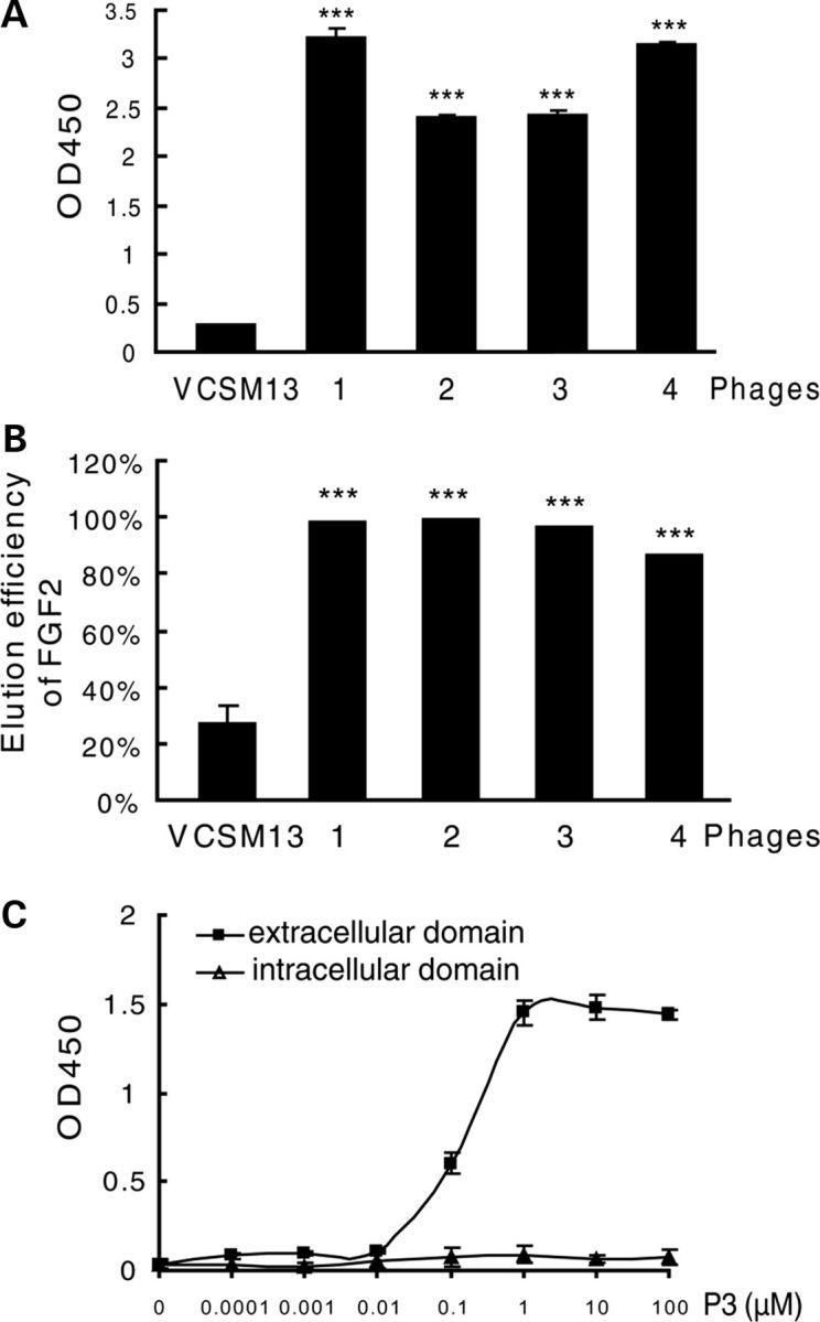
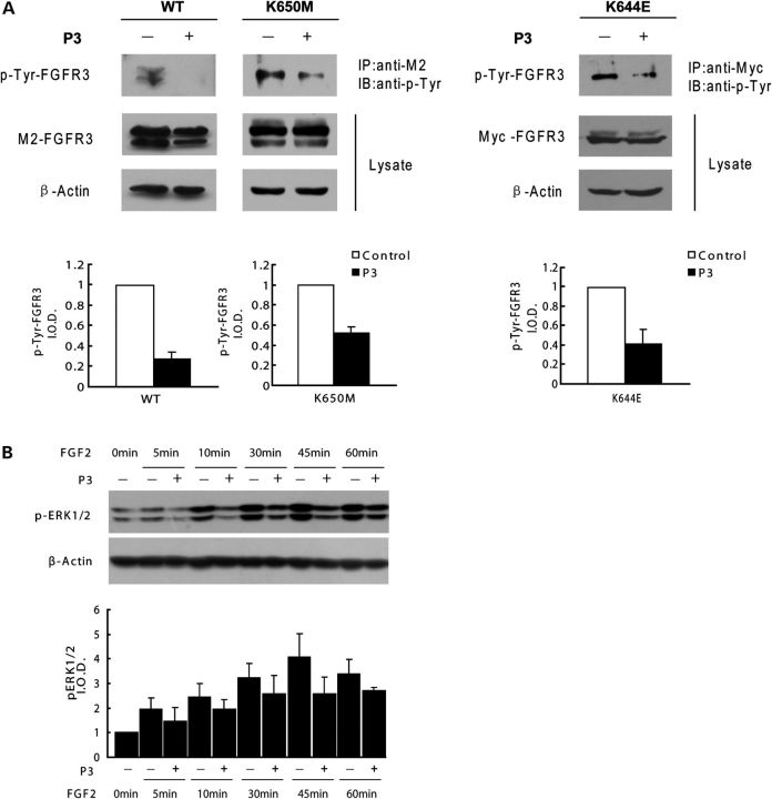
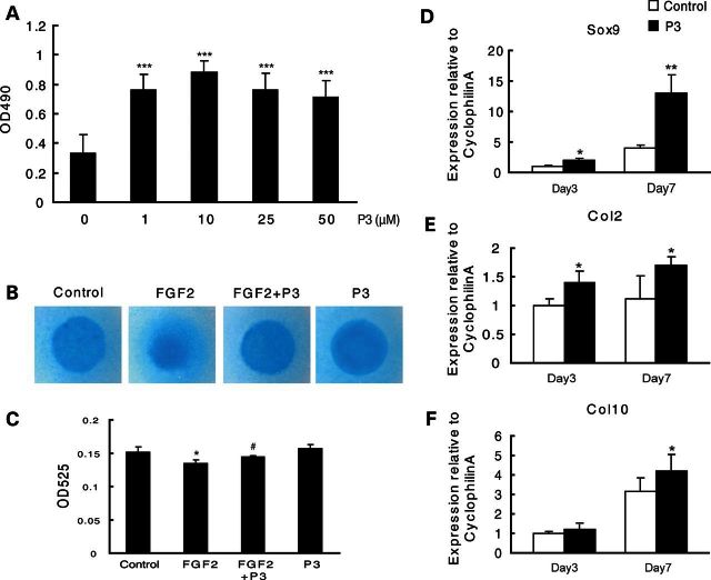
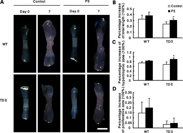
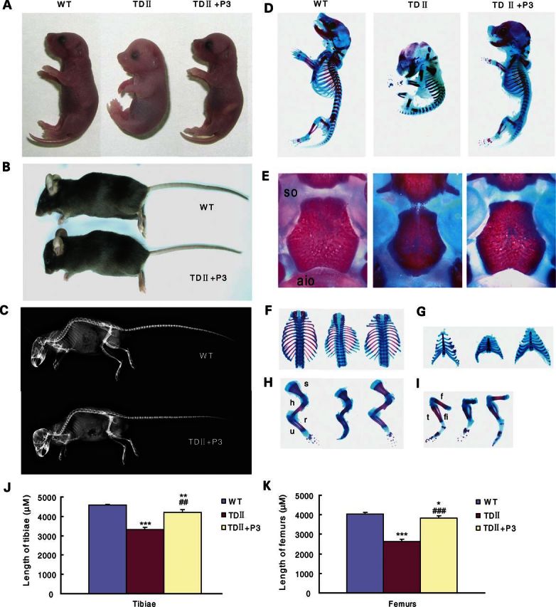
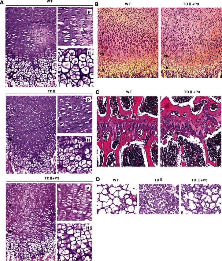
Gain-of-function mutations in fibroblast growth factor receptor-3 (FGFR3) lead to several types of human skeletal dysplasia syndromes including achondroplasia, hypochondroplasia and thanatophoric dysplasia (TD). Currently, there are no effective treatments for these skeletal dysplasia diseases. In this study, we screened, using FGFR3 as a bait, a random 12-peptide phage library and obtained 23 positive clones that share identical amino acid sequences (VSPPLTLGQLLS), named as peptide P3. This peptide had high binding specificity to the extracellular domain of FGFR3. P3 inhibited tyrosine kinase activity of FGFR3 and its typical downstream molecules, extracellular signal-regulated kinase/mitogen-activated protein kinase. P3 also promoted proliferation and chondrogenic differentiation of cultured ATDC5 chondrogenic cells. In addition, P3 alleviated the bone growth retardation in bone rudiments from mice mimicking human thanatophoric dysplasia type II (TDII). Finally, P3 reversed the neonatal lethality of TDII mice. Thus, this study identifies a novel inhibitory peptide for FGFR3 signaling, which may serve as a potential therapeutic agent for the treatment of FGFR3-related skeletal dysplasia.


Figures

Figure 1.
Figure 1.
Specific binding of the positive phage clones to FGFR3. (A) The binding affinity to FGFR3 of four selected positive phage clones and the control vcsM13 was determined by ELISA assay (n = 3, ***P < 0.001, versus VCSM13). (B) Detection of FGF2 elution efficiency to the four selected positive phage clones. The elution efficiency of FGF2 is calculated as follows: (the OD450 value of the phage binding to FGFR3 before competitive elution with FGF2 − the OD450 value of the phage remaining binding to FGFR3 after competitive elution with FGF2)/the OD450 value of the phage binding to FGFR3 before competitive elution with FGF2. (n = 3, ***P < 0.001, versus VCSM13). (C) Affinity detection of peptide P3 binding to FGFR3 by ELISA. Increasing amounts of P3 were immobilized and incubated with the extracellular region or the intracellular region of human FGFR3 protein. Specific binding was detected using antibodies against the extracellular region and the intracellular region of human FGFR3, respectively.
Figure 2.
Figure 2.
Peptide P3 inhibits FGFR3 signaling. (A) Peptide P3 inhibits tyrosine kinase activation of FGFR3. WT, K650M and K644E mutant of FGFR3 were transiently expressed in 293T cells, lysed and immunoprecipitated with an anti-M2 antibody or anti-Myc antibody. Blots were then incubated with an anti-phospho-tyrosine antibody. Phospho-tyrosine band signal intensity was quantified by densitometry. The error bars indicate the standard deviation (SD) from three separate experiments. (B) Peptide P3 inhibits the FGF2-mediated ERK/MAPK phosphorylation in FGFR3-expressing chondrocytic cell line ATDC5. Western blot analysis demonstrated that the levels of phospho-ERK1/2 induced by FGF2 (20 ng/ml) were down-regulated by P3 (10 μm) in ATDC5 cells. The levels of phospho-ERK1/2 were quantified by densitometry.
Figure 3.
Figure 3.
Peptide P3 promotes the proliferation and chondrogenic differentiation of ATDC5 cells. (A) MTT proliferation assay showing that P3 increases the proliferation of ATDC5 cells (n = 6, ***P < 0.01, versus control). (B) ATDC5 cells in micormass cultures were treated with FGF2 (20 ng/ml) and/or P3 (10 μm), and were stained with Alcian blue on Day 7. (C) DMMB assay for quantification of sulfated glycosaminoglycans in micormass culture (n = 3, *P < 0.05, versus control. #P < 0.05, versus FGF2). (DF) Relative mRNA expression levels of chondrogenic marker genes were measured by real-time PCR. The expression levels of Sox9, Col2 and Col10 mRNA in chondrogenic ATDC5 cells were significantly increased on Day 7 after induction in the presence of P3 (10 μm). (n = 3, *P < 0.05, **P < 0.01, versus control.)
Figure 4.
Figure 4.
Peptide P3 suppresses FGFR3-mediated growth inhibition in cultured murine metatarsal bones. (A) Representative photographs show the rescuing effects of peptide P3 on growth retarded of cultured metatarsal bones from TDII mice. Scale bar, 500 μm. (BD) Quantification of the percentage increases in total length, and the lengths of hypertrophic and proliferative zones of metatarsal bones from TDII or WT mice cultured for 7 days in the absence (control) or presence of P3 (10 μm). (n = 4, *P < 0.05, versus control.)
Figure 5.
Figure 5.
Peptide P3 rescues the lethal phenotype in thanatophoric dysplasia type II (TDII) mice. (A) WT mice and TDII mice treated with or without peptide P3 at P1. (B) TDII mouse treated with peptide P3 and the WT controls at P140. (C) X-ray images of the TDII mouse treated with peptide P3 and the WT controls at P140. (D) Alizarin red and Alcain blue staining of the skeletons of WT and TDII mice treated with or without peptide P3 at P1. (E) Cranial base stained with Alizarin red and Alcain blue in WT and TDII mice treated with or without peptide P3 at P1. (FI) Thoracic cages (F), forelimbs (H) and hindlimbs (I) of WT, TDII and P3-treated mice at P1. (J and K) Quantification of tibial and femoral length of WT mice, TDII mice and P3-treated mice at P1. (n = 3, *P < 0.05, **P < 0.01, ***P < 0.001, versus WT. ##P < 0.01, ###P < 0.001, versus TDII.) so, spheno-occipital; aio, anterior intraoccipital; s, scapula; h, humerus; r, radius; u, ulna; f, femur; t, tibia; fi, fibula.
Figure 6.
Figure 6.
Peptide P3 rescues the abnormal growth plate and the lung phenotypes in the TDII mice. (A) Histology of the growth plate of proximal tibia in WT, TDII and P3-treated mice at P1. (B and C) Histological analysis of proximal tibia in WT and P3-treated mice at P5 (B) and P140 (C). (D) Morphology of the lungs in WT, TDII and P3-treated mice at P1.

Similar articles

Cited by

References

    1. Xian C.J. Roles of epidermal growth factor family in the regulation of postnatal somatic growth. Endocr. Rev. 2007;28:284–296. - PubMed
    1. Shiang R., Thompson L.M., Zhu Y.Z., Church D.M., Fielder T.J., Bocian M., Winokur S.T., Wasmuth J.J. Mutations in the transmembrane domain of FGFR3 cause the most common genetic form of dwarfism, achondroplasia. Cell. 1994;78:335–342. - PubMed
    1. Horton W.A. Fibroblast growth factor receptor 3 and the human chondrodysplasias. Curr. Opin. Pediatr. 1997;9:437–442. - PubMed
    1. Bonaventure J., Rousseau F., Legeai-Mallet L., Le Merrer M., Munnich A., Maroteaux P. Common mutations in the fibroblast growth factor receptor 3 (FGFR 3) gene account for achondroplasia, hypochondroplasia, and thanatophoric dwarfism. Am. J. Med. Genet. 1996;63:148–154. - PubMed
    1. Rousseau F., el Ghouzzi V., Delezoide A.L., Legeai-Mallet L., Le Merrer M., Munnich A., Bonaventure J. Missense FGFR3 mutations create cysteine residues in thanatophoric dwarfism type I (TD1) Hum. Mol. Genet. 1996;5:509–512. - PubMed

Publication types

MeSH terms

Supplementary concepts