. 2012 Nov;464(5):481-92.
doi: 10.1007/s00424-012-1160-5. Epub 2012 Sep 27.

Airway smooth muscle STIM1 and Orai1 are upregulated in asthmatic mice and mediate PDGF-activated SOCE, CRAC currents, proliferation, and migration

Affiliations

Airway smooth muscle STIM1 and Orai1 are upregulated in asthmatic mice and mediate PDGF-activated SOCE, CRAC currents, proliferation, and migration

Amy M Spinelli et al. Pflugers Arch. 2012 Nov.

Abstract

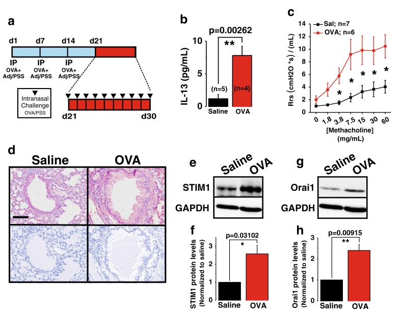
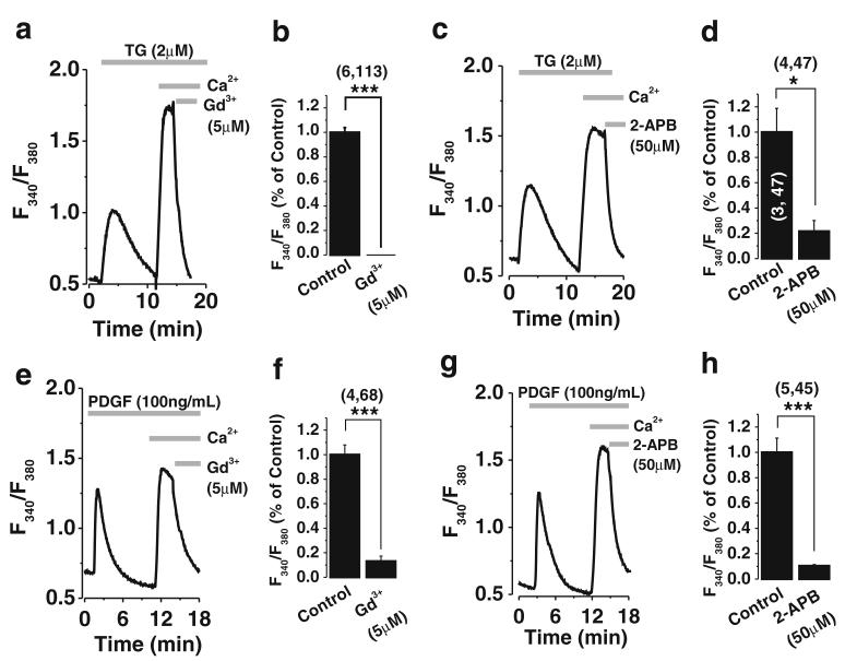
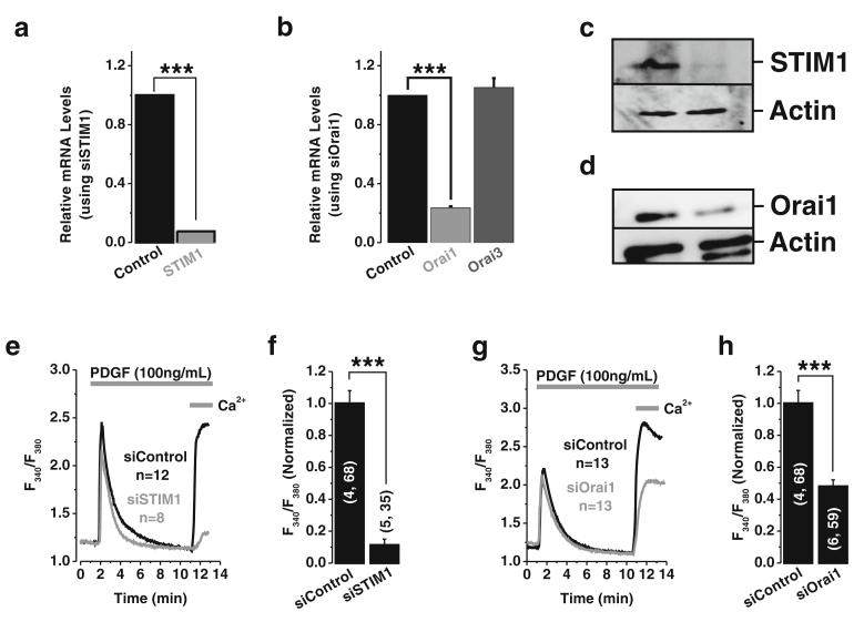
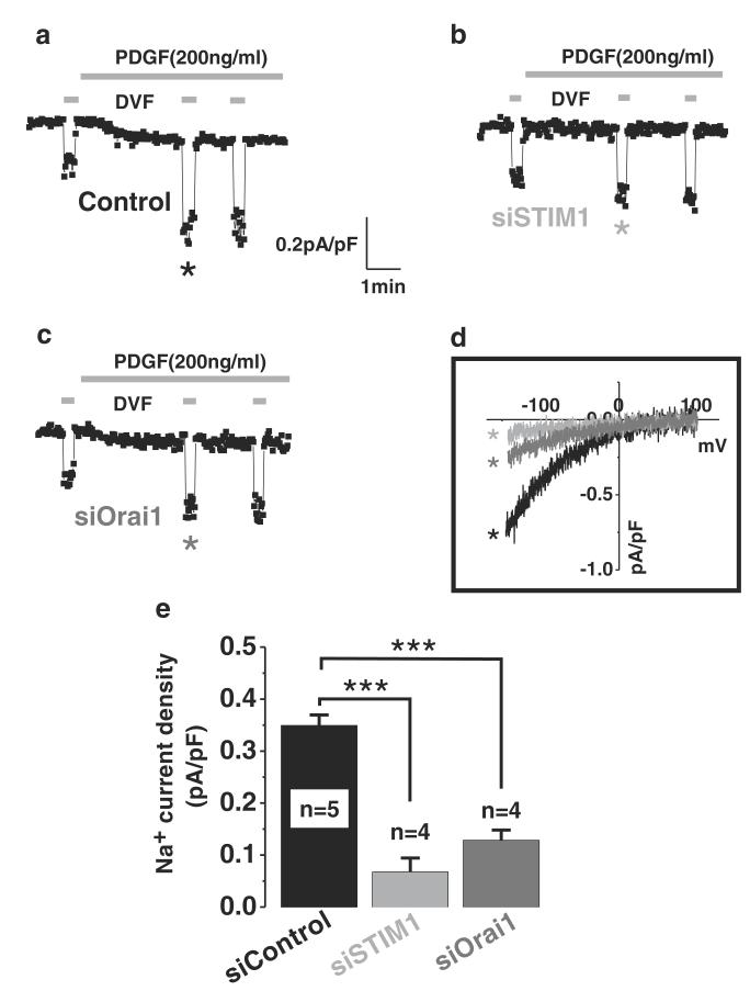
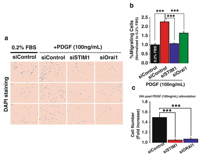
Airway smooth muscle cell (ASMC) remodeling contributes to the structural changes in the airways that are central to the clinical manifestations of asthma. Ca(2+) signals play an important role in ASMC remodeling through control of ASMC migration and hypertrophy/proliferation. Upregulation of STIM1 and Orai1 proteins, the molecular components of the store-operated Ca(2+) entry (SOCE) pathway, has recently emerged as an important mediator of vascular remodeling. However, the potential upregulation of STIM1 and Orai1 in asthmatic airways remains unknown. An important smooth muscle migratory agonist with major contributions to ASMC remodeling is the platelet-derived growth factor (PDGF). Nevertheless, the Ca(2+) entry route activated by PDGF in ASMC remains elusive. Here, we show that STIM1 and Orai1 protein levels are greatly upregulated in ASMC isolated from ovalbumin-challenged asthmatic mice, compared to control mice. Furthermore, we show that PDGF activates a Ca(2+) entry pathway in rat primary ASMC that is pharmacologically reminiscent of SOCE. Molecular knockdown of STIM1 and Orai1 proteins inhibited PDGF-activated Ca(2+) entry in these cells. Whole-cell patch clamp recordings revealed the activation of Ca(2+) release-activated Ca(2+) (CRAC) current by PDGF in ASMC. These CRAC currents were abrogated upon either STIM1 or Orai1 knockdown. We show that either STIM1 or Orai1 knockdown significantly inhibited ASMC proliferation and chemotactic migration in response to PDGF. These results implicate STIM1 and Orai1 in PDGF-induced ASMC proliferation and migration and suggest the potential use of STIM1 and Orai1 as targets for ASMC remodeling during asthma.


Figures

Fig. 1
Fig. 1
Orai1 and STIM1 proteins are upregulated in a mouse model of allergen-induced asthma. Schematic diagram of sensitization and challenge protocol for allergen-induced asthma (a). Three- to four-week-old C57BL/6J mice were sensitized to OVA by carrying out three weekly i.p. injections of 25 μg of OVA with adjuvant. One week after last sensitization, all mice were challenged intranasally with 100 μg of OVA for 10 consecutive days. A group of animals were sacrificed 24 h after final challenge, and BALF was collected to quantify airway IL-13 concentration as an indicator of airway inflammation (b; control n=5; OVA n=4). Twenty-four hours after final challenge, respiratory mechanics were performed to determine airway hyperresponsiveness by measuring systemic airway function (Rrs) with forced oscillations during challenges to increasing doses of methacholine (c; control n= 7; OVA n=6). Analysis of OVA-induced airway remodeling was examined by H&E staining of lung tissue harvested 1 week after final challenge and compared to saline control airways (d; for both experimental groups, n=3 mice with six airways each). Note in OVA-treated mice areas of extensive remodeling in the mucosa and submucosal layers of the lower airways. Images are taken at ×40; bar=40 μm. ImageJ plugin for color deconvolution was executed separating out the hematoxylin nuclear stain in order to visually distinguish the differences in cellular infiltrate surrounding control and OVA-treated airways (bottom images). Twenty-four hours after final challenge, Western blotting of ASM tissue derived from control saline-treated mice compared to OVA-treated animals shows increased protein expression of STIM1 (e, f) and Orai1 (g, h) in OVA-treated animals as compared to control animals. Quantification of band densitometry from n=3 independent mice/experiment is shown in f and h
Fig. 2
Fig. 2
PDGF-activated Ca2+ entry is pharmacologically identical to SOCE. Ca2+ signals were measured in rat ASMC using the ratiometric dye Fura2 in response to either 2 μM thapsigargin (TG; a, c) or 100 ng/ml PDGF (e, g) stimulation in nominally Ca2+-free bath solutions, followed by restoration of 2 mM Ca2+ to the bath, and subsequent addition of pharmacological inhibitors was indicated by the gray bars. The use of inhibitors known to inhibit SOCE 2-APB (50 μM) and Gd3+ (5 μM) inhibited thapsigargin-activated SOCE (a-d) as well as PDGF-activated Ca2+ entry (e-h). Traces represent averages from several cells assayed in a coverslip from a single experiment while bar graphs are averages of the total number of cells from several experiments. (x, y) next to bar graphs represent x=number of independent experiments and y=total number of cells
Fig. 3
Fig. 3
STIM1 and Orai1 mediate PDGF-activated Ca2+ entry in ASMC. SiRNA sequences targeting STIM1 or Orai1 were transfected into ASMCs, and their efficiency of knockdown was assessed by quantification of relative mRNA levels by qPCR, 96 h post transfection. Employment of siRNA targeting STIM1 and Orai1 significantly reduced STIM1 and Orai1 mRNA levels by ~93 and ~76 %, respectively, as shown in a and b. However, Orai3 mRNA levels were not changed when transfecting ASMC with Orai1 siRNA (b). The efficiency of protein knockdown was documented by Western blot analysis and is shown in c and d for STIM1 and Orai1, respectively. Representative Ca2+ imaging traces (e) and statistical analysis (f) from cells transfected with either control siRNA or STIM1 siRNA and stimulated with PDGF (100 ng/ml) showing abrogation of the PDGF-activated Ca2+ entry (from 1±0.08069 to 0.1128±0.03757) in cells transfected with STIM1 siRNA as compared to siRNA non-targeting control. Similarly, Orai1 knockdown significantly inhibited PDGF-activated Ca2+ entry (from 1±0.08069 to 0.47926±0.04198) as shown in g; statistical analysis for this experiment is also shown h. (x, y) next to each data bar: x=number of independent runs, y=total number of cells
Fig. 4
Fig. 4
Whole-cell CRAC currents are activated by PDGF and encoded by STIM1 and Orai1 in ASMC. Whole-cell patch clamp electrophysiology in ASMCs transfected with either control siRNA or siRNA sequences targeting STIM1 or Orai1. STIM1 knockdown completely abrogated both Ca2+ and Na+ (assessed in DVF bath solutions) CRAC currents activated by PDGF (b) as compared to control (sicontrol 0.3494±0.02018 vs 0.06775± 0.02675 siSTIM1). Similarly, Orai1 knockdown (c) led to significant reduction of Ca2+ and Na+ CRAC currents activated by PDGF as compared to control (sicontrol 0.3494± 0.02018 vs 0.1285±0.01967 siOrai1). Na+ CRAC I/V relationships (d) show the requirement of STIM1 and Orai1 for PDGF-activated CRAC currents in ASMC. Statistical analyses are shown in e
Fig. 5
Fig. 5
STIM1 and Orai1 are required for chemotactic migration of ASMC in response to PDGF. Rat ASMC were transfected with either control siRNA or siRNA sequences targeting STIM1 and Orai1. Sixteen hours before migration experiments, cells were serum-starved in media containing 0.2 % FBS. Cells were then harvested with trypsin, and the role of STIM1 and Orai1 in PDGF-induced chemotactic cell migration was determined in a Boyden chamber. Migrating cells were fixed and stained with the nuclear stain DAPI. Representative fields from membranes assayed in triplicates are depicted (a). ASMC chemotactic cell migration was significantly increased in control cells stimulated with 100 ng/ml of PDGF (a). This increase in PDGF-induced cell migration was attenuated in ASMC transfected with either STIM1 siRNA or Orai1 siRNA (a). Quantification of these differences are depicted in b where total number of migrating cells per condition was quantified by analyzing 10 fields per membrane, in triplicates from two independent experiments. c Proliferation assay was performed as described in the “Material and methods.” Rat ASMC were transfected with either siSTIM1, siOrai1, or siControl. Twenty-four hours after PDGF stimulation, ASMC were detached and counted. Cell numbers (after 24 h with 100 ng/ml PDGF) were normalized to control cells under basal conditions (24 h in 0.2%FBS). Values represent fold induction per condition normalized to basal growth conditions (0.2 % FBS)

References

    1. Abdullaev IF, Bisaillon JM, Potier M, Gonzalez JC, Motiani RK, Trebak M. Stim1 and Orai1 mediate CRAC currents and store-operated calcium entry important for endothelial cell proliferation. Circ Res. 2008;103(11):1289–1299. doi:10.1161/01.RES.0000338496.95579.56. - PMC - PubMed
    1. Bai TR. Evidence for airway remodeling in chronic asthma. Curr Opin Allergy Clin Immunol. 2010;10(1):82–86. - PubMed
    1. Beech DJ. Ion channel switching and activation in smoothmuscle cells of occlusive vascular diseases. Biochem Soc Trans. 2007;35(Pt 5):890–894. doi:10.1042/BST0350890. - PubMed
    1. Bentley JK, Hershenson MB. Airway smooth muscle growth in asthma: proliferation, hypertrophy, and migration. Proc Am Thorac Soc. 2008;5(1):89–96. - PMC - PubMed
    1. Berra-Romani R, Mazzocco-Spezzia A, Pulina MV, Golovina VA. Ca2+ handling is altered when arterial myocytes progress from a contractile to a proliferative phenotype in culture. Am J Physiol Cell Physiol. 2008;295(3):C779–C790. doi:10.1152/ajpcell.00173.2008. - PMC - PubMed

Publication types

MeSH terms