. 2012 Sep 26;32(39):13439-53.
doi: 10.1523/JNEUROSCI.2039-12.2012.

Spatially coordinated kinase signaling regulates local axon degeneration

Affiliations

Spatially coordinated kinase signaling regulates local axon degeneration

Mark Chen et al. J Neurosci. .

Abstract

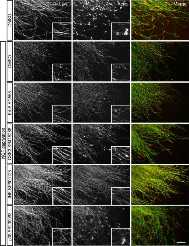
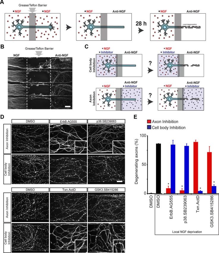
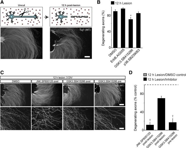
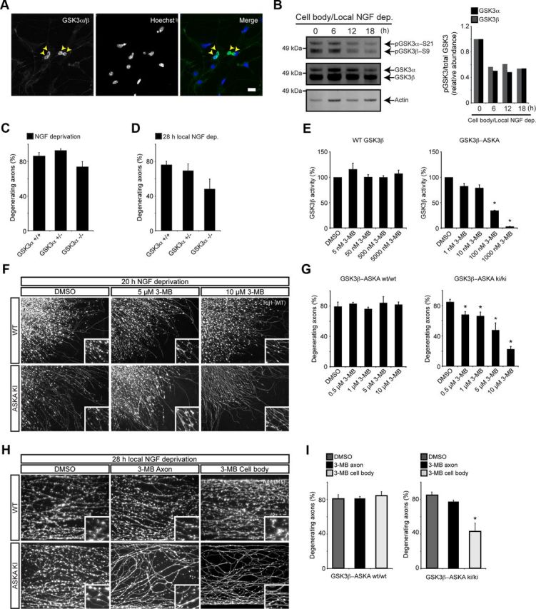
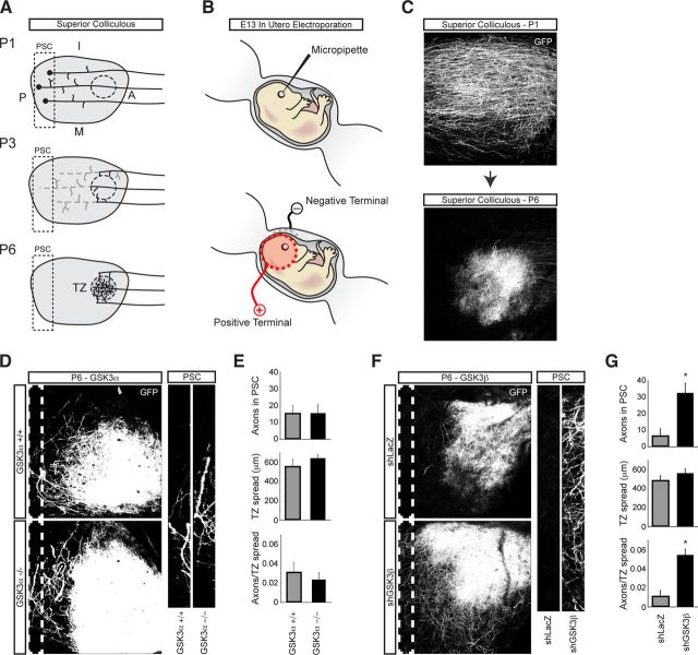
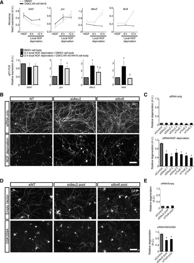
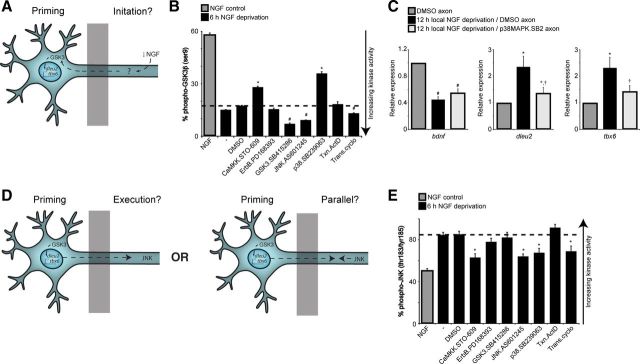
In addition to being a hallmark of neurodegenerative disease, axon degeneration is used during development of the nervous system to prune unwanted connections. In development, axon degeneration is tightly regulated both temporally and spatially. Here, we provide evidence that degeneration cues are transduced through various kinase pathways functioning in spatially distinct compartments to regulate axon degeneration. Intriguingly, glycogen synthase kinase-3 (GSK3) acts centrally, likely modulating gene expression in the cell body to regulate distally restricted axon degeneration. Through a combination of genetic and pharmacological manipulations, including the generation of an analog-sensitive kinase allele mutant mouse for GSK3β, we show that the β isoform of GSK3, not the α isoform, is essential for developmental axon pruning in vitro and in vivo. Additionally, we identify the dleu2/mir15a/16-1 cluster, previously characterized as a regulator of B-cell proliferation, and the transcription factor tbx6, as likely downstream effectors of GSK3β in axon degeneration.


Figures

Figure 1.
Figure 1.
Identification of chemical compounds that inhibit axon degeneration. Small-molecule inhibitors were applied to cultured E13.5 mouse DRG explants for 2 h before adding NGF neutralizing antibodies. After 20 h of NGF deprivation, cells were fixed and labeled for microtubules (Tuj1; green), which are enriched in the axon shaft, and actin (red), a major component of growth cones and an indicator of NGF signaling. Selected positive hits are shown, including ErbB inhibitor AG555 (10 μm), GSK3 inhibitor SB415286 (30 μm), JNK inhibitor SP600125 (20 μm), and p38MAPK inhibitor SB239063 (30 μm). Images are representative of results obtained from at least three independent experiments. Scale bar, 50 μm. High-magnification insets are 123 μm squares.
Figure 2.
Figure 2.
Compartment-specific roles for various pathways in axon degeneration. A, Diagram illustrating localized degeneration in the Campenot chamber. Dissociated DRGs neurons are plated in the “cell body” compartment, and axons extend under a “grease/Teflon” barrier into the “axon compartment.” Axons locally deprived of NGF degenerate over a period of 28 h. B, Low-magnification image showing degeneration of axons locally deprived of NGF. Proximal axons in the cell body compartment maintained in NGF and do not show signs of degeneration. C, Illustration of Campenot chamber assay used to determine functional localization of pathways. Concurrent with local NGF deprivation from axons, inhibitors were applied to either the axon or the cell body compartment. After 28 h, neurons were fixed and labeled for microtubules (Tuj1). D, Images of NGF-deprived axons with inhibitors applied to distal axons—at the site of degeneration—or in the cell body compartment. The p38MAPK and ErbB inhibitors are required at the site of degeneration, while GSK3 and transcription inhibitors are required at the cell body compartment to block axon degeneration. E, Quantification of axon degeneration in Campenot chambers (*p < 0.05, Student's t test; n = 3–7). Values represent mean ± SEM. Scale bars: B, 100 μm; D, 25 μm. High-magnification insets in D are 62 μm squares.
Figure 3.
Figure 3.
Long-term GSK3 inhibition reduces Wallerian degeneration. A, Diagram illustrating lesion experiment. DRG explants were cultured for 3 d, lesioned, and fixed after 12 h of degeneration. Lesioned axons show extensive degeneration at the microtubule level. B, Either DMSO, EGFR/ErbB inhibitor (10 μm AG555), GSK3 inhibitor (30 μm SB415286), or p38MAPK inhibitor (30 μm SP239063) were applied immediately after axon lesion, and evaluated for microtubule degeneration 12 h later. Values represent mean ± SEM (*p < 0.05 compared with DMSO control, Student's t test; n = 3–4). Of the tested inhibitors, only GSK3 inhibitor SB415286 reduces Wallerian degeneration. C, GSK3 inhibitor (30 μm SB415286) or JNK inhibitor (20 μm SP600125) were individually applied after axon lesion and compared with 18 h of GSK3 inhibitor (30 μm SB415286) treatment before axon lesion. Long-term inhibition of GSK3 robustly inhibits axon degeneration following lesion. D, Quantification of percentage degenerating axons, normalized to DMSO controls. Values represent mean ± SEM (p < 0.05 compared with GSK3 inhibition postlesion, Student's t test; n = 3). Scale bars: A, C, top panels, 400 μm; C, bottom panels, 200 μm.
Figure 4.
Figure 4.
GSK3β, and not GSK3α, is a key regulator axon degeneration in vitro. A, Cultured DRG immunocytochemistry shows GSK3 enrichment in the soma. The yellow arrowheads point to neuronal cell bodies. B, Phosphorylation state of GSK3 isoforms in the cell body compartment following distal axon NGF deprivation. Over a period of 18 h, both isoforms of GSK3 are dephosphorylated, indicating elevated kinase activity. Representative blot from three independent experiments is shown. Relative abundance of pGSK3α/total GSK3α and pGSK3β/total GSK3β are shown in the adjacent graph. Note the difference in total protein loaded for each lane. C, E13.5 DRG explants from GSK3α+/+, GSK3α+/−, and GSK3α−/− embryos were cultured overnight, and then deprived of NGF for 20 h. Genetic deletion of GSK3α has no effect on axon degeneration following NGF deprivation. Values represent mean ± SEM (n ≥ 3). D, Dissociated DRG neurons from GSK3α+/+, GSK3α+/−, and GSK3α−/− were cultured for 5 d in Campenot chambers and then deprived of NGF for 28 h. Genetic deletion of GSK3α does not significantly reduce local axon degeneration. Values represent mean ± SEM (n ≥ 5). E, HEK293 cells were transfected with wild-type GSK3β or GSK3β V110I/L132A/F175L. Kinase activities were determined in the presence of varying concentrations of 3MB-PP1 using anti-V5 immunoprecipitates and a continuous fluorescent kinase assay. 3MB-PP1 potently inhibits GSK3β-ASKA activity but not wild-type activity. Values represent mean ± SEM (*p < 0.05, Wilcoxon's test, 25 μg of lysate, 20 μm ATP, n = 3). F, Images of DRG axons after 20 h NGF deprivation, cultured from ASKA mutants or wild-type animals. In the presence of ASKA inhibitor 3MB-PP1, ASKA mutants show significantly less degeneration. Wild-type explants degenerate with 3MB-PP1, indicating that GSK3β inhibition is specific. G, Quantification of reduced axon degeneration of GSK3β-ASKA knock-in DRGs in the presence of ASKA inhibitors. Values represent mean ± SEM (*p < 0.05, Student's t test; n = 9). H, Compartment-specific inhibition of GSK3β using chemical genetics. DRGs from GSK3β-ASKA knock-in embryos were cultured in Campenot chambers for 5 d. 3MB-PP1 (10 μm) was added to either the cell body or axon compartment immediately following local NGF deprivation. 3MB-PP1 potently inhibits axon degeneration when applied to the cell body compartment alone, but not the axon compartment alone. I, Quantification of reduced local axon degeneration, specifically in GSK3β-ASKA knock-in neurons exposed to 3MB-PP1 in the cell body compartment. Values represent mean ± SEM (*p < 0.05, Student's t test; n ≥ 6). Scale bar: A, 60 μm. The high-magnification insets in F and H are 62 μm squares.
Figure 5.
Figure 5.
GSK3β, and not GSK3α, is a key regulator of naturally occurring developmental axon pruning in vivo. A, Diagram illustrating developmental axon pruning of RGC axons in the superior colliculus. At postnatal day 1 (P1), axons initially overshoot their target region and extend into the posterior superior colliculus (PSC). At P3, axons prune back through degeneration. By P6, axon terminals are confined to the termination zone (TZ), their final destination. B, Diagram illustrating in utero electroporation. Following DNA microinjection, DNA was electroporated by creating an electric field with paddle electrodes placed on either side of the embryo's head. C, Representative maximum intensity projection (MIP) images of GFP-positive axons within the SC at P1 and P6. D, Representative MIP images of GFP-positive axons within the SC at postnatal P6 from GSK3α+/+ and GSK3α−/− animals. The white dotted line outlines the region of the SC defined as the PSC, shown in higher magnification to the right. E, Quantification of axon pruning as measured by the number of GFP-positive axons in the PSC, size of the TZ, and the number of axons in the PSC normalized to the size of the TZ for GSK3α+/+ (n = 5) and GSK3α−/− (n = 6). Values represent mean ± SEM. No difference in RGC axon pruning was observed between GSK3α-null and wild-type littermate controls. F, GFP was coexpressed with either shLacZ or shGSK3β. Knockdown of GSK3β results in significantly more GFP-positive axons in the PSC compared with shLacZ control. G, Quantification of axon pruning as measured by the number of GFP-positive axons in the PSC, size of the TZ, and the number of axons in the PSC normalized to the size of the TZ for shLacZ (n = 15) and shGSK3β (n = 9) animals. Values represent mean ± SEM (*p < 0.05, Student's t test). Scale bar: B, C, 100 μm.
Figure 6.
Figure 6.
Time course microarray to identify candidate GSK3-driven axon degeneration genes. A, Expression profiling of genes following local NGF deprivation. RNA was isolated from neurons locally deprived of NGF for 6 and 12 h, with and without GSK3 inhibition (30 μm AR-A014418) and compared with neurons globally maintained in NGF. Values represent mean ± SEM (n = 5). bdnf mRNA decreases and jun mRNA increases after local NGF deprivation, as expected. Dleu2 and tbx6 meet the criteria for candidate axon degeneration genes. They are upregulated after 12 h of local NGF deprivation in a GSK3-dependent manner. Results from the microarray experiment were verified by qRT-PCR. Samples from NGF control were compared with 12 h NGF deprivation with and without GSK3 inhibition. Values represent mean ± SEM (*p < 0.05 compared with NGF control; p < 0.05 compared with 12 h NGF deprivation, Wilcoxon's test; n ≥ 8). B, DIV2 siRNA-transfected DRG neurons maintained in NGF (siRNA only) or following 20 h NGF deprivation (NGF deprivation). siRNA targeting dleu2 and tbx6 do not affect baseline degeneration of cultured DRGs, but do reduce degeneration following NGF deprivation. C, Automated quantification of degeneration in NGF control and after 20 h NGF deprivation. Values are relative to the siControl/NGF deprivation condition and represent mean ± SEM (*p < 0.05 compared with siControl, Student's t test; n = 3 wells from 1 representative experiment). D, Cultured hippocampal/cortical neurons were transfected on DIV5 with siRNA and GFP, plus either Control vector or constitutively active GSK3 (GSK3βS9A or S9A). Neurons were fixed after 3 d of expression (8 DIV). The siRNA have no effect on baseline neurite degeneration when combined with Control vector. Both siDleu2 and siTbx6 reduce relative degeneration when cotransfected with constitutively active GSK3β. E, Automated quantification of degeneration after cotransfection with dleu2 or tbx6 siRNA and either Control vector or S9A (*p < 0.05, Student's t test; n ≥ 12 wells from a representative experiment). Values represent mean ± SEM. Scale bars: B, D, 100 μm.
Figure 7.
Figure 7.
Concerted activity of kinases functioning in spatially distinct compartments is necessary for developmental degeneration. A, Pathways that are required for GSK3-dependent “priming” are described as “initiation” pathways. B, Dissociated DRG neurons were globally deprived of NGF for 6 h, with or without inhibitors of various pathways. Cell lysates were assayed for phospho-GSK3β (Ser9) and total GSK3β. Only CaMKK inhibitor (15 μm STO-609) and p38MAPK inhibitor (30 μm SB239063) reduce dephosphorylation of GSK3β following NGF deprivation. Values represent mean ± SEM (*,#p < 0.05 compared with NGF deprivation/DMSO control, Student's t test; n = 3). C, Local NGF deprivation/DMSO induces upregulation dleu2 and tbx6, and downregulation of bdnf. Applying the p38MAPK inhibitor (30 μm SB239063) in the axon compartment with local NGF deprivation prevents upregulation of dleu2 and tbx6 but does not reverse the downregulation of bdnf (*p < 0.05 compared with NGF control; p < 0.05 compared with NGF deprivation/DMSO, Wilcoxon's test; n = 5). D, Does JNK act downstream of GSK3 or in parallel? E, Cell lysates from B were assayed for pJNK activity. GSK3 inhibitor (30 μm SB415286) does not prevent JNK phosphorylation after NGF withdrawal. Values represent mean ± SEM (*,#p < 0.05 compared with NGF deprivation/DMSO, Student's t test; n = 3).

References

    1. Azoulay-Alfaguter I, Yaffe Y, Licht-Murava A, Urbanska M, Jaworski J, Pietrokovski S, Hirschberg K, Eldar-Finkelman H. Distinct molecular regulation of glycogen synthase kinase-3alpha isozyme controlled by its N-terminal region: functional role in calcium/calpain signaling. J Biol Chem. 2011;286:13470–13480. - PMC - PubMed
    1. Bain J, McLauchlan H, Elliott M, Cohen P. The specificities of protein kinase inhibitors: an update. Biochem J. 2003;371:199–204. - PMC - PubMed
    1. Bijur GN, Jope RS. Proapoptotic stimuli induce nuclear accumulation of glycogen synthase kinase-3 beta. J Biol Chem. 2001;276:37436–37442. - PMC - PubMed
    1. Bijur GN, Jope RS. Glycogen synthase kinase-3 beta is highly activated in nuclei and mitochondria. Neuroreport. 2003;14:2415–2419. - PubMed
    1. Bilimoria PM, de la Torre-Ubieta L, Ikeuchi Y, Becker EB, Reiner O, Bonni A. A JIP3-regulated GSK3beta/DCX signaling pathway restricts axon branching. J Neurosci. 2010;30:16766–16776. - PMC - PubMed

MeSH terms