. 2012 Sep 26;32(39):13568-81.
doi: 10.1523/JNEUROSCI.1364-12.2012.

Time-dependent cross talk between spinal serotonin 5-HT2A receptor and mGluR1 subserves spinal hyperexcitability and neuropathic pain after nerve injury

Affiliations

Time-dependent cross talk between spinal serotonin 5-HT2A receptor and mGluR1 subserves spinal hyperexcitability and neuropathic pain after nerve injury

Zigor Aira et al. J Neurosci. .

Abstract

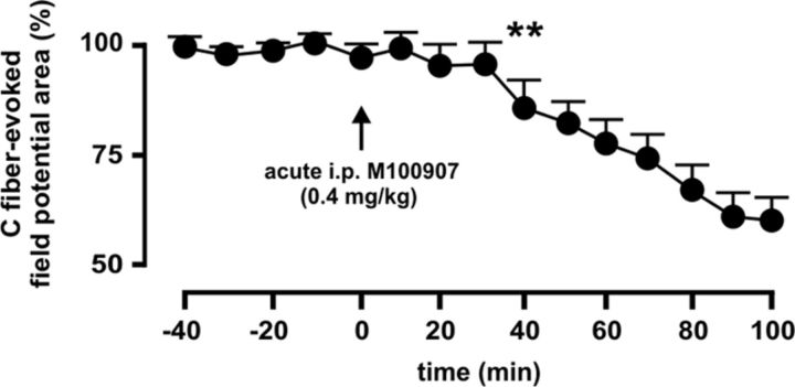
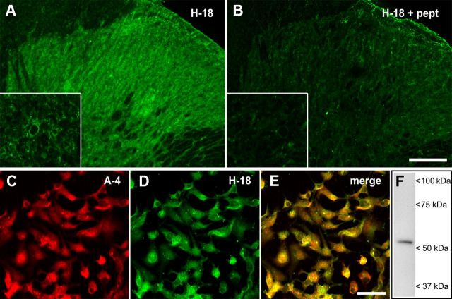
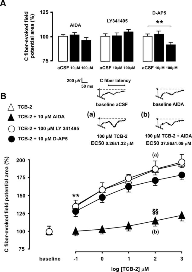
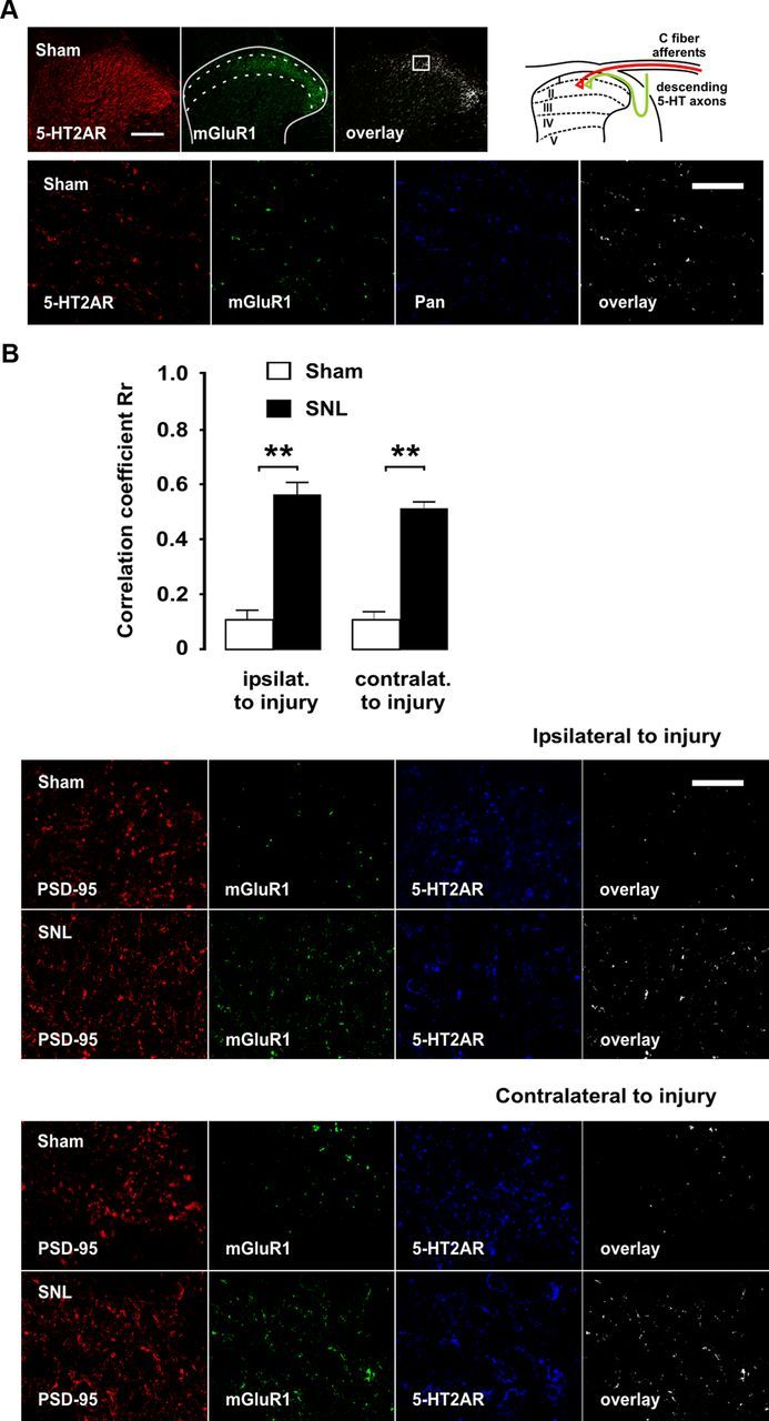
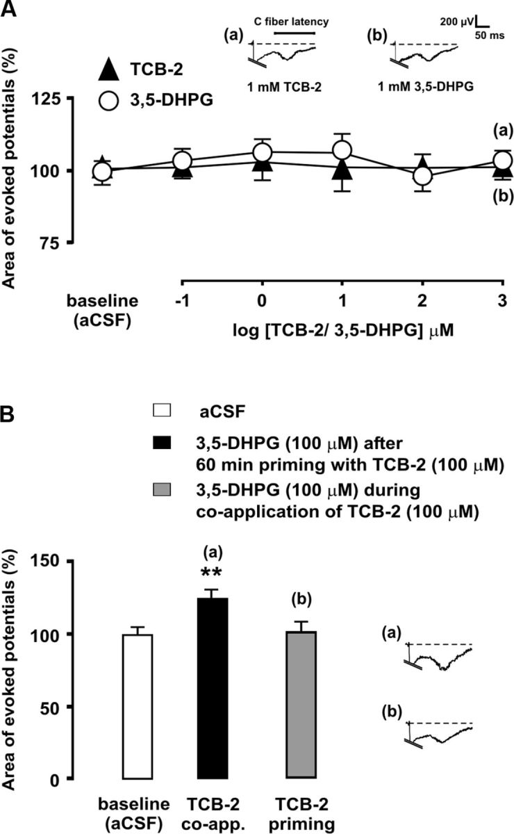
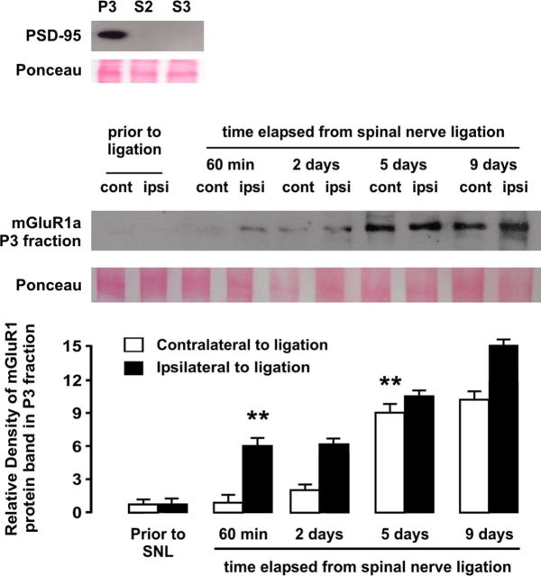
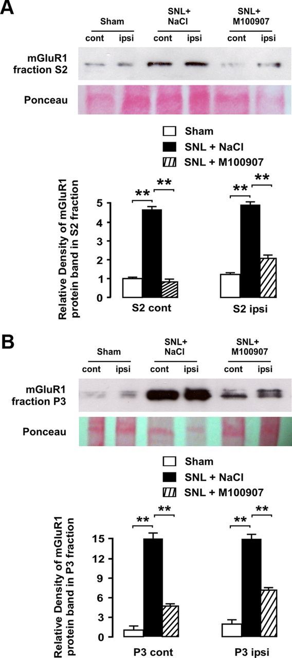
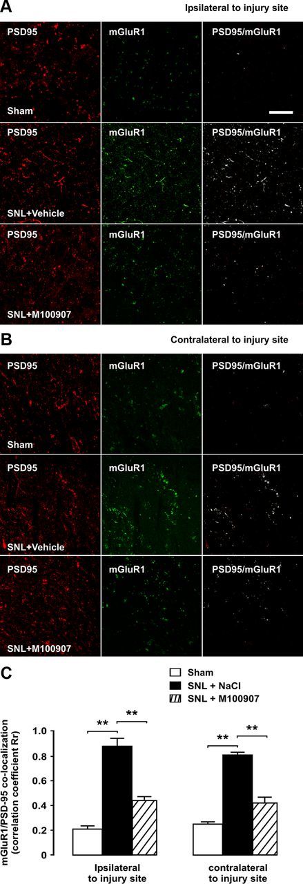
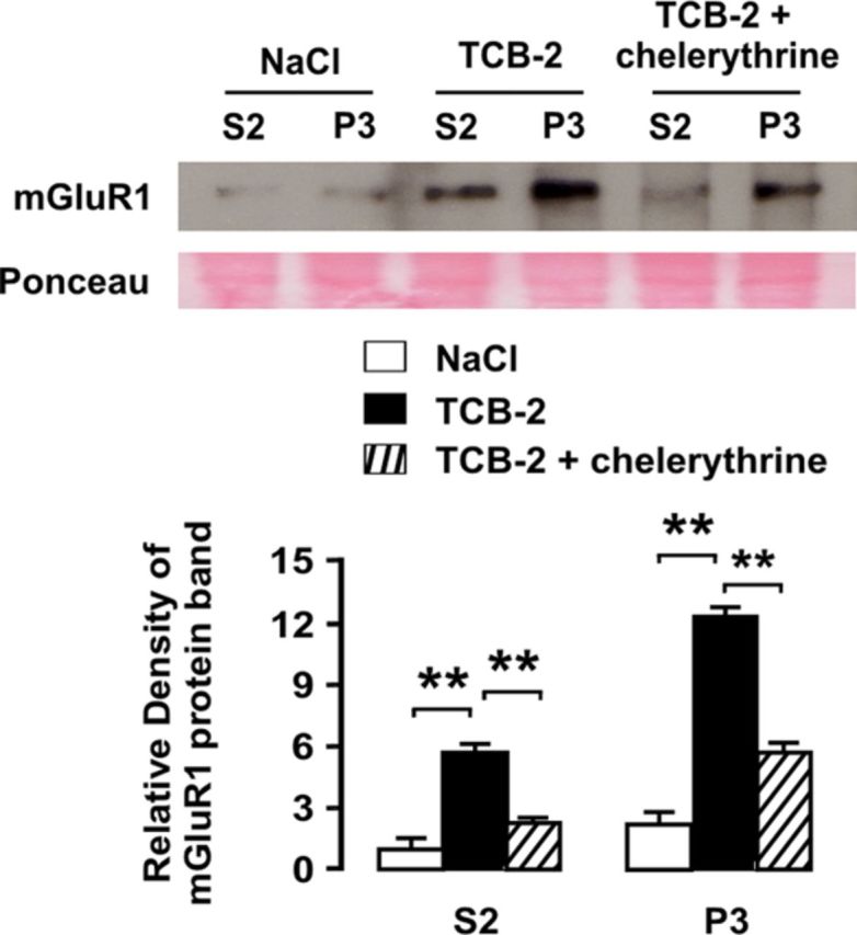
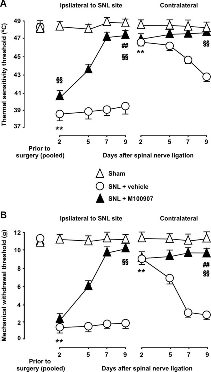
Emerging evidence implicates serotonergic descending facilitatory pathways from the brainstem to the spinal cord in the maintenance of pathologic pain. Upregulation of the serotonin receptor 2A (5-HT(2A)R) in dorsal horn neurons promotes spinal hyperexcitation and impairs spinal μ-opioid mechanisms during neuropathic pain. We investigated the involvement of spinal glutamate receptors, including metabotropic receptors (mGluRs) and NMDA, in 5-HT(2A)R-induced hyperexcitability after spinal nerve ligation (SNL) in rat. High-affinity 5-HT(2A)R agonist (4-bromo-3,6-dimethoxybenzocyclobuten-1-yl)methylamine hydrobromide (TCB-2) enhanced C-fiber-evoked dorsal horn potentials after SNL, which was prevented by mGluR1 antagonist AIDA [(RS)-1-aminoindan-1,5-dicarboxylic acid] but not by group II mGluR antagonist LY 341495 [(2S)-2-amino-2-[(1S,2S)-2-carboxycycloprop-1-yl]-3-(xanth-9-yl)propanoic acid] or NMDA antagonist d-AP5 [D-(-)-2-amino-5-phosphonopentanoic acid]. 5-HT(2A)R and mGluR1 were found to be coexpressed in postsynaptic densities in dorsal horn neurons. In the absence of SNL, pharmacological stimulation of 5-HT(2A)R with TCB-2 both induced rapid bilateral upregulation of mGluR1 expression in cytoplasmic and synaptic fractions of spinal cord homogenates, which was attenuated by PKC inhibitor chelerythrine, and enhanced evoked potentials during costimulation of mGluR1 with 3,5-DHPG [(RS)-3,5-dihydroxyphenylglycine]. SNL was followed by bilateral upregulation of mGluR1 in 5-HT(2A)R-containing postsynaptic densities. Upregulation of mGluR1 in synaptic compartments was partially prevented by chronic administration of selective 5-HT(2A)R antagonist M100907 [(R)-(+)-α-(2,3-dimethoxyphenyl)-1-[2-(4-fluorophenyl)ethyl]-4-pipidinemethanol], confirming 5-HT(2A)R-mediated control of mGluR1 upregulation triggered by SNL. Changes in thermal and mechanical pain thresholds following SNL were increasingly reversed over the days after injury by chronic 5-HT(2A)R blockade. These results emphasize a role for 5-HT(2A)R in hyperexcitation and pain after nerve injury and support mGluR1 upregulation as a novel feedforward activation mechanism contributing to 5-HT(2A)R-mediated facilitation.


Figures

Figure 1.
Figure 1.
Effect of proximal–distal displacement of stimulation electrodes over the sciatic trunk on the peak latencies of C-fiber-evoked field potentials. Shifting of the stimulation electrode pair 10 or 20 mm distally from the reference position located at the most proximal location possible (distant ∼100 mm from the dorsal root entry zone) results in a linear increase in the average peak latencies, confirming that the field potentials considered here to be mediated by C-fibers are actually attributable to this afferent input. A typical location of recording electrodes is indicated by a solid circle in the schematic on the right. Error bars indicate SEM.
Figure 2.
Figure 2.
Intraperitoneally delivered 5-HT2AR antagonist M100907 depresses C-fiber-evoked dorsal horn potentials. The effect of acute intraperitoneal administration of M100907 (0.4 mg/kg) on evoked potentials is shown 9 d after SNL. C-fiber-evoked potentials were decreased significantly 40 min after M100907 delivery, and depression increased with time at least until recordings were discontinued 100 min after administration. The asterisks indicate statistical significance (p < 0.01; n = 5) at Bonferroni test's when comparing mean potential areas to before drug treatment. Error bars indicate SEM.
Figure 3.
Figure 3.
Specificity of the 5-HT2AR H-18 antibody. Confocal scanning fluorescent micrographs of the spinal cord dorsal horn immunolabeled with the goat polyclonal H-18 antibody are shown before (A) and after (B) preabsorption with the immunizing peptide at a 3:1 ratio of peptide to antibody protein. The insets depict higher magnification images taken at the level of the lamina II of the dorsal horn. Dense 5-HT2AR immunostaining in the neuropil was almost abolished by preabsorption of the antibody with the antigen. Double immunofluorescence experiments in HEK293A cells using the mouse monoclonal A-4 (C) and goat polyclonal H-18 (D) antibodies generated against different epitopes of the 5-HT2AR revealed an almost identical distribution of the immunofluorescence signals (E), further supporting the specificity of the goat polyclonal H-18 antibody. Western immunoblot analysis of total lysate (20 μg) using the mouse monoclonal A-4 antibody confirmed the endogenous expression of 5-HT2AR in HEK293 cells (F). Scale bars: B, 100 μm; insets, 10 μm; E, 50 μm.
Figure 4.
Figure 4.
Enhancement of C-fiber-evoked spinal field potentials that is promoted by 5-HT2AR after SNL is mediated by mGluR1, but not by mGluR2/3 or NMDA. Preliminary experiments to determine the effects of pharmacological blockade of mGluR1, mGluR2/3, and the NMDA receptor in nerve-ligated rats are shown in A. Administration of NMDA receptor antagonist d-AP5 at 100 μm significantly decreased mean evoked potential areas, and thus a 10 μm concentration was considered as subclinical for further experiments, whereas 10 and 100 μm concentrations were selected for mGluR1 antagonist AIDA and for group II mGluR antagonist LY 341495, respectively. Mean areas of C-fiber-evoked potentials are shown in B during spinal superfusion with increasing, cumulative concentrations of 5-HT2AR agonist TCB-2 (100 μm) either alone (open triangles) or in combination with subclinical concentrations of AIDA (solid triangles), LY341495 (open circles), or d-AP5 (solid circles). Coadministration of neither LY341495 nor d-AP5 altered the ability of TCB-2 to augment evoked field potentials in nerve-ligated rats. In contrast, coadministration of AIDA increased the minimal effective concentration of TCB-2 by 3 orders of magnitude and dramatically increased the EC50 of TCB-2. Statistical significance (p < 0.01 at Bonferroni's test) of increases in field potential areas by effect of TCB-2 relative to baseline controls in the absence or presence of AIDA is denoted by asterisks or section signs, respectively. Representative recordings shown at the top illustrate the enhancing effect of 100 μm TCB-2 on C-fiber-evoked field potentials in the absence (a) or concurrent presence of 10 μm AIDA (b). Error bars indicate SEM.
Figure 5.
Figure 5.
Enrichment in mGluR1 in 5-HT2AR-containing postsynaptic sites in superficial laminae of the spinal dorsal horn after SNL. Micrographs of double- and triple-immunofluorescence labelings for confocal laser-scanning microscopy of a transverse section of the L5 segment are shown. Lower magnification micrographs in A provide an overview of the dorsal horn, where approximate boundaries of superficial laminae are shown as dashed lines and the region from which high-power micrograph below was taken is indicated by a box. Scale bar, 100 μm. At higher magnification, mGluR1/5-HT2AR colocalization overlay was found to be located in neuropilar elements reminiscent of dendritic process. Scale bar, 3 μm. The neuronal location of 5-HT2AR was confirmed by colocalization with the Pan neuronal marker. The schematic drawing at the top provides a spatial clue as to the convergence of descending serotonergic axons and primary C-fiber afferents in the dorsal horn. In B, coexistence of mGluR1 and 5-HT2AR is shown in postsynaptic densities in dorsal horn neurons ipsilateral and contralateral to injury, as identified by the postsynaptic marker PSD-95, as well as coexpression changes at day 9 after SNL. Scale bar, 3 μm. The asterisks in correlation coefficient graphs indicate statistical significance (p < 0.01) at Fisher's exact test for comparison of Pearson's correlation coefficients. Error bars indicate SEM.
Figure 6.
Figure 6.
Coactivation of 5-HT2AR and mGluR1 augments C-fiber-evoked dorsal horn potentials in sham-operated rats. In A, mean areas of C-fiber-evoked field potentials are shown during spinal superfusion with increasing, cumulative concentrations the mGluR1 agonist 3,5-DHPG (open circles) or the 5-HT2AR agonist TCB-2 (solid triangles). The bar diagram in B shows the magnitudes of evoked potentials during administration of 100 μm 3,5-DHPG, either during simultaneous application of 100 μm TCB-2 or after prior priming with TCB-2, which consisted in administration of 100 μm TCB-2 for 60 min and subsequent washout. The asterisks indicate statistical significance (p < 0.01 at Bonferroni's test) of mean evoked potential area comparisons to baseline (aCSF) controls. Representative recordings illustrate the effects of drug administration on evoked field potentials. Error bars indicate SEM.
Figure 7.
Figure 7.
Western blot analysis of the time course of upregulation of mGluR1 in synaptic fraction (P3) of dorsal horn homogenates in rats subjected to SNL, ipsilateral and contralateral to injury site. Relative density analyses from before injury revealed a significant ipsilateral increase of mGluR1 protein density 1 h after ligation, whereas increase in mGluR1 expression in dorsal horn contralateral to injury became significant at day 5 after ligation. The asterisks indicate statistical significance (p < 0.01) at Student's t test for density comparisons to before injury (total n was 20). As shown at the top, only the synaptic fraction but not cytoplasmic fraction S2 or crude synaptic vesicle fraction S3, was enriched in postsynaptic density protein PSD-95. Ponceau staining was used to confirm equal protein loading. Error bars indicate SEM.
Figure 8.
Figure 8.
Chronic blockade of 5-HT2AR attenuates SNL-induced upregulation of mGluR1 in dorsal horn neurons. Western blot analyses are shown illustrating significant bilateral increases in mGluR1 protein in S2 (A) and P3 fractions (B) from dorsal horn homogenates ipsilateral and contralateral to injury site after SNL. Daily intraperitoneal injections of 5-HT2AR antagonist M100907 (0.4 mg/kg) prevented and attenuated in mGluR1 upregulation in S2 fraction contralateral and ipsilateral to injury site, respectively. In synaptosomal (P3) fraction, mGluR1 upregulation was bilaterally and markedly attenuated by chronic M100907 treatment. As revealed by relative density analyses normalized to contralateral side in sham-operated rats, chronic 5-HT2AR blockade significantly attenuated SNL-induced upregulation of mGluR1 in both subcellular fractions and both sides of the body (asterisks indicate significance at p < 0.01 at Student's t test; total n was 12). Error bars indicate SEM.
Figure 9.
Figure 9.
Bilateral upregulation of mGluR1 in postsynaptic densities after SNL is dependent on 5-HT2AR activation. Micrographs of double-immunofluorescence labelings for mGluR1 and the postsynaptic density marker PSD-95 are shown of a transverse section of L5. All pictures were taken from superficial layers of the dorsal horn as indicated in Figure 2. Scale bar, 5 μm. Coexpression of mGluR1 and PSD-95, as shown by white overlay and confirmed by correlation coefficient analyses, is increased after SNL both ipsilaterally (A) and contralaterally (B) to injury, indicating increased presence of mGluR1 in postsynaptic densities. Chronic blockade of 5-HT2AR by daily injections of antagonist M100907 significantly and bilaterally reduced SNL-induced upregulation of mGluR1. The asterisks in correlation coefficient graphs (C) indicate statistical significance (p < 0.01) at Fisher's exact test for comparison of Pearson's correlation coefficients. Error bars indicate SEM.
Figure 10.
Figure 10.
Upregulation of mGluR1 is induced by 5-HT2AR via activation of PKC. As shown by Western blot analyses, 5-HT2AR stimulation by spinal superfusion with agonist TCB-2 can upregulate mGluR1 protein in the spinal dorsal horn both in S2 and P3 fractions. For quantification, Western blot bands were normalized to S2 fraction band density in the control condition (superfusion with NaCl). Coadministration of PKC inhibitor chelerythrine markedly reduced TCB-2-induced upregulation of mGluR1. The asterisks indicate statistical significance at p < 0.01 at Student's t test. Ponceau staining was used to confirm equal protein loading. Error bars indicate SEM.
Figure 11.
Figure 11.
5-HT2AR mediates late-onset thermal and mechanical allodynia after SNL. Heat pain thresholds assessed in the hot-plate test (A), as well as the 50% paw withdrawal threshold to a static mechanical stimulus with von Frey monofilaments (B) are shown over multiple time points after SNL. After ligation of L5, both thermal and mechanical threshold were significantly lowered both ipsilaterally and contralaterally to injury site in vehicle-treated animals. The latter were mild and evolved over a few days after surgery, whereas profound ipsilateral changes were already present at day 2 after surgery and lasted for the entire evaluation period (9 d). No changes in mechanical and thermal thresholds were found in sham-operated animals. The asterisks indicate statistical significance at p < 0.01 at the nonparametric Kruskal–Wallis one-way ANOVA and Mann–Whitney rank tests when comparing mean threshold values to before surgery in vehicle-treated animals (for the sake of clarity, only the earliest time point demonstrating significant changes is labeled). Chronic 5-HT2AR blockade with M100907 progressively decreased SNL-induced mechanical and thermal allodynia over the days following injury. Antiallodynic effects of M100907 were conspicuous from day 5 onward and mechanical and thermal thresholds were reverted by M100907 to near-basal levels by day 9 after injury. The section signs and pound signs denote statistical significance (same α and statistical test as above) when comparing mean thresholds values in M100907-treated rats to values in vehicle-treated rats and to values in sham-operated rats, respectively (for clarity, only data from day 9 after injury are labeled for statistical significance). Error bars indicate SEM.

References

    1. Aira Z, Buesa I, Salgueiro M, Bilbao J, Aguilera L, Zimmermann M, Azkue JJ. Subtype-specific changes in 5-HT receptor-mediated modulation of C fibre-evoked spinal field potentials are triggered by peripheral nerve injury. Neuroscience. 2010;168:831–841. - PubMed
    1. Aira Z, Buesa I, García del Caño G, Salgueiro M, Mendiable N, Mingo J, Aguilera L, Bilbao J, Azkue JJ. Selective impairment of spinal mu opioid receptor-mechanism by plasticity of serotonergic facilitation mediated by 5-HT2A and 5-HT2B receptors. Pain. 2012;153:1418–1425. - PubMed
    1. Aldridge GM, Podrebarac DM, Greenough WT, Weiler IJ. The use of total protein stains as loading controls: an alternative to high-abundance single-protein controls in semi-quantitative immunoblotting. J Neurosci Methods. 2008;172:250–254. - PMC - PubMed
    1. Almási R, Pethö G, Bölcskei K, Szolcsányi J. Effect of resiniferatoxin on the noxious heat threshold temperature in the rat: a novel heat allodynia model sensitive to analgesics. Br J Pharmacol. 2003;139:49–58. - PMC - PubMed
    1. Alvarez FJ, Kavookjian AM, Light AR. Synaptic interactions between GABA-immunoreactive profiles and the terminals of functionally defined myelinated nociceptors in the monkey and cat spinal cord. J Neurosci. 1992;12:2901–2917. - PMC - PubMed

Publication types

MeSH terms

Substances