. 2013 May;101(5):1255-64.
doi: 10.1002/jbm.a.34420. Epub 2012 Sep 27.

3D bioprinting of heterogeneous aortic valve conduits with alginate/gelatin hydrogels

Affiliations

3D bioprinting of heterogeneous aortic valve conduits with alginate/gelatin hydrogels

Bin Duan et al. J Biomed Mater Res A. 2013 May.

Abstract

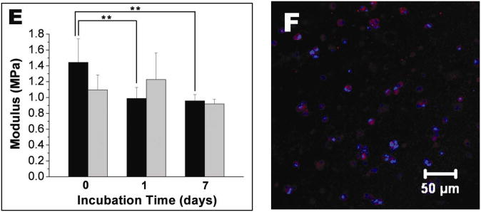
Heart valve disease is a serious and growing public health problem for which prosthetic replacement is most commonly indicated. Current prosthetic devices are inadequate for younger adults and growing children. Tissue engineered living aortic valve conduits have potential for remodeling, regeneration, and growth, but fabricating natural anatomical complexity with cellular heterogeneity remain challenging. In the current study, we implement 3D bioprinting to fabricate living alginate/gelatin hydrogel valve conduits with anatomical architecture and direct incorporation of dual cell types in a regionally constrained manner. Encapsulated aortic root sinus smooth muscle cells (SMC) and aortic valve leaflet interstitial cells (VIC) were viable within alginate/gelatin hydrogel discs over 7 days in culture. Acellular 3D printed hydrogels exhibited reduced modulus, ultimate strength, and peak strain reducing slightly over 7-day culture, while the tensile biomechanics of cell-laden hydrogels were maintained. Aortic valve conduits were successfully bioprinted with direct encapsulation of SMC in the valve root and VIC in the leaflets. Both cell types were viable (81.4 ± 3.4% for SMC and 83.2 ± 4.0% for VIC) within 3D printed tissues. Encapsulated SMC expressed elevated alpha-smooth muscle actin, while VIC expressed elevated vimentin. These results demonstrate that anatomically complex, heterogeneously encapsulated aortic valve hydrogel conduits can be fabricated with 3D bioprinting.


Figures

Figure 1
Figure 1
Live/Dead assay for encapsulated VIC within alginate/gelatin hydrogel discs. (A) 1 day; (B) 7 days; (C) cell viability measured based on Live/Dead images; (D) average cell area measured based on Live/Dead images. (n=6, *p <0.05)
Figure 2
Figure 2
Live/Dead assay for encapsulated SMC within alginate/gelatin hydrogel discs. (A) 1 day; (B) 7 days; (C) cell viability measured based on Live/Dead images; (D) average cell area measured based on Live/Dead images. (n=6, *p <0.05)
Figure 3
Figure 3
Fluorescent staining for encapsulated VIC and SMC after 7 day culture. (A, C) Alexa Fluor 488 conjugated phalloidin staining for F-actin (green) and Draq 5 counterstaining for cell nuclei (blue); (B, D) αSMA and vimentin staining intensity for encapsulated VIC and SMC within hydrogel discs. (inset: representative image of immunohistochemical staining for αSMA (green) and vimentin (red), and Draq 5 counterstaining for cell nuclei (blue), n=6, **p<0.01). (A, B) Encapsulated VIC; (C, D) encapsulated SMC.
Figure 4
Figure 4
Hydrogel tensile biomechanics. (A) Stress-strain curves of alginate/gelatin samples after incubation in culture medium (inset: as-printed alginate/gelatin hydrogel sample for tensile test); (B) Typical stress-strain curves of alginate/gelatin samples encapsulated with VIC; (C) change of ultimate strength with incubation time; (D) change of failure strain with incubation time (black column: cell-free hydrogel samples; gray column: cell laden hydrogel samples); (E) change of modulus with incubation time; (black column: cell-free hydrogel samples; gray column: cell laden hydrogel samples; n=4-6 for cell-free hydrogel samples, n=3 for cell-laden hydrogel samples; *p<0.05, **p<0.01) (F) immunohistochemical staining of VIC encapsulated dumbbell shaped hydrogel sample for Col1A2 (red) and Draq 5 counterstaining for cell nuclei after 7 day culture (blue).
Figure 4
Figure 4
Hydrogel tensile biomechanics. (A) Stress-strain curves of alginate/gelatin samples after incubation in culture medium (inset: as-printed alginate/gelatin hydrogel sample for tensile test); (B) Typical stress-strain curves of alginate/gelatin samples encapsulated with VIC; (C) change of ultimate strength with incubation time; (D) change of failure strain with incubation time (black column: cell-free hydrogel samples; gray column: cell laden hydrogel samples); (E) change of modulus with incubation time; (black column: cell-free hydrogel samples; gray column: cell laden hydrogel samples; n=4-6 for cell-free hydrogel samples, n=3 for cell-laden hydrogel samples; *p<0.05, **p<0.01) (F) immunohistochemical staining of VIC encapsulated dumbbell shaped hydrogel sample for Col1A2 (red) and Draq 5 counterstaining for cell nuclei after 7 day culture (blue).
Figure 5
Figure 5
Bioprinting of 3D grid pattern structure. (A) Schematic illustration of the bioprinting process with single cell type and single syringe; (B) bioprinted alginate/gelatin hydrogel structure after ionic crosslinking; (C) fluorescent image of printed 2D structure with encapsulation of cell tracker red labeled VIC; (D) close view of (C); (E) Live/Dead staining of VIC within bioprinted hydrogel after 7 days culture (the dashed line indicates the pore area).
Figure 6
Figure 6
Bioprinting of aortic valve conduit. (A) Aortic valve model reconstructed from micro-CT images. The root and leaflet regions were identified with intensity thresholds and rendered separately into 3D geometries into STL format (green color indicates valve root and red color indicates valve leaflets); (B, C) schematic illustration of the bioprinting process with dual cell types and dual syringes; (B) root region of first layer generated by hydrogel with SMC; (C) leaflet region of first layer generated by hydrogel with VIC; (D) fluorescent image of first printed two layers of aortic valve conduit; SMC for valve root were labeled by cell tracker green and VIC for valve leaflet were labeled by cell tracker red. (E) as-printed aortic valve conduit.
Figure 7
Figure 7
Fluorescent staining for bioprinted aortic valve conduit with encapsulation of VIC and SMC after 7 day culture. (A, C) Live/Dead assay for encapsulated VIC in the leaflet and SMC in valve root after 7 day culture; (B, D) αSMA and vimentin staining intensity for encapsulated VIC and SMC within the bioprinted conduit. (inset: representative image of immunohistochemical staining for αSMA (green) and vimentin (red), and Draq 5 counterstaining for cell nuclei (blue), n=6, **p<0.01). (A, B); staining for VIC in the leaflet; (C, D) staining for SMC in the root.

References

    1. Butcher JT, Mahler GJ, Hockaday LA. Aortic valve disease and treatment: The need for naturally engineered solutions. Adv Drug Deliv Rev. 2011;63:242–268. - PubMed
    1. Hinton RB, Yutzey KE. Heart Valve Structure and Function in Development and Disease. Annu Rev Physiol. 2011;73:29–46. - PMC - PubMed
    1. Filova E, Straka F, Mirejovsky T, Masin J, Bacakova L. Tissue-engineered heart valves. Physiol Res. 2009;58:S141–S158. - PubMed
    1. Vesely I. Heart valve tissue engineering. Circ Res. 2005;97:743–755. - PubMed
    1. Brown JW, Ruzmetov M, Rodefeld MD, Mahomed Y, Turrentine MW. Incidence of and risk factors for pulmonary autograft dilation after ross aortic valve replacement. Ann Thorac Surg. 2007;83:1781–1789. - PubMed

Publication types

LinkOut - more resources